Mastitis or breast cancer
High yield
Red, swollen breast: fever and lactation favour mastitis; no fever, peau d'orange and antibiotic failure mean inflammatory cancer.
Surgery · Breast surgerySurgery · Surgical oncologyExam Prep · State exam
1. Definition
- Mastitis → inflammation of the breast tissue, infective or non-infective; abscess → localised collection of pus
- Inflammatory breast cancer (IBC) → aggressive breast cancer with tumour cells blocking the dermal lymphatics; red, oedematous breast, often with no discrete mass
- The red breast → common presentation of both; cancer and inflammatory cancer can mimic infection
2. Why the difference matters
- Mastitis → treated in primary care or the emergency department with antibiotics
- IBC → rare, 1–5% of breast cancers, but the most aggressive subtype
- IBC treated as infection → weeks of antibiotics, delayed diagnosis, higher stage
- Neglected breast cancer → secondary inflammation and ulceration; looks infected, but is not classed as IBC
- Cancer can also sit under a true abscess → residual mass after drainage
3. Who gets it
Mastitis
- Lactational → breastfeeding women, mostly in the first 3 months; Staphylococcus aureus
- Periductal → younger women who smoke; mixed aerobic and anaerobic flora
- Duct ectasia → peri- and postmenopausal women
- Granulomatous → young parous women, oral contraceptive use
Breast cancer
- Lifetime risk → about 1 in 9 women in the UK; 1% of cases in men
- Risk rises with age; most over 50 years
- IBC → any age, including young women; any histology or receptor status
- Breast cancer in pregnancy and lactation → rare, presents late because changes are blamed on lactation
4. Clinical picture
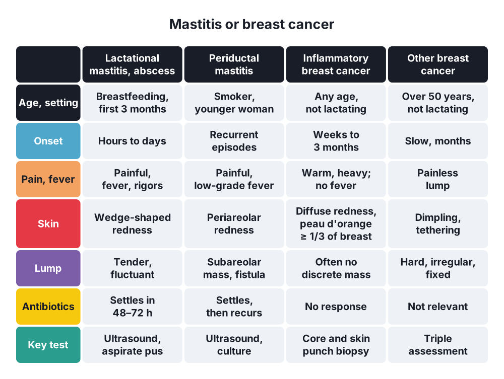
Features that favour mastitis or abscess
- Lactating, cracked nipple, milk stasis
- Fever, rigors, malaise and raised white cell count
- Rapid onset over hours to days
- Wedge-shaped, tender, hot area
- Fluctuant tender mass → abscess
- Subareolar mass, seropurulent discharge, fistula at the areolar margin → periductal mastitis in a smoker
- Responds to antibiotics in 48–72 h
Features that favour inflammatory breast cancer
- Not lactating, no fever, white cell count normal
- Onset over weeks to 3 months
- Diffuse redness, warmth and swelling; erythema over a third of the breast or more
- Peau d'orange → skin oedema with dimpling at the hair follicles
- Breast feels heavy, firm, enlarged; pain may be mild
- Often no discrete lump
- Nipple flattening, inversion or crusting
- Hard, fixed axillary or supraclavicular nodes
- No response to antibiotics
Features of other breast cancer
- Painless, hard, irregular lump; tethered or fixed
- Skin dimpling, ulceration
- Nipple → Paget's disease (eczema-like change starting on the nipple), new inversion, bloody discharge
- Systemic → weight loss, bone pain, breathlessness
5. Signs that overlap
- Peau d'orange → seen in both IBC and acute mastitis
- Redness and warmth → both
- Tender axillary nodes → mastitis; hard, fixed nodes → cancer
- Nipple retraction → cancer, or slit-like retraction in periductal mastitis
- Lump → abscess (fluctuant, tender) or cancer (hard, irregular)
6. Investigations
Mastitis
- Full blood count, C-reactive protein
- Culture of milk or pus; MRSA (methicillin-resistant S. aureus) screen
- Ultrasound → abscess, size, loculation; guides aspiration
- Avoid mammography in the acute painful phase
Suspected cancer
- Triple assessment → clinical examination + imaging + core biopsy; accuracy about 99.6%
- Mammography and ultrasound → mass, calcifications, skin thickening; IBC may show only skin thickening
- Core biopsy → histology and oestrogen receptor, progesterone receptor and HER2 (human epidermal growth factor receptor 2) status
- Skin punch biopsy → tumour cells in the dermal lymphatics; a negative biopsy does not exclude IBC
- Ultrasound and biopsy of the axillary nodes
- IBC → staging with CT of chest, abdomen and pelvis and a bone scan, or PET-CT; IBC is stage III (T4d) at least
- MRI → breast extent when imaging is unclear
7. Management pathway
Mastitis
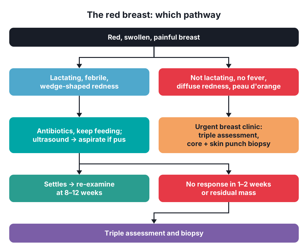
- Lactational → flucloxacillin, keep breastfeeding or expressing, analgesia
- Non-lactational → co-amoxiclav, or a macrolide plus metronidazole
- Abscess → ultrasound-guided aspiration, repeated until dry; incision and drainage if aspiration fails or the skin is thin
- No improvement in 48 h → ultrasound for abscess
When to suspect cancer
- No response to 1–2 courses of antibiotics
- Not lactating and no fever
- Residual mass after the infection settles
- Woman over 35–40 years with a first non-lactational "infection"
- Recurrent infection at the same site
- Hard, fixed axillary nodes
Cancer
- Urgent referral to a breast clinic for triple assessment
- IBC → trimodal treatment:
- Neoadjuvant chemotherapy (plus anti-HER2 therapy if HER2 positive)
- Modified radical mastectomy with axillary node clearance, no immediate reconstruction
- Post-mastectomy radiotherapy to the chest wall and nodes
- IBC → sentinel node biopsy not used (high false-negative rate); breast conservation not offered
- Endocrine therapy → oestrogen receptor positive tumours
Follow-up rule
- Re-examine every treated breast infection at 8–12 weeks
- Residual mass → triple assessment
8. Pitfalls
- Two courses of antibiotics in a non-lactating woman without a review → missed IBC
- Negative skin biopsy → IBC still possible; diagnosis is clinical
- Normal mammogram → does not exclude IBC
- Abscess in an older woman → biopsy the wall or the residual mass
- Lactating woman with a hard mass that does not settle → biopsy; pregnancy does not protect
Show as a list
Mastitis or breast cancer
- Mastitis
- Lactating
- Fever, rigors
- Wedge redness
- Fluctuant mass
- Settles in 48–72 h
- Inflammatory cancer
- No fever
- Peau d'orange
- No discrete lump
- Weeks to months
- No antibiotic response
- Overlap
- Redness
- Peau d'orange
- Nipple retraction
- Tests
- Ultrasound
- Triple assessment
- Skin punch biopsy
- Management
- Antibiotics, aspiration
- Review at 8–12 weeks
- Neoadjuvant chemotherapy
- Mastectomy, radiotherapy
Source
- Oxford Handbook of Clinical Surgery (McLatchie, Borley, Chikwe) → chapter 6, Breast and endocrine surgery
- Sheehan L, Skene A. Benign breast disorders → Surgery (Oxford) 2024; 42(12)
- Sabiston Textbook of Surgery, 22nd edition → chapter 68, Diseases of the Breast