
36. Breast carcinoma
High yield
Most common cancer in women; triple assessment, TNM and receptor status guide surgery, radiotherapy, endocrine, anti-HER2 and chemotherapy.
Exam Prep · State examSurgery · Breast surgerySurgery · Surgical oncology
1. Definition
- Breast carcinoma → malignant epithelial tumour of the terminal duct lobular unit (TDLU) of the breast
- In situ carcinoma → malignant cells confined by an intact basement membrane: ductal (DCIS) or lobular (LCIS)
- Invasive carcinoma → cells breach the basement membrane and infiltrate the stroma
2. Epidemiology
- Most common cancer in women in the UK and worldwide; 2.3 million new cases worldwide in 2020
- Lifetime risk → 1 in 7 to 1 in 9 women in the UK; about 55 000 women and 370 men a year
- Men → about 1% of cases
- Almost half diagnosed over 65 years; about three-quarters of women survive 10 years or more
- Presentation → about two-thirds symptomatic, one-third through screening
3. Aetiology and risk factors
Non-modifiable
- Female sex, increasing age
- Family history → one first-degree relative diagnosed at ≤ 50 years doubles the risk
- Genetic → about 5% of cancers:
- BRCA1 → 60–80% lifetime breast cancer risk, 40–50% ovarian
- BRCA2 → 40–85% breast, 10–25% ovarian; male breast, prostate and pancreatic cancer
- TP53 (Li-Fraumeni), PTEN (Cowden), CDH1, PALB2, CHEK2
- Under 30 years with breast cancer → 25% chance of a genetic cause
- Early menarche, late menopause → long oestrogen exposure
- Dense breasts
- Previous breast cancer, atypical hyperplasia (risk × 4), LCIS
- Chest radiotherapy in childhood or youth (Hodgkin's lymphoma)
Modifiable
- Nulliparity, first birth after 35 years
- Breastfeeding → risk falls 4% for every 12 months
- Combined hormone replacement therapy (HRT), oral contraceptive pill
- Postmenopausal obesity → aromatisation of androgens in fat
- Alcohol, physical inactivity
4. Pathogenesis
- Normal TDLU epithelium
- Hyperplasia → atypical hyperplasia
- Carcinoma in situ (DCIS)
- Invasive carcinoma
- Spread:
- Local → skin, nipple, pectoral fascia, chest wall
- Lymphatic → axillary nodes (levels I–III), internal mammary, supraclavicular
- Blood → bone (most common), lung, liver, brain
- Skin dimpling → tumour shortens Cooper's ligaments
- Peau d'orange → blocked dermal lymphatics, skin oedema
5. Classification
Histological
- In situ:
- DCIS → 80% found on screening as microcalcifications; 10–50% become invasive if untreated; graded low, intermediate, high
- LCIS → classical LCIS is a risk marker (risk × 7–8); pleomorphic LCIS treated as DCIS
- Invasive:
- No special type (NST, formerly ductal) → 75–80%
- Invasive lobular → 15%; multifocal, bilateral, poorly seen on mammography
- Good prognosis special types → tubular, mucinous, cribriform
- Poor prognosis → metaplastic, micropapillary
- Special presentations → Paget's disease of the nipple, inflammatory carcinoma
Grade
- Nottingham grade → gland formation + nuclear pleomorphism + mitoses, each scored 1–3
- Grade 1 → total 3–5
- Grade 2 → total 6–7
- Grade 3 → total 8–9
Molecular (receptor) subtypes
- Luminal A → oestrogen receptor (ER) positive, low Ki-67; about 40%; best prognosis
- Luminal B → ER positive, high Ki-67, HER2 normal or amplified
- HER2-enriched → human epidermal growth factor receptor 2 overexpressed in 15–20%; poorer outcome without anti-HER2 therapy
- Triple negative → ER, progesterone receptor (PR) and HER2 negative; basal-like; BRCA1; worst prognosis
TNM staging (key points)
- T1 ≤ 2 cm, T2 > 2–5 cm, T3 > 5 cm
- T4 → chest wall (T4a), skin ulceration, satellite nodules or peau d'orange (T4b), both (T4c), inflammatory carcinoma (T4d)
- N1 → mobile ipsilateral axillary nodes; N2 → fixed or matted axillary nodes, or internal mammary; N3 → infraclavicular or supraclavicular nodes
- M1 → distant metastases
- Stage I-II → early breast cancer; stage III → locally advanced; stage IV → metastatic
6. Clinical picture
Symptoms
- Breast lump → most common; painless unless inflammatory carcinoma
- Nipple change → retraction, inversion, eczema-like rash (Paget's disease)
- Bloody nipple discharge from one duct
- Change in breast shape or contour
- Axillary lump
- Metastatic → bone pain, breathlessness, jaundice, headache, weight loss
Signs
- Hard, irregular, ill-defined lump
- Immobile, tethered to skin or fixed to chest wall
- Skin → dimpling, puckering, peau d'orange, ulceration, fungation, satellite nodules
- Nipple → destroyed, inverted, deviated
- Axillary or supraclavicular nodes → hard, matted or fixed
- Inspection with arms raised and pectorals tensed → shows subtle dimpling
7. Diagnosis
Triple assessment
- Every lump or area of localised nodularity; each part scored 1–5 (P, M or U, B or C):
- Normal (or inadequate sample)
- Benign
- Indeterminate
- Suspicious, probably malignant
- Malignant
- Discordance between the three parts → further assessment
Imaging
- Mammography → women over 40 years; masses, distortion, microcalcifications
- Ultrasound → first choice under 40 years; all ages for a focal area; routine assessment of the axilla
- MRI → lobular carcinoma before breast conservation, dense breasts, high-risk screening, implants, discordant results
- Scoring → BI-RADS (Breast Imaging Reporting and Data System)
Pathology
- Core biopsy (14G, 3–6 cores) → invasive versus in situ, grade, ER, PR, HER2
- Vacuum-assisted biopsy → calcifications, B3 lesions
- Punch biopsy of skin → Paget's disease, inflammatory carcinoma
- Axillary node → ultrasound-guided core biopsy or fine-needle aspiration (FNA)
- Marker clip placed at the biopsy site
Staging
- Early, node-negative → staging scans not routine
- Node-positive, locally advanced or symptoms → CT chest, abdomen and pelvis + bone scan
- Bloods → liver function tests, calcium
Screening
- NHS Breast Screening Programme → women 50–70 years, two-view mammography every 3 years
- Per 1000 screened → about 5 lives saved, 17 overdiagnosed
- High risk → annual MRI from a younger age
8. Differential diagnosis
- Fibroadenoma, phyllodes tumour
- Cyst
- Fat necrosis → after trauma; hard, tethered, calcified
- Abscess, periductal mastitis, granulomatous mastitis
- Sclerosing adenosis, radial scar
- Duct ectasia → nipple retraction
- Eczema of the nipple → starts on the areola; Paget's starts on the nipple
9. Treatment
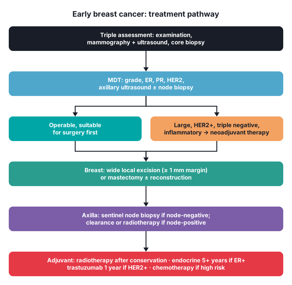
- Planned at a multidisciplinary team (MDT) meeting
Surgical: breast
- Breast-conserving surgery (wide local excision) → most cancers:
- Margin → ≥ 1 mm clear on histology
- Re-excision rate → about 20%
- Always with whole-breast radiotherapy → survival equal to mastectomy
- Impalpable lesion → wire, magnetic seed or radiofrequency tag localisation; specimen X-ray
- Oncoplastic techniques → volume displacement or replacement
- Mastectomy:
- Large tumour in a small breast, multifocal disease, central tumour, extensive DCIS
- Previous chest radiotherapy
- Patient preference
- Reconstruction → immediate or delayed: implant, latissimus dorsi flap, deep inferior epigastric perforator (DIEP) flap
Surgical: axilla
- Clinically and ultrasound node-negative → sentinel lymph node biopsy (SLNB):
- Radioisotope + Patent Blue dye (anaphylaxis 0.1%)
- Less lymphoedema than clearance
- Biopsy-proven positive nodes → axillary node clearance (levels I–III) or axillary radiotherapy, per MDT
- Positive sentinel node → clearance, radiotherapy or no further treatment, depending on disease burden
- DCIS → no axillary surgery; SLNB only with mastectomy, mass-forming DCIS, microinvasion or DCIS > 4 cm
Conservative: radiotherapy and systemic therapy
- Radiotherapy:
- After breast conservation → 26 Gy in 5 fractions over 1 week (FAST-Forward)
- After mastectomy → T3–T4 or node-positive disease
- Nodal fields → supraclavicular fossa, axilla
- Omission → selected women ≥ 65 years with small, node-negative, ER-positive, low-grade tumours on endocrine therapy
- Endocrine therapy → ER-positive tumours, at least 5 years:
- Premenopausal → tamoxifen (risk of endometrial cancer, venous thromboembolism)
- Postmenopausal → aromatase inhibitor (anastrozole, letrozole, exemestane); bone loss
- High-risk node-positive → add abemaciclib (CDK4/6 inhibitor)
- Anti-HER2 therapy → trastuzumab ± pertuzumab for 1 year; cardiotoxicity, monitor with echocardiography
- Chemotherapy → anthracycline and taxane; high grade, node-positive, HER2-positive, triple negative, young patients
- Neoadjuvant therapy → before surgery to downstage; HER2-positive, triple-negative, locally advanced, inflammatory
- Prognostic tools → Nottingham Prognostic Index (NPI) = 0.2 × size (cm) + grade (1–3) + node score (1–3); Oncotype DX for ER-positive, node-negative cancers with NPI ≥ 3.4; PREDICT
- Germline BRCA, high risk → olaparib
Special situations
- Inflammatory carcinoma → neoadjuvant chemotherapy, modified radical mastectomy, post-mastectomy radiotherapy
- Paget's disease → underlying carcinoma in 82–94%; central excision of nipple-areola or mastectomy
- Elderly, unfit, ER-positive → primary endocrine therapy
- Pregnancy → surgery in any trimester; chemotherapy from the 2nd trimester; no radiotherapy or tamoxifen in pregnancy
- Metastatic → palliative endocrine, chemotherapy, anti-HER2; radiotherapy for bone pain; toilet mastectomy for a fungating tumour
10. Complications
Of surgery
- Seroma, haematoma, wound infection
- Positive margins → re-excision
- Lymphoedema → mainly after axillary clearance or axillary radiotherapy
- Injury to the long thoracic nerve (winged scapula), thoracodorsal nerve, intercostobrachial nerve (numb inner arm)
- Flap necrosis, implant complications
Of disease and treatment
- Local and regional recurrence
- Metastases → spinal cord compression, hypercalcaemia, pathological fracture, malignant pleural effusion
- Radiotherapy → skin reaction, fibrosis, cardiac and lung damage
- Chemotherapy → neutropenic sepsis, cardiotoxicity (anthracyclines, trastuzumab), early menopause
11. Prognosis
- Prognostic factors → axillary node status (strongest), tumour size, grade, receptor status, lymphovascular invasion
- 5-year survival → stage I about 99%, stage IV about 25–30%
- NPI → < 3.4 good, 3.4–5.4 moderate, > 5.4 poor
Show as a list
Breast carcinoma
- Risk factors
- Age
- BRCA1, BRCA2
- Oestrogen exposure
- Combined HRT
- Pathology
- DCIS, LCIS
- No special type
- Lobular
- Nottingham grade
- Receptors
- Luminal A, B
- HER2-enriched
- Triple negative
- Diagnosis
- Triple assessment
- Mammography
- Core biopsy
- Staging CT, bone scan
- Treatment
- Wide local excision
- Mastectomy
- Sentinel node biopsy
- Radiotherapy
- Endocrine therapy
- Trastuzumab
- Chemotherapy
- Prognosis
- Node status
- NPI


