
35. Benign breast neoplasms and precancerous conditions
High yield
Fibroadenoma, cysts, intraductal papilloma and phyllodes tumour; atypical hyperplasia, LCIS and DCIS raise cancer risk.
Exam Prep · State examSurgery · Breast surgerySurgery · Surgical oncology
1. Definition
- Benign breast disease → non-malignant lesions of the breast: aberrations of normal development, benign tumours, inflammation
- Precancerous conditions → lesions that mark or carry an increased risk of invasive breast cancer
- Triple assessment → clinical examination + imaging + needle biopsy; applied to every breast lump; accuracy about 99.6%
2. Aetiology and risk factors
- Oestrogen and progesterone → cyclical changes, fibroadenoma growth, cysts
- Age → each lesion has a typical age
- Hormone replacement therapy (HRT) → new cysts after the menopause
- Family history, genetic mutations → precancerous lesions
3. Pathogenesis
- Aberrations of normal development and involution (ANDI):
- Development (15–25 years) → fibroadenoma, juvenile hypertrophy
- Cyclical change (25–40 years) → cyclical mastalgia, nodularity (fibrocystic change)
- Involution (35–55 years) → cysts, duct ectasia, sclerosing adenosis
- Precancer → epithelial proliferation → atypia → carcinoma in situ → invasive cancer (not every lesion progresses)
4. Classification
Benign breast lesions
- Fibroadenoma
- Breast cyst
- Fibrocystic change
- Intraductal papilloma
- Phyllodes tumour → benign, borderline or malignant
- Hamartoma (fibroadenolipoma), adenoma, lipoma
- Fat necrosis, galactocele
Cancer risk of benign lesions
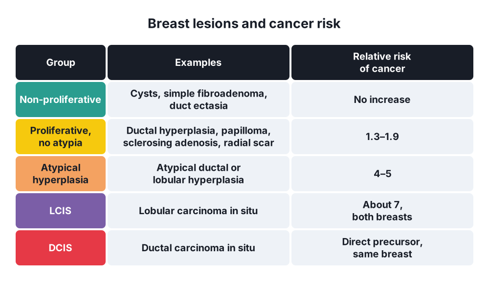
- Non-proliferative (cysts, simple fibroadenoma, duct ectasia) → little or no increased risk (relative risk about 1.3)
- Proliferative without atypia (usual ductal hyperplasia, sclerosing adenosis, papilloma, radial scar) → relative risk 1.3–1.9; radial scar 1.8–3.0, excise
- Proliferative with atypia (atypical ductal or lobular hyperplasia) → relative risk 4–5
- Lobular carcinoma in situ (LCIS) → relative risk about 7
- Ductal carcinoma in situ (DCIS) → non-invasive cancer; direct precursor
5. Benign neoplasms
A. Fibroadenoma
- Most common tumour under 30 years; about 10% of women
- Stromal and epithelial elements, encapsulated
- Firm, smooth or lobulated, painless, highly mobile ("breast mouse")
- Ultrasound → well defined, oval, hypoechoic
- Natural history → 50% resolve, 25% stay the same, 25% grow
- Complex fibroadenoma → relative risk about 3
- Malignant change → rare, about 0.2%
- Treatment:
- Core biopsy if over 25 years
- Confirmed on core biopsy, < 3 cm → reassure, no excision needed
- Excision → > 3 cm, rapid growth, suspected phyllodes, patient preference
- Enlarging fibroadenoma → phyllodes found in about 2.4%
- Giant (> 5 cm) and juvenile fibroadenoma → excision
B. Breast cyst
- Women over 35 years to menopause; at least 1 in 14 women
- Smooth, well-defined, may be tender; multiple or recurrent in 50%
- Ultrasound → anechoic with posterior enhancement
- Simple cyst → aspirate if symptomatic; cytology only if blood-stained
- Core biopsy or excision → blood-stained fluid, residual mass, irregular wall, refilling within 2 weeks, complex cyst, new cyst after the menopause
- Intracystic carcinoma → about 0.1%
C. Intraductal papilloma
- Polyp of a large subareolar duct
- Most common cause of bloody nipple discharge → papilloma in 60–80% of single-duct bloody discharge
- Within 5 cm of the nipple; palpable in about 30%
- Unilateral single-duct discharge → malignancy in up to 20%
- Diagnosis → ultrasound, mammography, ductography or ductoscopy, core biopsy
- Treatment → microdochectomy (excision of the duct)
- Atypia → upgrade to cancer in about 50%; excise
- Multiple papillomas (papillomatosis) → increased cancer risk
D. Phyllodes tumour
- Fibroepithelial tumour with cellular stroma, leaf-like pattern
- Age 35–50 years, about 10 years older than fibroadenoma
- Malignant → under 5%
- Large, rapidly growing, smooth mass; may stretch the skin
- Core biopsy may not separate it from fibroadenoma
- Treatment → wide local excision; benign → clear margin; borderline or malignant → 5–10 mm or more; mastectomy for large tumours
- No axillary surgery → spread is haematogenous
- Local recurrence if margins are inadequate
E. Other benign lesions
- Hamartoma → "breast within a breast"; looks like fibroadenoma
- Lipoma
- Fat necrosis → after trauma, surgery or radiotherapy; hard mass, skin tethering, calcifications; mimics cancer
- Galactocele → milk retention cyst after lactation; aspiration diagnostic and curative
6. Precancerous conditions
A. Atypical ductal hyperplasia (ADH)
- Found on core biopsy for calcifications
- Upgrade to DCIS or cancer at excision in 10–20% → surgical excision
- Risk reduction → tamoxifen or aromatase inhibitor, screening
B. Atypical lobular hyperplasia (ALH) and LCIS
- Lobular neoplasia → incidental finding, no mass, no calcifications
- LCIS → marker of risk in both breasts, not a direct precursor
- Classic LCIS → observation, screening, chemoprevention (tamoxifen → 59% fewer cancers in a trial)
- Pleomorphic or florid LCIS → excision to clear margins
C. Ductal carcinoma in situ (DCIS)
- Malignant cells within the ducts, basement membrane intact
- Usually found as microcalcifications on screening mammography
- Direct precursor of invasive ductal carcinoma → 10–50% become invasive if untreated
- Treatment → breast-conserving surgery + radiotherapy, or mastectomy; no axillary clearance; sentinel node biopsy only with mastectomy
- Endocrine therapy → oestrogen receptor-positive DCIS
D. High-risk states
- BRCA1, BRCA2 mutations → lifetime risk up to 70%
- Previous chest radiotherapy (Hodgkin's lymphoma)
- Strong family history
- Management → MRI screening, chemoprevention, risk-reducing mastectomy
7. Clinical picture
Symptoms
- Breast lump
- Nipple discharge → single duct, bloody
- Mastalgia → cyclical or non-cyclical
- No symptoms → found on screening (calcifications, LCIS, DCIS)
Signs
- Mobile, smooth, rubbery lump → fibroadenoma
- Fluctuant, smooth lump → cyst
- Large, bosselated lump, stretched shiny skin → phyllodes tumour
- Discharge from one duct on pressure at one point of the areola → papilloma
8. Diagnosis
Imaging
- Ultrasound → first-line under 40 years; solid versus cystic
- Mammography → over 40 years; calcifications
- MRI → high-risk women, dense breasts, implants
- Scoring → BI-RADS (Breast Imaging Reporting and Data System)
Pathology
- Core needle biopsy → preferred over fine-needle aspiration
- Vacuum-assisted biopsy → small lesions, calcifications, removal of benign lesions
- Excision biopsy → discordant triple assessment
9. Differential diagnosis
- Breast carcinoma
- Inflammatory lesions → abscess, granulomatous mastitis
- Fat necrosis
- Lipoma, sebaceous cyst
10. Complications
- Phyllodes tumour → local recurrence, malignant change
- Missed carcinoma → discordant triple assessment not followed up
- Excision → haematoma, infection, cosmetic deformity
Show as a list
Benign breast disease
- ANDI
- Development
- Cyclical change
- Involution
- Benign tumours
- Fibroadenoma
- Cyst
- Intraductal papilloma
- Phyllodes tumour
- Precancer
- ADH, ALH
- LCIS
- DCIS
- BRCA carriers
- Diagnosis
- Triple assessment
- Ultrasound
- Core biopsy
- Treatment
- Reassure
- Excision
- Microdochectomy
- Wide excision


