8. Wound healing
High yield
Healing runs through haemostasis, inflammation, proliferation and remodelling; by primary, secondary or tertiary intention to a scar.
Exam Prep · State examSurgery · General surgery
1. Definition
- Wound healing → restoration of tissue continuity after injury, by regeneration or repair
- Regeneration → replacement with identical tissue (epidermis, liver, bone)
- Repair → replacement with connective tissue → scar
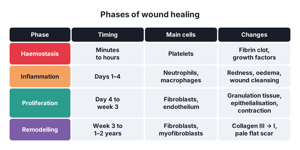
2. Phases of healing: tissue processes
A. Haemostasis
- Minutes to hours
- Vasoconstriction, platelet plug, fibrin clot
- Platelets release growth factors → platelet-derived growth factor (PDGF), transforming growth factor beta (TGF-β)
B. Inflammation (days 1–4)
- Vasodilatation, increased permeability → redness, heat, swelling, pain
- Neutrophils → first 24–48 h; kill bacteria, remove debris
- Macrophages → days 2–4; main cell of healing; phagocytosis, growth factors
- Lymphocytes
- Macroscopic → oedema and hyperaemia of the edges, exudate
- Result → wound cleansed of dead tissue (biological cleansing)
C. Proliferation (day 4 to week 3)
- Fibroblasts → collagen type III
- Angiogenesis → new capillary loops
- Granulation tissue → new capillaries, fibroblasts, macrophages in a loose matrix
- Granulation tissue appearance → red, moist, granular, bleeds easily on touch
- Epithelialisation → keratinocytes migrate from the edges and skin appendages; 1 mm per day
- Wound contraction → myofibroblasts pull the edges together
D. Remodelling (maturation) (week 3 to 1–2 years)
- Collagen type III replaced by type I
- Collagen cross-linking, realignment along tension lines
- Fewer vessels and cells → scar turns from red to pale
- Tensile strength:
- 20% of normal at 3 weeks
- 80% maximum at 3 months; never reaches 100%
3. Types of healing
Primary intention (per primam)
- Clean wound, edges apposed (sutured surgical incision)
- Minimal granulation tissue
- Epithelial bridge within 24–48 h
- Heals in 6–8 days → thin, linear scar
Secondary intention (per secundam)
- Wound left open: tissue loss, infection, gaping edges
- Cavity fills with granulation tissue
- Marked wound contraction
- Slow epithelialisation from the edges
- Result → wide, irregular, weak scar; contracture risk
Tertiary intention (delayed primary closure)
- Contaminated wound left open 3–5 days, then sutured once clean
Healing under a scab (sub crusta)
- Superficial wounds, abrasions, superficial burns
- Epithelium grows under the dried crust
4. Scar
- Normal scar → flat, pale, within the wound borders after maturation
- Hypertrophic scar → raised, within the wound borders; appears within weeks; regresses with time
- Keloid → grows beyond the wound borders; does not regress; recurs after excision; more common in dark skin, earlobe, sternum, shoulder
- Contracture → shortening of a scar across a joint
- Marjolin's ulcer → squamous cell carcinoma in a chronic scar or ulcer
- Treatment of abnormal scars → pressure, silicone gel, steroid injection, excision with radiotherapy (keloid)
5. Factors affecting healing
Local
- Infection → most common cause of delayed healing
- Poor blood supply, ischaemia
- Haematoma, seroma, foreign body, necrotic tissue
- Tension, movement
- Previous radiotherapy
- Wound site → head and neck heal fastest
General
- Age
- Malnutrition → protein, vitamin C (collagen cross-linking), zinc
- Diabetes mellitus
- Anaemia, hypoxia, smoking
- Steroids, chemotherapy, immunosuppression
- Jaundice, uraemia, malignancy
- Obesity
6. Complications of healing
- Wound infection
- Dehiscence → burst abdomen
- Incisional hernia
- Chronic wound → ulcer, sinus, fistula
- Hypertrophic scar, keloid, contracture
Show as a list
Wound healing
- Haemostasis
- Platelet plug
- Inflammation
- Neutrophils
- Macrophages
- Proliferation
- Granulation tissue
- Epithelialisation
- Contraction
- Remodelling
- Collagen III to I
80% strength at 3 months
- Types
- Primary intention
- Secondary intention
- Tertiary intention
- Under a scab