49. Varicose veins of the lower limbs
High yield
Dilated, tortuous superficial veins from valve failure; duplex shows reflux; endothermal ablation is first-line treatment.
Exam Prep · State examSurgery · General surgery
1. Definition
- Varicose veins → dilated (≥ 3 mm), tortuous superficial veins of the leg with valve incompetence and reflux
- Reticular veins → 1–3 mm subdermal veins
- Telangiectasia (thread or spider veins) → < 1 mm intradermal veins
- Chronic venous insufficiency (CVI) → skin changes and ulcers from long-standing venous hypertension
2. Epidemiology
- About one-third of adults
- Women slightly more than men; rises with age
- Venous ulcers → about 1% of adults; 70% recur
3. Aetiology and risk factors
Anatomy (revision)
- Superficial veins:
- Great saphenous vein (GSV) → in front of the medial malleolus → medial leg and thigh → saphenofemoral junction (SFJ) 2–3 cm below and lateral to the pubic tubercle
- Small saphenous vein (SSV) → behind the lateral malleolus → back of the calf → saphenopopliteal junction (SPJ) in the popliteal fossa
- Nerves → saphenous nerve with the GSV below the knee; sural nerve with the SSV
- Deep veins → under the deep fascia, with the arteries
- Perforators → connect superficial to deep; valves direct blood inwards
- Calf muscle pump → soleus and gastrocnemius squeeze the deep veins; valves stop backflow
Causes
- Primary (most) → weak vein wall and valves:
- Family history
- Female sex, pregnancy (progesterone, pressure)
- Prolonged standing, obesity, age
- Secondary:
- Previous DVT → deep valve damage (post-thrombotic)
- Pelvic masses → pregnancy, fibroids, ovarian tumour
- Arteriovenous fistula
- Congenital → Klippel-Trénaunay syndrome, absent valves
4. Pathogenesis
- Vein wall weakens, valves fail at the SFJ, SPJ or perforators
- Reflux → blood flows back down when standing
- Venous hypertension in the superficial veins
- Veins dilate, lengthen and twist → more valves fail
- Raised pressure in capillaries → leakage of fluid, red cells and fibrinogen
- Haemosiderin → brown pigment; white cell trapping and inflammation → lipodermatosclerosis
- Skin breaks down → venous ulcer
5. Classification
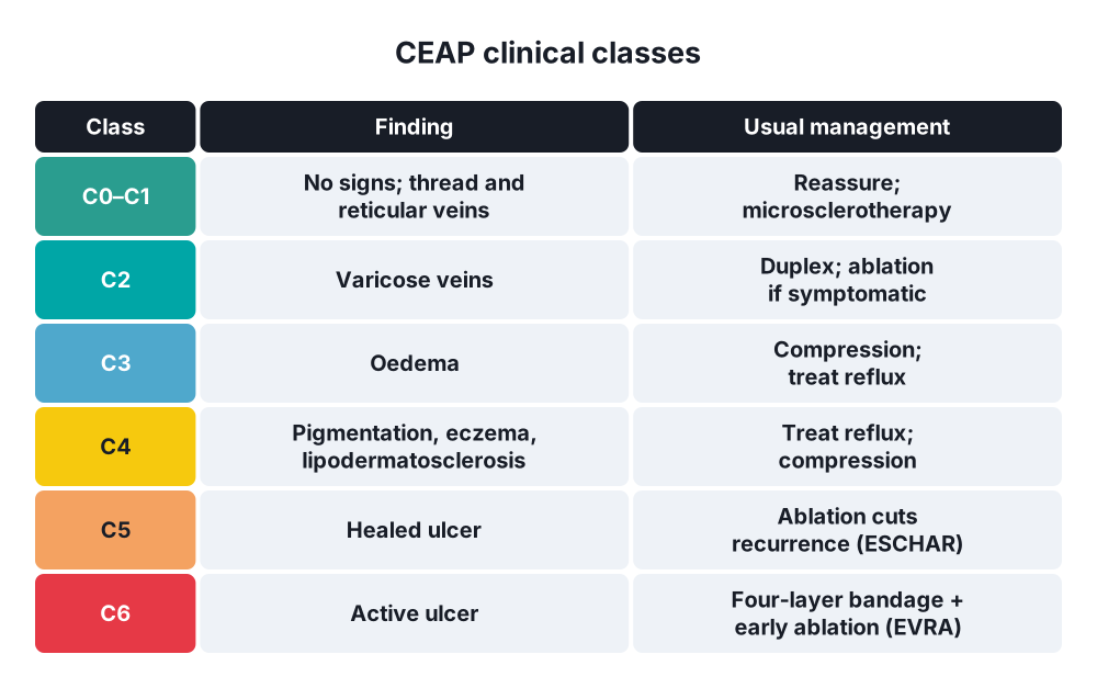
CEAP (2020)
- C (clinical):
- C0 → no signs
- C1 → telangiectasia, reticular veins
- C2 → varicose veins
- C3 → oedema
- C4 → skin changes: a pigmentation, eczema; b lipodermatosclerosis, atrophie blanche; c corona phlebectatica
- C5 → healed ulcer
- C6 → active ulcer
- E (aetiology) → congenital, primary, secondary
- A (anatomy) → superficial, deep, perforator
- P (pathophysiology) → reflux, obstruction
By vein
- Truncal (GSV, SSV), tributary, reticular, telangiectasia
6. Clinical picture
Symptoms
- Cosmetic concern
- Aching, heaviness, tiredness, itching
- Swelling, worse at the end of the day, in hot weather and before periods
- Night cramps
- Restless legs
Signs
- Examine standing
- Tortuous veins along the GSV (medial) or SSV (back of calf)
- Saphena varix → soft bluish groin lump at the SFJ; disappears lying down; cough impulse and thrill → mimics femoral hernia
- Cough impulse at the SFJ
- Skin changes at the gaiter area (above the medial malleolus):
- Oedema
- Brown haemosiderin pigmentation, varicose eczema
- Lipodermatosclerosis → hard, tight skin; "inverted champagne bottle" leg
- Atrophie blanche → white scars
- Venous ulcer → medial gaiter area, shallow, sloping edges, granulating base, not very painful
Bedside tests (now mostly replaced by duplex)
- Trendelenburg (tourniquet) test:
- Lie down, raise the leg, empty the veins
- Tourniquet high on the thigh (or press on the SFJ)
- Stand up
- Veins stay empty, then fill on release → SFJ incompetence
- Veins fill with the tourniquet on → incompetence below it (repeat lower)
- Perthes' test → tourniquet on, patient walks; veins empty → deep veins patent; veins fill with pain → deep vein obstruction
- Tap test → impulse felt below → incompetent valves (unreliable)
- Handheld Doppler → reflux > 1 s at SFJ or SPJ
7. Diagnosis
- Venous duplex ultrasound → gold standard before any treatment:
- Reflux > 0.5 s in superficial veins (> 1 s in deep veins)
- Site of reflux → SFJ, SPJ, perforators
- Deep vein patency and previous DVT
- Vein diameter, maps the treatment
- ABPI → before compression (exclude arterial disease; compression unsafe if ABPI < 0.8)
- CT or MR venography → pelvic or iliac vein obstruction
- Varicography → recurrent veins, rarely
8. Differential diagnosis
- Saphena varix versus femoral hernia, lymph node, femoral aneurysm, psoas abscess
- Leg swelling → DVT, lymphoedema, heart failure, hypoproteinaemia
- Leg ulcer → arterial, mixed, diabetic, vasculitic, Marjolin's ulcer (squamous cell carcinoma in a long-standing ulcer)
- Venous malformation (young patient, Klippel-Trénaunay)
9. Treatment
- Indications → symptoms, skin changes, ulcers (C2 symptomatic to C6), bleeding, superficial thrombophlebitis; cosmetic treatment is usually not funded
Conservative
- Lifestyle → weight loss, exercise, leg elevation
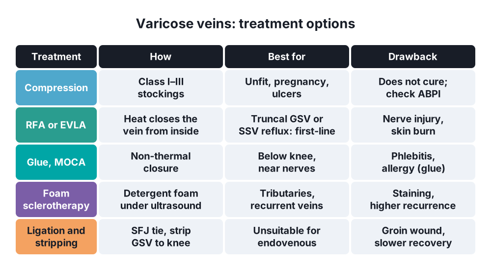
- Graduated compression stockings:
- Class I (< 25 mmHg) → mild symptoms
- Class II (25–35 mmHg) → varicose veins, CVI
- Class III (35–45 mmHg) → severe CVI, after ulcers
- Check ABPI first
- Venous ulcer → four-layer compression bandaging if ABPI > 0.8 (heals about 70% in 12 weeks)
Surgical: endovenous (first-line for truncal reflux)
- Endothermal ablation → first choice:
- Radiofrequency ablation (RFA) or endovenous laser ablation (EVLA)
- Catheter into the GSV under ultrasound, tumescent local anaesthetic, vein heated from inside → closes
- Done as a day case; quick return to work
- Non-thermal → cyanoacrylate glue, mechanochemical ablation
- Ultrasound-guided foam sclerotherapy → sodium tetradecyl sulphate or polidocanol foam; tributaries, recurrent veins
- Ambulatory phlebectomy (stab avulsions) → removes branch varicosities at the same time
- Microsclerotherapy → thread and reticular veins
Surgical: open
- High ligation and stripping → if endovenous treatment is not suitable:
- Groin incision over the SFJ
- Flush ligation of the GSV at the SFJ with all tributaries (superficial epigastric, superficial circumflex iliac, external pudendal)
- Strip the GSV to the knee only (to protect the saphenous nerve)
- Stab avulsions of varicosities
- SSV → SPJ ligation in the popliteal fossa (mark it with duplex); protect the sural and common peroneal nerves
- Perforators → subfascial endoscopic perforator surgery (SEPS), now rarely
- ESCHAR trial → superficial surgery + compression reduces ulcer recurrence; EVRA trial → early ablation heals ulcers faster
10. Complications
Of disease
- Superficial thrombophlebitis
- Bleeding → from a thin vein at the ankle; lie flat, raise the leg, press
- Eczema, lipodermatosclerosis, venous ulcer
- DVT (raised risk)
Of treatment
- Bruising, pain
- Recurrence → about 20–30% at 5 years (up to 50% at 10 years after surgery)
- Nerve injury → saphenous (ablation, stripping), sural (SSV)
- Thermal skin burn, skin staining (sclerotherapy)
- DVT, endovenous heat-induced thrombosis
- Groin wound infection, lymphocoele
- Femoral vein or artery injury (rare, serious)
Show as a list
Varicose veins
- Anatomy
- GSV: SFJ
- SSV: SPJ
- Perforators
- Calf pump
- Causes
- Primary valve failure
- Pregnancy, standing
- Post-DVT
- Clinical
- Aching, swelling
- Saphena varix
- Skin changes
- Venous ulcer
- CEAP C0-C6
- Tests
- Trendelenburg
- Duplex: gold standard
- ABPI
- Treatment
- Compression
- RFA, EVLA
- Foam sclerotherapy
- Ligation, stripping


