
55. Umbilical hernia, umbilical cysts and fistulas
High yield
Infantile hernias close by themselves; adult paraumbilical hernias need repair; vitelline and urachal remnants form cysts, sinuses and fistulas.
Exam Prep · State examSurgery · General surgerySurgical Anatomy · Abdominal wall and hernia
1. Definition
- Umbilical hernia → hernia through the umbilical ring
- Infantile (congenital) → through the unclosed umbilical ring after birth
- Paraumbilical (adult) → through the linea alba just above or below the umbilicus
- Umbilical cyst, sinus or fistula → remains of the vitelline (omphalomesenteric) duct or urachus that did not close
2. Epidemiology
- Infantile umbilical hernia → about 10–20% of newborns; more in African-Caribbean, premature and low-birth-weight babies
- Most close by 2–3 years (over 80%)
- Paraumbilical hernia → adults over 35; women 3–5 times more than men; obese, multiparous
- Umbilical and paraumbilical → about 10% of primary abdominal wall hernias in adults
- Meckel's diverticulum → 2% of people ("rule of 2s")
- Urachal remnants → rare; more in boys
3. Aetiology and risk factors
Embryology (revision)
- Umbilical cord carries:
- Two umbilical arteries → become the medial umbilical ligaments
- One umbilical vein → becomes the ligamentum teres (in the falciform ligament)
- Vitelline duct → joins the midgut to the yolk sac; normally closes by week 7–9
- Allantois → urachus → joins the bladder to the umbilicus; becomes the median umbilical ligament
- After birth → the ring closes by scarring
Umbilical hernia
- Infant → umbilical ring not fully closed; prematurity, Down's syndrome, hypothyroidism, Beckwith-Wiedemann, mucopolysaccharidoses
- Adult → raised pressure on a weak linea alba:
- Obesity, multiple pregnancies
- Ascites (cirrhosis), peritoneal dialysis
- Chronic cough, straining
- Divarication of the recti
- Laparoscopic port at the umbilicus
Cysts and fistulas
- Vitelline duct → whole duct open, part open, or fibrous band
- Urachus → whole or part open; bladder outlet obstruction keeps it open
4. Pathogenesis
- Infantile → true umbilical defect; wide neck, symmetric; strangulation very rare
- Paraumbilical → defect in the linea alba next to the umbilicus; narrow neck with fibrous edge; sac contains omentum or bowel; higher risk of incarceration and strangulation
- Ascites → hernia stretches thin; skin may ulcerate and rupture → ascitic leak, peritonitis (Flood syndrome)
- Infected remnants → abscess, discharge at the umbilicus
5. Classification
Umbilical hernia
- Infantile (true umbilical)
- Paraumbilical (supra- or infraumbilical)
- Adult true umbilical
- Exomphalos → congenital defect with a sac (see abdominal wall defects)
- EHS ventral hernia → M3 (umbilical); width W1 < 2 cm, W2 2–4 cm, W3 > 4 cm
Vitelline duct remnants
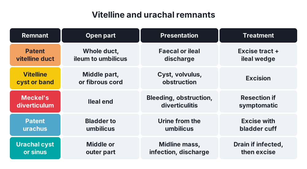
- Patent vitelline duct (umbilical-ileal fistula) → whole duct open; faecal or ileal discharge from the umbilicus
- Umbilical sinus → outer end open
- Vitelline (enterocystoma) cyst → middle part open
- Fibrous band → umbilicus to ileum; volvulus, obstruction
- Meckel's diverticulum → inner end open (most common)
- Umbilical polyp (granuloma) → red mucosal tag
Urachal remnants
- Patent urachus → urine leaks from the umbilicus
- Urachal cyst → middle part; lower midline mass, infection
- Urachal sinus → outer end; discharge
- Urachal (vesicourachal) diverticulum → bladder end; urinary infection, stones
- Urachal adenocarcinoma → rare; at the bladder dome
6. Clinical picture
Symptoms
- Infant → soft bulge at the umbilicus, larger on crying; painless
- Adult paraumbilical → lump beside the umbilicus, pain, dragging, colic; may become irreducible
- Large hernia → skin changes, intertrigo, ulceration
- Strangulation → painful, tense, red, irreducible lump, vomiting
- Remnants:
- Discharge from the umbilicus → faeces or bowel content (vitelline) or urine (urachus), mucus, pus
- Red moist polyp
- Lower abdominal midline mass, fever, dysuria (infected urachal cyst)
- Bowel obstruction (band, volvulus)
Signs
- Infant → central, symmetric bulge; defect felt with a fingertip; reduces easily
- Paraumbilical → crescent-shaped umbilicus (sac displaces the umbilicus to one side); firm neck; cough impulse
- Ascites → tense, thin-skinned, shiny hernia
- Granuloma → small pink tissue after the cord separates (no mucosa)
- Polyp → mucosa (bowel); does not respond to silver nitrate
7. Diagnosis
- Hernia → clinical
- Ultrasound → small or obese patients, contents
- CT → obese patients, multiple defects, complications, divarication
- Remnants:
- Ultrasound of the umbilicus and bladder → cysts, urachal tract
- Fistulogram (sinogram) → contrast fills ileum (vitelline) or bladder (urachus)
- Micturating cystourethrogram → patent urachus; exclude bladder outlet obstruction
- Discharge analysis → creatinine (urine), bile, bowel content
- Meckel's scan (technetium pertechnetate) → bleeding Meckel's
- Biopsy of a polyp → gastric or intestinal mucosa
8. Differential diagnosis
- Umbilical granuloma versus polyp
- Paraumbilical hernia versus lipoma, divarication of the recti (no defect)
- Sister Mary Joseph nodule → umbilical metastasis (stomach, colon, ovary, pancreas)
- Umbilical endometriosis (cyclical bleeding)
- Omphalitis (infection of the cord stump in newborns)
- Caput medusae → portal hypertension
- Pilonidal sinus of the umbilicus
- Cullen's sign → periumbilical bruising (pancreatitis, ruptured ectopic)
9. Treatment
Conservative
- Infantile umbilical hernia → wait; most close by 2–3 years
- No strapping or coins (no benefit, skin damage)
- Repair if → persists after 3–4 years, defect > 1.5–2 cm, growing, incarcerated, or symptomatic
- Umbilical granuloma → silver nitrate cautery, or salt application, or ligation
- Ascites → control the ascites first (diuretics, paracentesis, TIPS); repair after
Surgical: umbilical hernia
- Infant:
- Curved infraumbilical skin-crease incision
- Dissect and reduce the sac
- Close the fascia with absorbable sutures
- Fix the umbilical skin down to the repair (keeps the navel shape)
- Adult paraumbilical → repair almost always (risk of strangulation):
- Defect < 1 cm → sutured repair with non-absorbable sutures
- Defect ≥ 1 cm → mesh (preperitoneal flat mesh or patch) → lower recurrence than suture (EHS guidelines)
- Large defects, obese, recurrent, multiple defects → laparoscopic IPOM or open retromuscular (Rives-Stoppa) mesh
- Mayo "vest-over-pants" repair → historical; no better than simple closure
- Very large hernia with skin damage → excise the umbilicus (omphalectomy)
- Strangulated → emergency repair; open the sac, resect dead omentum or bowel; sutured or biological mesh if contaminated
- Cirrhosis with ascites → emergency repair for rupture or strangulation; elective repair after ascites is controlled
Surgical: remnants
- Vitelline remnants → excise the whole tract with a wedge of ileum, or short ileal resection with anastomosis, via an umbilical or small laparotomy incision
- Umbilical polyp → excise; explore for a deeper remnant
- Urachal remnants → complete excision of the tract down to the bladder dome, with a cuff of bladder (prevents recurrence and cancer)
- Infected urachal cyst → drain first, antibiotics, then excise later
- Correct any bladder outlet obstruction
10. Complications
- Hernia → incarceration, strangulation, skin ulceration, rupture (ascites)
- Repair → seroma, wound infection, recurrence (higher with suture repair, obesity, smoking), mesh infection, umbilical skin necrosis
- Vitelline → bleeding (ectopic gastric mucosa), obstruction, volvulus, intussusception, Meckel's diverticulitis
- Urachal → infection, abscess, stones, urachal carcinoma
Show as a list
Umbilicus
- Embryology
- Umbilical vein: ligamentum teres
- Arteries: medial ligaments
- Vitelline duct
- Urachus
- Infantile hernia
- Closes by 3 years
- Repair after 3-4 years
- Paraumbilical hernia
- Obese women
- Narrow neck
- Strangulation
- Mesh if 1 cm or more
- Vitelline remnants
- Patent duct: faeces
- Cyst, sinus, band
- Meckel's
- Urachal remnants
- Patent urachus: urine
- Cyst, sinus
- Excise with bladder cuff
- Differential
- Granuloma
- Sister Mary Joseph


