23. Ulcers
High yield
Chronic skin or mucosal defect with no tendency to heal; mainly venous, arterial, neuropathic or pressure ulcers.
Exam Prep · State examSurgery · General surgery
1. Definition
- Ulcer → defect of skin or mucosa with loss of epithelium, slow healing and little tendency to heal spontaneously
- Erosion → loss of epithelium only, heals without scar
- Chronic wound → no healing after 6 weeks
2. Aetiology: causes
Vascular
- Venous → chronic venous insufficiency, varicose veins, post-thrombotic syndrome; 70% of leg ulcers
- Arterial → peripheral arterial disease, atherosclerosis
- Mixed arterial and venous
- Lymphoedema
- Vasculitis → rheumatoid arthritis, lupus
- Hypertensive (Martorell) ulcer
Neuropathic (trophic)
- Diabetic neuropathy → plantar ulcers
- Leprosy, spinal cord injury, spina bifida, alcohol, syringomyelia
Pressure
- Pressure ulcers → immobile patients over sacrum, heels, hips
Traumatic, physical, chemical
- Burns, frostbite, radiation, chemicals
Infective
- Tuberculosis, syphilis (gumma), Buruli ulcer, fungal
Neoplastic
- Basal cell carcinoma (rodent ulcer), squamous cell carcinoma, melanoma
- Marjolin's ulcer → squamous cell carcinoma in a chronic scar or ulcer
Other
- Pyoderma gangrenosum → inflammatory bowel disease
- Haematological → sickle cell disease
- Self-inflicted (artefact)
3. Pathological characteristics
- Poor blood supply, oedema, fibrosis around the ulcer
- Chronic inflammation, bacterial colonisation, biofilm
- Granulation tissue → pale, flabby, poor quality
- Wound bed stuck in the inflammatory phase → high protease levels, senescent fibroblasts
- Surrounding skin → induration, pigmentation (venous), atrophy (arterial)
4. Evolution of an ulcer
- Spreading stage → inflamed edge, slough, purulent discharge, pain
- Stage of cleansing (transition) → slough separates, red granulations appear
- Healing (repair) stage → healthy granulation tissue, bluish line of epithelium at the edge, wound contraction
- Callous ulcer → stuck stage; pale granulations, thick indurated edge, no healing
5. Clinical manifestations: examination of an ulcer
History
- Duration, pain, previous ulcers, trauma, varicose veins, deep vein thrombosis, diabetes, smoking
Local examination
- Site → gaiter area (venous), toes and pressure points (arterial), sole (neuropathic)
- Size, shape, number
- Edge:
- Sloping → healing, venous ulcer
- Punched out → arterial, neuropathic, syphilitic
- Undermined → tuberculosis, pressure ulcer
- Rolled (pearly) → basal cell carcinoma
- Everted → squamous cell carcinoma
- Floor → granulation tissue, slough, bone, tendon
- Base → induration, fixation to deep structures
- Discharge → serous, purulent, blood-stained
- Surrounding skin → pigmentation, eczema, lipodermatosclerosis, hair loss, shiny skin
- Regional lymph nodes → infection or malignancy
- Pulses, sensation (monofilament), ABPI
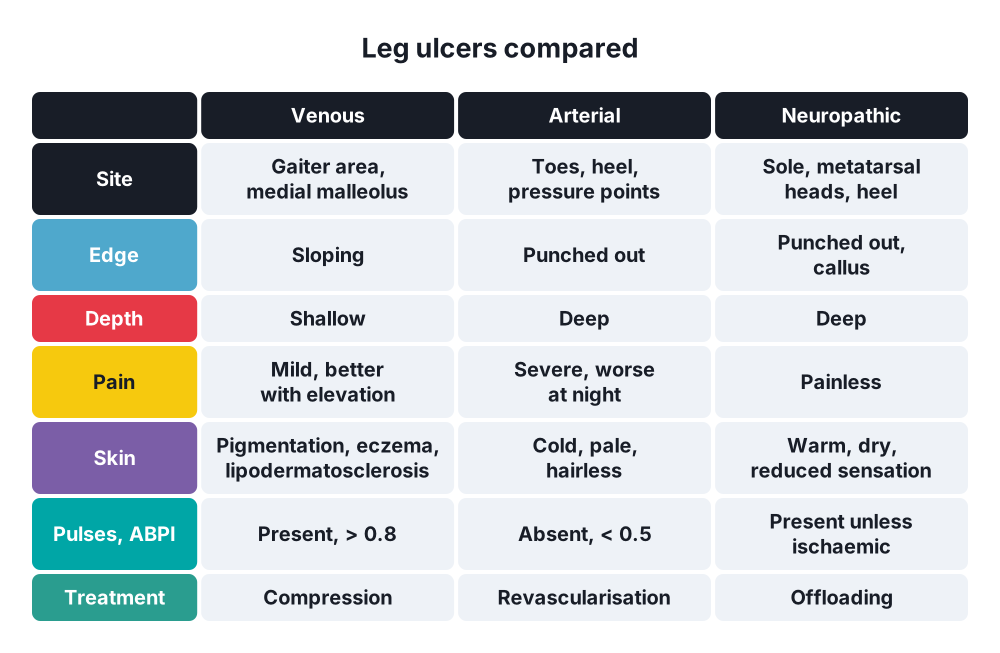
6. Main types compared
A. Venous ulcer
- Medial malleolus, gaiter area
- Large, shallow, sloping edge, moderate pain relieved by elevation
- Haemosiderin pigmentation, varicose eczema, lipodermatosclerosis
- Pulses present, ABPI > 0.8
B. Arterial ulcer
- Toes, heel, shin, pressure points
- Small, deep, punched out, very painful, worse with elevation and at night
- Cold, pale, hairless skin; absent pulses; ABPI < 0.5
C. Neuropathic ulcer
- Sole, under metatarsal heads, heel
- Painless, punched out, surrounded by callus
- Loss of sensation, warm foot, bounding pulses (if no arterial disease)
D. Pressure ulcer
- Sacrum, heels, greater trochanter, occiput
- Graded 1–4 (European Pressure Ulcer Advisory Panel):
- Non-blanching erythema, intact skin
- Partial-thickness skin loss, blister
- Full-thickness skin loss, subcutaneous fat visible
- Full-thickness tissue loss with bone, tendon or muscle exposed
7. Diagnosis
Laboratory
- Full blood count, glucose, HbA1c, CRP
- Swab → only if clinically infected
Investigations
- ABPI → before compression
- Duplex ultrasound → venous reflux, arterial disease
- Angiography → arterial ulcers before revascularisation
- X-ray, MRI → osteomyelitis
- Biopsy → atypical ulcer, raised or rolled edge, no healing after 3 months (exclude malignancy)
8. Principles of treatment
General
- Treat the cause
- Control diabetes, nutrition, stop smoking
- Treat anaemia, oedema
- Analgesia
Local
- Cleaning, debridement of slough and necrotic tissue
- Moist wound healing → appropriate dressings
- Treat infection → systemic antibiotics for cellulitis
- Negative pressure wound therapy
- Skin grafts, flaps → clean ulcer bed
By type
- Venous → multilayer compression bandaging (ABPI > 0.8), elevation, exercise; treat superficial venous reflux (endovenous ablation, surgery)
- Arterial → revascularisation (angioplasty, bypass); no compression; amputation if not salvageable
- Neuropathic → offloading (total contact cast, special footwear), debridement of callus, foot care
- Pressure → relieve pressure (turning every 2 h, pressure-relieving mattress), nutrition, debridement, flap reconstruction
- Malignant → wide excision, Mohs surgery, radiotherapy
9. Complications
- Infection, cellulitis, osteomyelitis
- Haemorrhage
- Contracture, deformity
- Marjolin's ulcer
- Amputation
Show as a list
Ulcers
- Causes
- Venous
- Arterial
- Neuropathic
- Pressure
- Infective
- Neoplastic
- Evolution
- Spreading
- Cleansing
- Healing
- Edge
- Sloping
- Punched out
- Undermined
- Rolled
- Everted
- Treatment
- Treat cause
- Debridement
- Compression
- Revascularisation
- Offloading