50. Thrombophlebitis, phlebothrombosis and post-phlebitic syndrome
High yield
Superficial vein thrombosis, deep vein thrombosis from Virchow's triad, and the chronic swollen, ulcerated leg it leaves.
Exam Prep · State examSurgery · General surgerySurgery · Emergency and trauma
1. Definition
- Superficial thrombophlebitis (superficial vein thrombosis, SVT) → thrombus with inflammation in a superficial vein
- Phlebothrombosis (deep vein thrombosis, DVT) → thrombus in a deep vein, often with little inflammation; can break off → pulmonary embolism (PE)
- Venous thromboembolism (VTE) → DVT and PE together
- Post-phlebitic (post-thrombotic) syndrome (PTS) → chronic venous insufficiency after DVT: pain, swelling, skin changes, ulcers
2. Epidemiology
- VTE → about 1–2 per 1,000 people a year; rises sharply with age
- Without prophylaxis → DVT after major general surgery in 20–30%, after hip or knee replacement in 40–60%
- PE → a leading cause of preventable death in hospital
- SVT → about 25% have a DVT at diagnosis
- PTS → 20–50% after DVT; severe in 5–10%
3. Aetiology and risk factors
Virchow's triad
- Stasis → immobility, surgery, long travel, paralysis, heart failure, pregnancy, varicose veins
- Hypercoagulability:
- Acquired → cancer, surgery, trauma, oestrogen (pill, HRT), pregnancy, sepsis, nephrotic syndrome, antiphospholipid syndrome, polycythaemia
- Inherited → factor V Leiden (most common), prothrombin gene mutation, protein C, protein S and antithrombin deficiency
- Vein wall injury → trauma, surgery, central lines, previous DVT
Superficial thrombophlebitis
- Varicose veins (most common in the leg)
- IV cannula, infusions, drug injection (arm)
- Pregnancy, pill, cancer
- Migratory (thrombophlebitis migrans) → Trousseau's sign of cancer (pancreas, lung), Buerger's disease, Behçet's
4. Pathogenesis
DVT
- Starts at the valve cusps of calf veins (stasis, low oxygen)
- Platelets and fibrin build up → red thrombus
- Grows upwards → popliteal, femoral, iliac veins
- Free-floating tail → breaks off → PE
- Thrombus organises; vein recanalises over months, but valves are destroyed
Post-thrombotic syndrome
- Destroyed valves → reflux; residual clot → obstruction
- Venous hypertension when standing and walking (failed calf pump)
- Capillary leak → oedema, haemosiderin, fibrosis
- Skin breaks down → venous ulcer
5. Classification
By site
- Distal (calf) DVT → below the popliteal vein; lower PE risk
- Proximal DVT → popliteal and above; higher PE risk
- Iliofemoral → worst swelling and highest PTS risk
- Upper limb → catheters, Paget-Schroetter syndrome (effort thrombosis of the subclavian vein, thoracic outlet)
By cause
- Provoked → major transient risk (surgery, trauma, immobility)
- Unprovoked → no clear cause; higher recurrence; look for cancer
Severe forms
- Phlegmasia alba dolens → white, swollen, painful leg (iliofemoral DVT)
- Phlegmasia caerulea dolens → blue, very swollen leg; arterial flow blocked → venous gangrene
PTS: Villalta score
- 5 symptoms (pain, cramps, heaviness, itching, paraesthesia) + 6 signs (oedema, skin induration, pigmentation, redness, venous ectasia, calf pain on compression), each 0–3
- Ulcer → severe PTS
- 5–9 mild, 10–14 moderate, ≥ 15 or ulcer → severe
6. Clinical picture
Symptoms
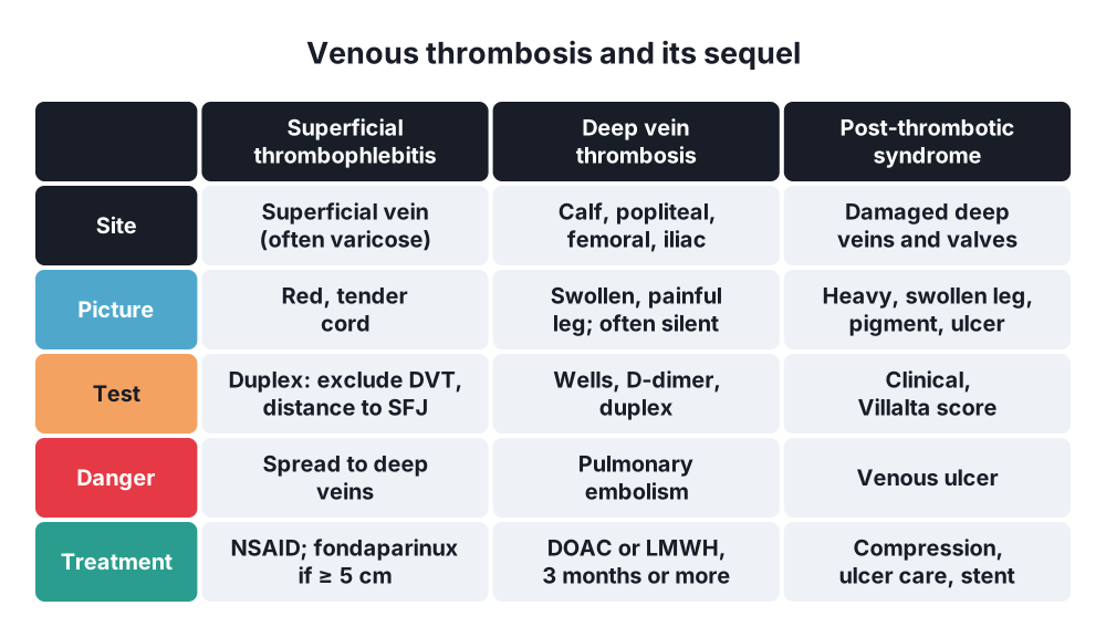
- SVT → painful, red, hot, tender cord along a superficial vein
- DVT:
- Often no symptoms
- Calf pain, swelling of the leg
- Warmth, redness
- Mild fever
- PE → sudden dyspnoea, pleuritic pain, haemoptysis, collapse
- PTS → heavy, aching, swollen leg, worse at the end of the day; itching; ulcers
Signs
- SVT → palpable tender cord, redness along the vein; check that it is not near the SFJ
- DVT:
- Unilateral pitting oedema
- Calf swelling > 3 cm compared with the other leg (10 cm below the tibial tuberosity)
- Tenderness along the deep veins
- Distended superficial veins
- Homans' sign (calf pain on dorsiflexion) → unreliable; not used
- Phlegmasia caerulea dolens → massive swelling, blue, cold, no pulses
- PTS → oedema, pigmentation, eczema, lipodermatosclerosis, medial gaiter ulcer
7. Diagnosis
DVT pathway
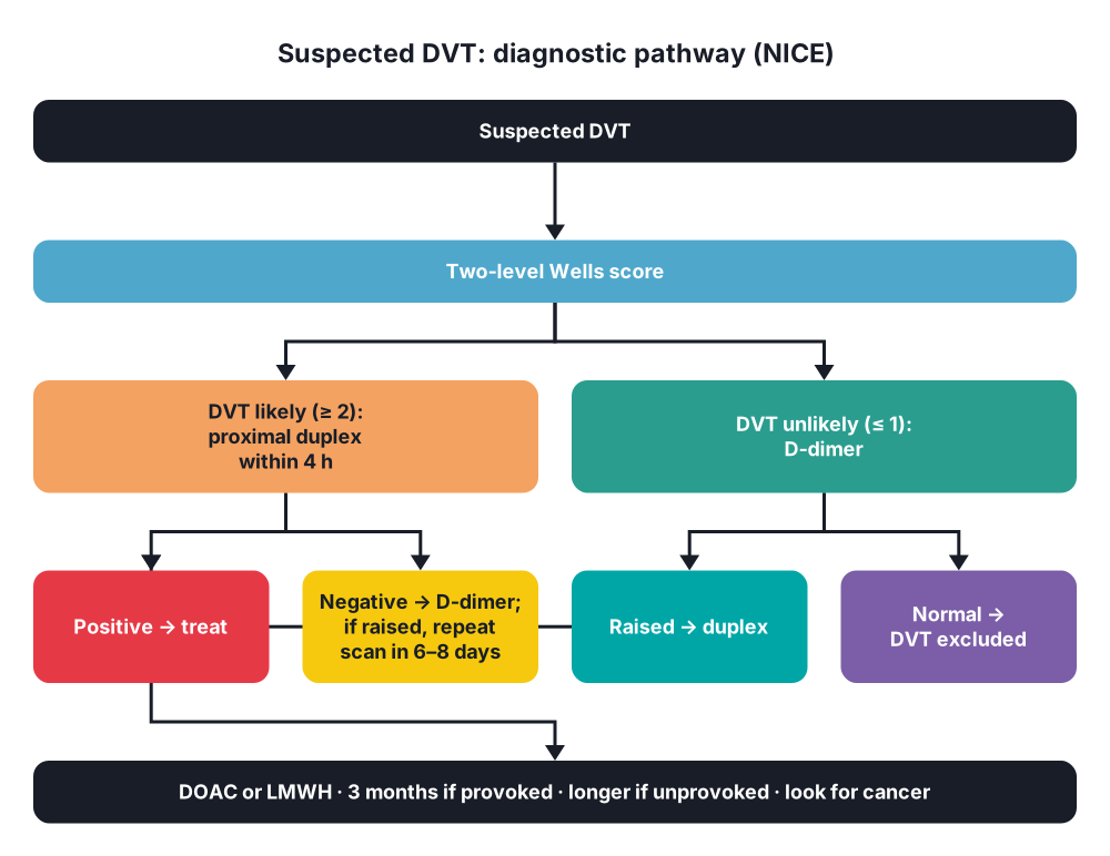
- Two-level Wells score for DVT:
- Active cancer, paralysis or plaster, bedridden > 3 days or surgery < 12 weeks, tenderness along deep veins, whole leg swollen, calf > 3 cm bigger, pitting oedema, collateral veins, previous DVT → +1 each
- Alternative diagnosis as likely → −2
- ≥ 2 → DVT likely; ≤ 1 → DVT unlikely
- DVT likely → proximal leg duplex ultrasound within 4 h
- DVT unlikely → D-dimer; negative → DVT excluded; positive → duplex
- Duplex ultrasound → test of choice; vein does not compress, no flow
- Venography → rarely
- D-dimer → sensitive, not specific (raised after surgery, cancer, infection, pregnancy, age)
- CT pulmonary angiography → suspected PE
Other tests
- FBC, clotting, U&E, liver tests (before anticoagulation)
- Unprovoked DVT → history and examination for cancer; thrombophilia testing only in selected patients
- SVT → duplex to look for DVT and distance from the SFJ
8. Differential diagnosis
- Ruptured Baker's (popliteal) cyst
- Cellulitis
- Calf muscle tear or haematoma
- Lymphoedema
- Heart failure, hypoalbuminaemia (both legs)
- Compartment syndrome
- Superficial thrombophlebitis
9. Treatment
Conservative: prevention
- Risk assessment for all admissions
- Mechanical → early mobilisation, hydration, graduated stockings, intermittent pneumatic compression
- Drugs → low molecular weight heparin (LMWH), fondaparinux, DOACs after joint replacement
- Extended prophylaxis (28 days) → major abdominal or pelvic cancer surgery, hip replacement
Conservative: superficial thrombophlebitis
- Analgesia, NSAIDs, warm compresses, compression, keep walking
- ≥ 5 cm long, or near the SFJ → fondaparinux 2.5 mg daily for 45 days (or prophylactic LMWH, rivaroxaban 10 mg)
- Within 3 cm of the SFJ → treat as a DVT (full anticoagulation)
- Cannula-related → remove the cannula
- Later → treat the varicose veins
Conservative: DVT
- Anticoagulation:
- DOAC → apixaban or rivaroxaban first-line (no injections, no monitoring)
- LMWH → in pregnancy (warfarin and DOACs not used) and in some patients with cancer
- Warfarin → INR 2–3, started with LMWH for at least 5 days until INR ≥ 2 for 24 h
- Unfractionated heparin → kidney failure, high bleeding risk, before procedures (APTT 1.5–2.5 × normal)
- Duration:
- Provoked → 3 months
- Unprovoked → 3–6 months, then consider long-term
- Cancer → at least 6 months
- Calf DVT → anticoagulate, or repeat scan at 1 week
- Elevation, early walking
- Heparin-induced thrombocytopenia (HIT) → platelet fall day 5–14; stop heparin, use argatroban or fondaparinux
Surgical: DVT
- Catheter-directed thrombolysis or pharmacomechanical thrombectomy → selected iliofemoral DVT in young, fit patients with recent symptoms (< 14 days) and low bleeding risk
- Phlegmasia caerulea dolens → urgent thrombolysis or surgical venous thrombectomy (Fogarty catheter) ± fasciotomy
- Inferior vena cava (IVC) filter → only when anticoagulation is contraindicated or PE recurs despite it; remove when possible
- May-Thurner syndrome → venous stent of the left common iliac vein
- Venous bypass (Palma operation: crossover saphenous vein graft for iliac occlusion), valve repair → rarely
Conservative: post-thrombotic syndrome
- Compression → class II–III stockings for life; bandaging for ulcers
- Elevation, exercise, weight loss, skin care (emollients, treat eczema)
- Ulcer → four-layer compression bandaging, dressings, skin graft
- Treat superficial reflux (ablation)
- Iliac vein stenting → deep vein obstruction
- Deep valve repair or transposition → specialist centres only
10. Complications
- Pulmonary embolism → death, chronic thromboembolic pulmonary hypertension
- Post-thrombotic syndrome, venous ulcer
- Recurrent DVT
- Venous gangrene
- Paradoxical embolism → stroke (patent foramen ovale)
- Anticoagulation → bleeding, HIT, osteoporosis (long-term heparin)
- SVT → spreads into deep veins (DVT or PE)
Show as a list
Venous thrombosis
- Superficial
- Varicose veins
- Cannula
- Migratory: cancer
- Fondaparinux
- DVT
- Virchow's triad
- Calf to proximal
- Wells, D-dimer
- Duplex
- DOAC, LMWH
- 3 months provoked
- Severe
- Phlegmasia
- Thrombolysis
- IVC filter
- Complications
- Pulmonary embolism
- Post-thrombotic syndrome
- PTS
- Venous hypertension
- Villalta score
- Compression
- Ulcer care


