108. Thromboembolic complications in surgery
High yield
Deep vein thrombosis and pulmonary embolism after surgery; assess every patient, prevent it, and treat it fast.
Exam Prep · State examSurgery · General surgery
1. Definition
- Venous thromboembolism (VTE) → deep vein thrombosis (DVT) and pulmonary embolism (PE), two stages of one disease
- DVT → thrombus in the deep veins, usually of the leg or pelvis
- Distal → calf veins below the popliteal vein
- Proximal → popliteal, femoral, iliac veins (higher risk of PE)
- PE → blockage of the pulmonary arteries by thrombus, usually from the leg or pelvic veins
- Superficial thrombophlebitis → thrombosis and inflammation of a superficial vein (great or small saphenous)
- Other emboli in surgery → fat embolism (long-bone fractures), air embolism (central lines, neurosurgery, laparoscopy), amniotic fluid, tumour, arterial (cardiac) emboli
- This question covers mainly postoperative VTE
2. Epidemiology
- VTE affects about 1–2 per 1000 people a year; risk rises steeply with age (1 in 100 over 80 years each year)
- Without prophylaxis → DVT in about 20–30% of general surgical patients and 40–60% after hip or knee replacement
- Hospital-acquired VTE causes about two-thirds of all VTE; much of it is preventable
- Untreated proximal DVT → about 50% embolise
- PE → mortality about 5–25%; the highest risk of fatal postoperative PE is 3–7 days after surgery
- About 10% of symptomatic PEs kill within 1 hour
- Over half of postoperative VTE occurs after discharge; risk lasts up to 3 months
3. Aetiology and risk factors
Virchow's triad
- Stasis → immobility, anaesthesia and paralysis, long operations, heart failure, pneumoperitoneum
- Endothelial injury → surgery, trauma, central lines, previous DVT
- Hypercoagulability → surgery itself, cancer, sepsis, pregnancy, oestrogen, thrombophilia
Patient factors
- Age over 60, obesity (BMI over 30)
- Active cancer or cancer treatment
- Previous VTE, family history in a first-degree relative
- Thrombophilia → factor V Leiden (most common, about 5% of Europeans), prothrombin G20210A, protein C, protein S or antithrombin deficiency, antiphospholipid syndrome
- Oestrogen → combined oral contraceptive, HRT; pregnancy and up to 6 weeks after birth
- Varicose veins with phlebitis, dehydration, heart or respiratory failure, inflammatory bowel disease, nephrotic syndrome, myeloproliferative disease
Surgical factors
- Major abdominal and pelvic surgery, especially for cancer
- Hip and knee replacement, hip fracture, spinal cord injury, major trauma
- Operation over 90 minutes (or over 60 minutes in the pelvis or lower limb)
- Emergency surgery with an inflammatory or intra-abdominal condition
- Reduced mobility for 3 days or more, plaster cast
4. Pathogenesis
- Most DVTs start in the soleal sinuses and valve pockets of the calf veins, often during the operation
- About half of intraoperative calf thrombi lyse within 72 hours; about 20% extend proximally
- Red thrombus (red cells in fibrin) grows into a free-floating tail → breaks off → embolus
- PE → emboli lodge in the pulmonary arteries:
- Increased pulmonary vascular resistance → right ventricular strain and dilatation
- Ventricular septum bows into the left ventricle → low cardiac output, hypotension
- Dead space (ventilation without perfusion) and shunting → hypoxaemia
- Small peripheral emboli → pulmonary infarction (pleuritic pain, haemoptysis)
- Long-term → valve damage → post-thrombotic syndrome; unresolved emboli → chronic thromboembolic pulmonary hypertension (CTEPH)
5. Classification
DVT
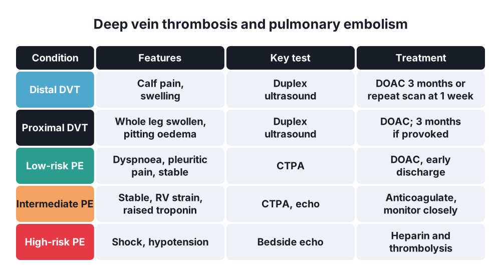
- Distal (calf) and proximal (popliteal and above)
- Iliofemoral DVT → whole leg swollen
- Phlegmasia alba dolens → white, swollen, painful leg (iliofemoral DVT)
- Phlegmasia caerulea dolens → blue, massively swollen leg with arterial compromise; may progress to venous gangrene
PE (European Society of Cardiology)
- High risk (massive) → shock or hypotension (systolic under 90 mmHg)
- Intermediate risk (submassive) → stable but right ventricular dysfunction on echo or CT, raised troponin
- Low risk → stable, no RV strain (PESI class I–II or sPESI 0)
By timing
- Provoked (transient risk factor such as surgery) and unprovoked
6. Clinical picture
DVT
- Often silent; the first sign may be PE
- Pain and tenderness along the deep veins of the calf or thigh
- Unilateral swelling (calf over 3 cm larger, measured 10 cm below the tibial tuberosity), pitting oedema
- Warmth, redness, dilated superficial collateral veins, low-grade fever
- Homans' sign (calf pain on dorsiflexion) is unreliable and not recommended
PE
- Sudden breathlessness (most common), tachypnoea, tachycardia
- Pleuritic chest pain, cough, haemoptysis (infarction)
- Collapse, syncope, hypotension, raised JVP, cyanosis → massive PE
- Low-grade fever, signs of DVT
- Unexplained hypoxia, AF or tachycardia in a postoperative patient → think of PE
- Cardiac arrest with pulseless electrical activity
7. Diagnosis
Suspected DVT
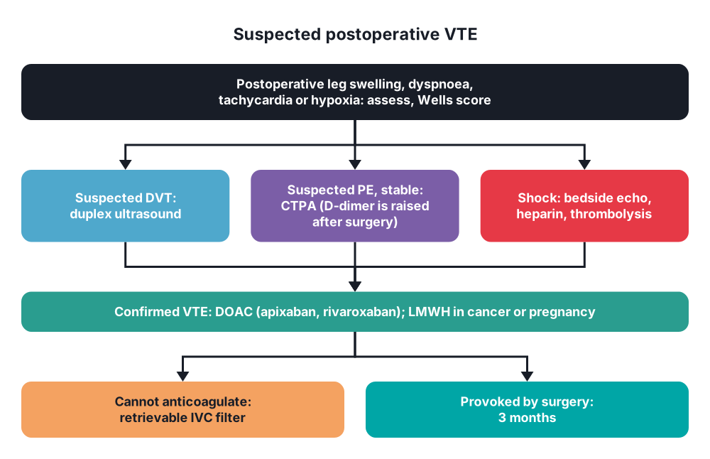
- Two-level Wells score for DVT → DVT likely (2 or more points) or unlikely (1 or fewer)
- DVT likely → proximal leg vein ultrasound within 4 hours; if negative, D-dimer, and repeat ultrasound in 6–8 days if D-dimer is positive
- DVT unlikely → D-dimer; if negative, DVT is excluded; if positive, ultrasound
- D-dimer is sensitive but not specific; raised after surgery, in cancer, infection, pregnancy and old age, so it is of little use in the first weeks after surgery
- Duplex ultrasound (compression) → test of choice (sensitivity 94–99% for proximal DVT)
- CT or MR venography → iliac and caval thrombosis
Suspected PE
- Two-level Wells score for PE → PE likely (over 4 points) or unlikely (4 or fewer); PERC rule to avoid tests in very low-risk patients
- PE likely → CT pulmonary angiography (CTPA) at once; if delayed, start anticoagulation
- PE unlikely → D-dimer; if positive, CTPA
- V/Q scan → if contrast allergy, renal failure, or pregnancy with normal chest X-ray
- Bedside echo in the unstable patient → right ventricular dilatation, McConnell's sign
- Supporting tests:
- ECG → sinus tachycardia (most common), right heart strain, new RBBB, S1Q3T3 (uncommon)
- Chest X-ray → usually normal; excludes other causes
- ABG → hypoxaemia, hypocapnia
- Troponin and BNP → right ventricular strain, risk grading
- Pregnancy → leg ultrasound first, then V/Q or CTPA
- Unprovoked VTE → history, examination and basic tests for cancer; thrombophilia testing only in selected patients and not while on anticoagulants
8. Differential diagnosis
- Of DVT → cellulitis, ruptured Baker's cyst, muscle tear or haematoma, superficial thrombophlebitis, lymphoedema, heart or kidney failure oedema, compartment syndrome, sarcoma
- Of PE → myocardial infarction, pneumonia, pneumothorax, aortic dissection, pericarditis, pulmonary oedema, atelectasis, sepsis
- Of postoperative breathlessness → atelectasis, pneumonia, fluid overload, anastomotic leak with sepsis
9. Treatment
Conservative
- Prevention (thromboprophylaxis):
- Assess every patient for VTE and bleeding risk on admission and again after 24 hours (NICE NG89 risk tool)
- Early mobilisation, leg exercises, good hydration
- Stop combined oral contraceptives or HRT 4 weeks before major elective surgery
- Mechanical → intermittent pneumatic compression (IPC) during and after surgery; anti-embolism stockings if no contraindication
- Contraindications to stockings → peripheral arterial disease, neuropathy, skin grafts, severe leg oedema
- The GAPS trial (2020) found no extra benefit of stockings on top of LMWH in elective surgery
- Pharmacological → low-molecular-weight heparin (LMWH) such as enoxaparin 40 mg or dalteparin 5000 units once daily, starting 6–12 hours after surgery
- Unfractionated heparin in severe renal failure
- Fondaparinux as an alternative
- Extended prophylaxis for 28 days → major abdominal or pelvic cancer surgery
- Orthopaedics → after hip replacement 28–35 days (LMWH, DOAC or aspirin in selected cases); after knee replacement 14 days
- Neuraxial anaesthesia → time LMWH doses around spinal or epidural catheter insertion and removal
- Treatment of DVT and PE:
- Start anticoagulation at once when VTE is confirmed, or suspected and tests are delayed
- First choice → a direct oral anticoagulant (DOAC) such as apixaban or rivaroxaban (no injection or monitoring needed)
- Alternatives → LMWH for at least 5 days followed by dabigatran or edoxaban; or LMWH with warfarin until INR is 2–3 for 2 days in a row
- LMWH (or a DOAC) in cancer-associated VTE; LMWH in pregnancy (warfarin is teratogenic, DOACs are not used)
- Unfractionated heparin infusion → severe renal failure, high bleeding risk, possible thrombolysis or surgery (short half-life, reversible with protamine)
- Duration:
- 3 months after a provoked VTE (surgery is a strong transient risk factor)
- At least 3–6 months, often long-term, after unprovoked VTE if bleeding risk is low
- As long as cancer is active
- Isolated calf DVT → anticoagulate for 3 months, or repeat ultrasound in 1 week if bleeding risk is high
- Early mobilisation is safe once anticoagulated; compression stockings do not prevent post-thrombotic syndrome
- Superficial thrombophlebitis over 5 cm → fondaparinux 2.5 mg for 45 days; ultrasound to exclude DVT if near the saphenofemoral junction
- Heparin-induced thrombocytopenia (HIT) → platelets fall by over 50% at days 5–10 with new thrombosis → stop all heparin and give a non-heparin anticoagulant (argatroban, fondaparinux, DOAC)
- Massive PE → oxygen, careful fluids, vasopressors, unfractionated heparin and systemic thrombolysis (alteplase) unless contraindicated
Surgical
- Catheter-directed thrombolysis or mechanical thrombectomy → massive PE with contraindication to or failure of thrombolysis; extensive iliofemoral DVT in young fit patients (less post-thrombotic syndrome); phlegmasia caerulea dolens
- Surgical pulmonary embolectomy (cardiopulmonary bypass) → massive PE when thrombolysis fails or is contraindicated
- Inferior vena cava (IVC) filter → proximal DVT or PE with a contraindication to anticoagulation (for example, recent major surgery or active bleeding), or recurrent PE despite proper anticoagulation
- Use retrievable filters and remove them when anticoagulation can start
- Filters do not treat the DVT and raise the risk of later DVT
- Venous thrombectomy → phlegmasia caerulea dolens with threatened limb when thrombolysis is not possible
- Pulmonary endarterectomy → selected patients with CTEPH
10. Complications
Of VTE
- Fatal PE
- Recurrent VTE → 7–13% in the first year after stopping treatment, higher after unprovoked VTE
- Post-thrombotic syndrome (20–50% after DVT) → chronic swelling, pain, heaviness, skin changes, venous ulcers
- CTEPH (about 2–4% after PE) → breathlessness, right heart failure
- Venous gangrene in phlegmasia caerulea dolens
- Paradoxical embolism (stroke) through a patent foramen ovale
Of treatment
- Major bleeding → about 1–3% a year on anticoagulants; higher with warfarin and in the elderly
- Reversal agents → vitamin K and prothrombin complex concentrate (warfarin), protamine (heparin), idarucizumab (dabigatran), andexanet alfa or prothrombin complex concentrate (factor Xa inhibitors)
- HIT, osteoporosis with long-term heparin, wound haematoma
- IVC filter thrombosis, migration, fracture
Prognosis
- Most patients with treated DVT or PE recover; PE with shock still carries over 25% mortality
Show as a list
Thromboembolic complications
- Virchow's triad
- Stasis
- Vessel injury
- Hypercoagulability
- Risk factors
- Age, cancer, previous VTE
- Major pelvic surgery
- Hip, knee replacement
- Prevention
- Risk assessment
- Mobilise early
- IPC, stockings
- LMWH, 28 days cancer
- DVT
- Swelling, pain
- Wells, D-dimer
- Duplex ultrasound
- PE
- Dyspnoea, tachycardia
- CTPA
- Massive: thrombolysis
- Treatment
- DOAC first
- LMWH in cancer, pregnancy
- 3 months if provoked
- IVC filter if no anticoagulation


