
79. Splenic injury
High yield
Most injured abdominal organ in blunt trauma; stable patients get non-operative care or embolisation, unstable patients splenectomy.
Exam Prep · State examSurgery · Emergency and traumaSurgery · General surgeryAnatomy · Abdomen
1. Definition
- Splenic injury → injury to the spleen (capsule, parenchyma, hilar vessels) from blunt, penetrating or iatrogenic force
- Delayed splenic rupture → bleeding days to weeks after injury (subcapsular haematoma that ruptures, or pseudoaneurysm)
- Spontaneous (atraumatic) rupture → in an enlarged diseased spleen (infectious mononucleosis, malaria, leukaemia)
2. Epidemiology
- Most commonly injured organ in blunt abdominal trauma (with the liver; 40–50% of solid organ injuries in some series)
- Causes → road traffic collisions, falls, sport, assaults; left lower rib fractures in up to 25–40%
- Non-operative management (NOM) → succeeds in about 85–90% of stable adults and over 95% of children
- Iatrogenic → colonic surgery (splenic flexure mobilisation), gastric and left kidney surgery, colonoscopy
- Overwhelming post-splenectomy infection (OPSI) → lifetime risk about 0.5–5%; mortality up to 50%
3. Aetiology and risk factors
Anatomy (revision)
- Left upper quadrant, under ribs 9–11, long axis along the 10th rib; weighs about 150 g; not palpable when normal
- Relations → stomach (front), left kidney (back), splenic flexure (below), diaphragm (above), tail of the pancreas at the hilum
- Ligaments → gastrosplenic (short gastric and left gastroepiploic vessels), splenorenal (splenic vessels and pancreatic tail), splenocolic, phrenicosplenic
- Splenic artery (from the coeliac trunk) → tortuous along the upper border of the pancreas; segmental end arteries (allows partial resection)
- Splenic vein → behind the pancreas, joins the SMV to form the portal vein
- Accessory spleens (splenunculi) → in 10–30%; hilum, ligaments
Functions
- Filters old red cells, bacteria, platelets
- Immune → removes encapsulated bacteria (pneumococcus, Haemophilus, meningococcus); makes IgM, opsonins (tuftsin, properdin)
- Reservoir of platelets (one-third)
Risk factors
- High-energy blunt trauma, left lower rib fractures
- Splenomegaly → infectious mononucleosis (avoid contact sport for 3–4 weeks), malaria, haematological disease, portal hypertension
- Anticoagulants, coagulopathy
- Children → relatively larger spleen, thinner chest wall
4. Pathogenesis
- Direct blow, compression against the ribs, or deceleration (tear at the hilum or ligaments)
- Capsular tear → bleeding into the peritoneum (haemoperitoneum)
- Subcapsular haematoma → may expand and rupture later (delayed rupture; "latent period of Baudet")
- Pseudoaneurysm → may rupture days later
- Blood under the left diaphragm → irritates the phrenic nerve → left shoulder tip pain (Kehr's sign)
- After splenectomy → loss of filtering and opsonisation → OPSI from encapsulated bacteria; thrombocytosis
5. Classification
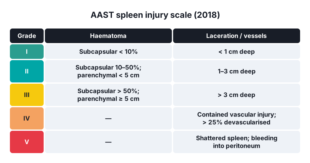
AAST spleen injury scale (2018)
- Grade I → subcapsular haematoma < 10% surface; laceration < 1 cm deep; capsular tear
- Grade II → subcapsular haematoma 10–50%; intraparenchymal haematoma < 5 cm; laceration 1–3 cm deep
- Grade III → subcapsular haematoma > 50% or expanding; ruptured haematoma; intraparenchymal haematoma ≥ 5 cm; laceration > 3 cm deep
- Grade IV → any vascular injury (pseudoaneurysm, AV fistula) or active bleeding confined to the capsule; laceration involving segmental or hilar vessels with > 25% devascularisation
- Grade V → shattered spleen; any vascular injury with active bleeding beyond the spleen into the peritoneum
WSES classification
- Minor → AAST I–II, stable
- Moderate → AAST III, stable
- Severe → AAST IV–V stable, or any grade unstable
6. Clinical picture
Symptoms
- Left upper quadrant pain
- Left shoulder tip pain (Kehr's sign) → diaphragmatic irritation
- Generalised abdominal pain if haemoperitoneum is large
- Dizziness, thirst, collapse → blood loss
- Delayed rupture → sudden pain and collapse days after a minor injury
Signs
- Haemorrhagic shock → tachycardia, hypotension, pallor, cool peripheries
- LUQ tenderness, guarding; distension
- Left lower rib fractures, bruising
- Ballance's sign → fixed dullness in the left flank (clotted blood) and shifting dullness on the right
- Peritonism with a large haemoperitoneum
- Children → may stay stable until sudden collapse
7. Diagnosis
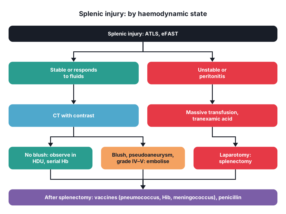
- ATLS primary survey (ABCDE); bloods: FBC, crossmatch, coagulation, lactate, base deficit, blood gas
- eFAST → free fluid in the splenorenal recess and pelvis; in unstable patients → theatre
- CT with IV contrast (arterial and portal venous phases) → stable patients:
- AAST grade
- Contrast blush (active bleeding), pseudoaneurysm → angioembolisation
- Haemoperitoneum volume, associated injuries (left kidney, pancreatic tail, diaphragm, colon)
- Angiography → diagnosis and embolisation
- Repeat CT → only for symptoms, a fall in haemoglobin, or high-grade injuries (pseudoaneurysm check at 48–72 hours in some centres)
- Diagnostic peritoneal lavage → rarely (no FAST)
8. Differential diagnosis
- Left kidney injury, pancreatic tail injury
- Liver injury, diaphragm rupture
- Rib fractures alone, haemothorax
- Gastric or colonic injury
- Retroperitoneal haematoma (pelvic fracture)
9. Treatment
Conservative
- Non-operative management (NOM) → standard for haemodynamically stable patients, any grade:
- Conditions → stable (or responds to fluids), no peritonitis, no other injury needing laparotomy, CT available, HDU or ICU monitoring, theatre and surgeon available 24 hours
- Serial examination and haemoglobin every 6 hours for 24–48 hours
- Bed rest (short), early mobilisation; VTE prophylaxis after 24–48 hours if no bleeding
- Avoid contact sport for 6–12 weeks (grades III–V) or 2–3 months for children
- Failure → mostly within 72 hours; risk higher with grade IV–V, large haemoperitoneum, age over 55
- Angioembolisation → improves NOM success:
- Contrast blush on CT, pseudoaneurysm, AV fistula in a stable patient
- Consider for grades IV–V in stable patients, even without a blush
- Proximal (main splenic artery coils, lowers perfusion pressure) or distal (selective, bleeding branch)
- Vaccination after embolisation of the main artery → debated; many give it as for splenectomy if a large part is lost
Surgical
- Indications for laparotomy → haemodynamic instability (non-responder), peritonitis, other injuries needing surgery, failed NOM, no CT or ICU available
- Splenectomy (trauma):
- Massive transfusion protocol, tranexamic acid, permissive hypotension
- Midline laparotomy; pack the four quadrants
- Mobilise the spleen medially → divide the splenorenal and splenophrenic ligaments, lift the spleen into the wound with the tail of the pancreas
- Compress the hilum by hand
- Divide the short gastric vessels (gastrosplenic ligament) close to the spleen; avoid the stomach wall
- Ligate the splenic artery and vein separately at the hilum; protect the tail of the pancreas
- Check haemostasis; look for accessory spleens (in elective cases for haematological disease)
- Drain only if the pancreatic tail is injured
- Splenic conservation (splenorrhaphy) → stable patients at surgery with grade I–III injuries: haemostatic agents, suture repair over pledgets, mesh wrap (absorbable), partial splenectomy (segmental end arteries)
- Laparoscopic splenectomy → elective (ITP, haemolytic anaemia), selected delayed trauma cases in stable patients
- Damage control → in the lethal triad; splenectomy is fast and definitive (not a time for conservation)
- Splenic autotransplantation (omental pouch) → possible, uncertain value
After splenectomy (or loss of splenic function)
- Vaccinations → pneumococcal (PCV then PPSV23), Haemophilus influenzae type b, meningococcal (ACWY and B), yearly influenza
- Timing → 14 days before elective splenectomy; 14 days after emergency splenectomy (or before discharge)
- Antibiotic prophylaxis → phenoxymethylpenicillin (or erythromycin/amoxicillin) for at least 1–2 years after surgery (lifelong in high risk); standby course at home for fever
- Patient card and education → seek help early for fever; malaria prophylaxis; animal bites (Capnocytophaga)
- Thrombocytosis → aspirin if platelets over 1,000 × 10⁹/L (debated)
10. Complications
Of the injury and NOM
- Delayed rupture, rebleeding
- Pseudoaneurysm
- Splenic abscess, splenic infarction (after embolisation)
- Missed injuries (hollow viscus, pancreatic tail, diaphragm)
Of splenectomy
- Bleeding (short gastrics, hilum)
- Pancreatic tail injury → pancreatic fistula, pseudocyst
- Gastric wall injury (short gastrics) → gastric fistula
- Left subphrenic abscess, left basal atelectasis, left pleural effusion
- Thrombocytosis, VTE, portal or splenic vein thrombosis
- Overwhelming post-splenectomy infection (OPSI):
- Streptococcus pneumoniae (50–90%), Haemophilus influenzae, Neisseria meningitidis; Capnocytophaga canimorsus (dog bites), malaria, babesiosis
- Highest risk → children under 5, first 2 years after surgery, splenectomy for haematological disease (thalassaemia, sickle cell)
- Starts with flu-like illness → septic shock, DIC, Waterhouse-Friderichsen syndrome within hours
- Treatment → immediate IV antibiotics (ceftriaxone ± vancomycin), ICU
- Blood film → Howell-Jolly bodies, target cells, Pappenheimer bodies
Show as a list
Splenic injury
- Anatomy
- Ribs 9-11
- Splenorenal ligament
- Short gastrics
- Pancreatic tail at hilum
- Mechanism
- Blunt: commonest organ
- Left rib fractures
- Splenomegaly: EBV
- Clinical
- LUQ pain
- Kehr's sign
- Shock
- Tests
- eFAST: unstable
- CT: stable
- AAST I-V
- Stable
- Non-operative
- Angioembolisation for blush
- Serial Hb
- Unstable
- Massive transfusion
- Splenectomy
- Splenorrhaphy if stable
- After splenectomy
- Vaccines: pneumo, Hib, meningo
- Penicillin
- OPSI
- Howell-Jolly bodies


