
102. Rectal prolapse
High yield
Full-thickness rectum protruding through the anus; fix it from the abdomen if fit, from the perineum if frail.
Exam Prep · State examSurgery · General surgerySurgery · Colorectal
1. Definition
- Rectal prolapse (procidentia) → full-thickness intussusception of the rectal wall that protrudes through the anus
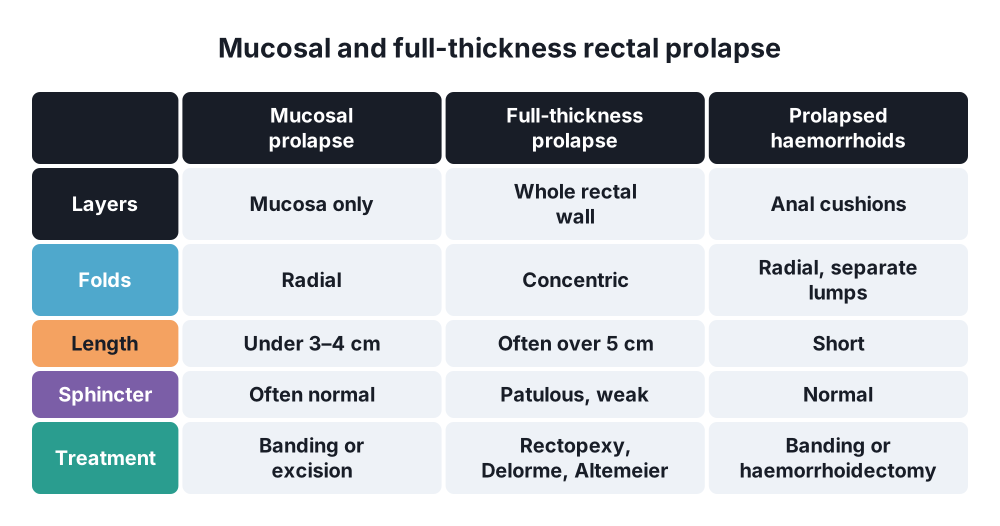
- Mucosal prolapse → only the mucosa protrudes (usually under 3–4 cm); radial folds; often with haemorrhoids
- Internal rectal intussusception (occult prolapse) → the rectum telescopes into itself but does not pass the anus
- Incarcerated prolapse → oedematous prolapse that cannot be reduced; may become strangulated and necrotic
2. Epidemiology
- About 2.5 per 100 000 people a year
- Women → about 6 times more often than men; peak in the 7th decade
- Children → peak at 1–3 years; usually mucosal; often settles with growth
- Young men → often linked with psychiatric illness, chronic constipation or learning disability
- Many women have associated pelvic floor disorders:
- Urinary incontinence in 20–35%
- Vaginal vault or uterine prolapse in 15–30%
3. Aetiology and risk factors
- Chronic straining → constipation, obstructed defecation
- Weak pelvic floor → multiparity, vaginal delivery, ageing, pudendal nerve damage
- Anatomical features:
- Deep pouch of Douglas (rectovaginal or rectovesical pouch)
- Lax lateral ligaments, poor fixation of the rectum to the sacrum
- Redundant (long, mobile) sigmoid colon
- Loss of the normal sacral curve, patulous anus, weak sphincters
- Connective tissue disease → Ehlers–Danlos, Marfan (younger patients, more recurrence)
- Children → cystic fibrosis, diarrhoea, malnutrition, whooping cough, spina bifida
- Neurological disease → spinal injury, multiple sclerosis, cauda equina lesions
4. Pathogenesis
- Two theories, now combined:
- Sliding hernia theory (Moschcowitz) → the deep pouch of Douglas pushes the anterior rectal wall through a weak pelvic floor
- Intussusception theory (Broden and Snellman) → circumferential intussusception starts 6–8 cm above the anal verge and slides down
- Defecography supports the intussusception theory
- Repeated prolapse stretches the sphincters and damages the pudendal nerves → faecal incontinence (in 50–75%)
- The prolapsed mucosa is exposed → mucus, bleeding, ulceration (solitary rectal ulcer)
- Venous congestion in an irreducible prolapse → oedema → incarceration → strangulation and necrosis
5. Classification
By depth
- Mucosal (partial) prolapse → radial folds; under 3–4 cm
- Full-thickness (complete) prolapse → concentric folds; often over 5 cm; double wall felt between the fingers
- Internal intussusception → seen only on defecography
Oxford grading of rectal prolapse (internal to external)
- Grade I–II → high or low rectorectal intussusception
- Grade III–IV → rectoanal intussusception
- Grade V → external prolapse
Clinical stages (Bulgarian and Russian schools)
- Stage I → prolapse only on defecation, reduces by itself
- Stage II → prolapse on straining or exertion, needs manual reduction
- Stage III → prolapse when standing or walking; sphincter weakness and incontinence
6. Clinical picture
- A lump coming out of the anus on straining, coughing or walking
- Early → reduces by itself; later → must be pushed back; finally → stays out
- Mucus discharge and soiling, perianal itching
- Bleeding from exposed, ulcerated mucosa
- Faecal incontinence (50–75%) or constipation and incomplete emptying (25–50%)
- Tenesmus, rectal pressure, pelvic pain
- Incarcerated prolapse → painful, swollen, dark red or black mass; may ulcerate or become gangrenous; urinary retention
- Associated urinary incontinence or vaginal prolapse in women
Examination
- Inspection → may show nothing at rest; ask the patient to strain on a commode to reproduce it
- Concentric mucosal folds → full-thickness prolapse
- Radial folds → mucosal prolapse or prolapsing haemorrhoids
- DRE → patulous anus, weak resting tone and squeeze
- Check for vaginal or uterine prolapse and cystocele
7. Diagnosis
- Clinical → if the prolapse is seen, no other test is needed to confirm it
- Colonoscopy (or at least sigmoidoscopy) before surgery → excludes a tumour as a lead point and synchronous disease; solitary rectal ulcer may be seen
- Defecography (fluoroscopic or MRI) → occult intussusception, rectocele, enterocele, sigmoidocele; multi-compartment prolapse
- Anorectal manometry → resting and squeeze pressures (predicts continence after repair)
- Endoanal ultrasound → sphincter defects
- Colonic transit study → severe constipation (slow transit changes the choice of operation)
- Urodynamics and urogynaecology review → if urinary or vaginal symptoms (combined repair)
- Children → sweat test for cystic fibrosis
8. Differential diagnosis
- Prolapsing haemorrhoids → radial folds, separate cushions, normal sphincter tone
- Mucosal prolapse
- Prolapsing rectal polyp or tumour (lead point)
- Intussusception of the colon through the anus (in children, ileocolic intussusception can reach the anus)
- Anal warts, hypertrophied papilla
- Solitary rectal ulcer syndrome (often coexists with internal intussusception)
9. Treatment
Conservative
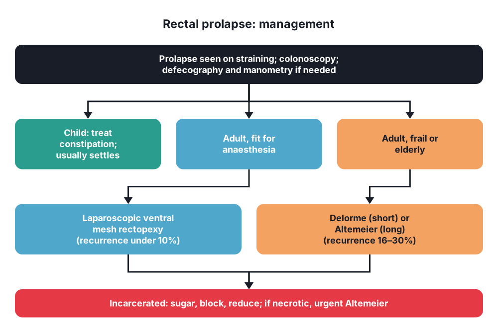
- Children → most resolve without surgery: treat constipation or diarrhoea, avoid long sitting on the potty, manual reduction; sclerotherapy or surgery rarely
- Adults:
- Fibre, stool softeners, avoid straining
- Pelvic floor physiotherapy and biofeedback → for incontinence or obstructed defecation, before and after surgery
- Conservative care alone does not cure a full-thickness prolapse in adults; it is for patients unfit for any operation
- Incarcerated prolapse:
- Sedation or anal block, sugar sprinkled on the prolapse (osmotic) to reduce the oedema, cold packs, then gentle manual reduction
- If it cannot be reduced or is necrotic → urgent perineal rectosigmoidectomy (Altemeier)
Surgical
- Choice of approach:
- Abdominal approach → fit patients; lower recurrence (under 10% at 10 years)
- Perineal approach → elderly or frail patients; can be done under spinal or regional anaesthesia; higher recurrence (16–30%)
- Abdominal operations (usually laparoscopic or robotic):
- Laparoscopic ventral mesh rectopexy (D'Hoore) → dissect only in front of the rectum in the rectovaginal septum, fix a mesh to the anterior rectum and to the sacral promontory
- Spares the lateral ligaments and the autonomic nerves → less new constipation
- Treats internal intussusception and rectocele as well
- Mesh erosion is rare (about 1–2%); biological mesh is an option
- Posterior suture rectopexy → full mobilisation of the rectum, sutures to the presacral fascia
- Dividing the lateral ligaments may worsen constipation
- Resection rectopexy (Frykman–Goldberg) → sigmoid resection plus rectopexy for patients with long sigmoid and constipation; risk of anastomotic leak
- Older operations → Ripstein (anterior sling, causes obstruction), Wells (posterior Ivalon sponge), now rarely used
- Laparoscopic ventral mesh rectopexy (D'Hoore) → dissect only in front of the rectum in the rectovaginal septum, fix a mesh to the anterior rectum and to the sacral promontory
- Perineal operations:
- Delorme's procedure → strip the mucosa off the prolapsed rectum and plicate the muscle wall (concertina); for short prolapse (under 3–4 cm) and frail patients; recurrence 10–30%
- Altemeier's perineal rectosigmoidectomy → full-thickness resection of the prolapsed rectum and sigmoid with coloanal anastomosis; often with levatorplasty; best for long or incarcerated prolapse
- Thiersch's anal encirclement → a band around the anus; historical; only for very frail patients
- Mucosal prolapse → banding, sclerotherapy, or excision as in haemorrhoidectomy
- Persistent incontinence after repair (needs 6–12 months to recover) → biofeedback, sacral nerve stimulation, sphincter repair
10. Complications
Of the disease
- Faecal incontinence (sphincter stretching, pudendal neuropathy)
- Ulceration and bleeding, solitary rectal ulcer
- Incarceration, strangulation, gangrene, rarely rupture with small bowel evisceration through the anus
Of surgery
- Recurrence → abdominal under 10%, perineal 16–30%, Delorme up to 30%
- Constipation after posterior rectopexy (division of lateral ligaments)
- Anastomotic leak (resection rectopexy, Altemeier)
- Mesh erosion or infection, presacral bleeding
- Sexual and bladder dysfunction from pelvic nerve injury (rare with ventral dissection)
Prognosis
- Continence improves in most patients after repair, but not in all
Show as a list
Rectal prolapse
- Types
- Mucosal: radial folds
- Full-thickness: concentric
- Internal intussusception
- Causes
- Elderly women
- Straining, weak pelvic floor
- Deep pouch of Douglas
- Long sigmoid
- Symptoms
- Lump, mucus, bleeding
- Incontinence
- Incarceration
- Tests
- Strain on commode
- Colonoscopy
- Defecography
- Treatment
- Children: conservative
- Fit: ventral mesh rectopexy
- Frail: Delorme, Altemeier
- Incarcerated: sugar, reduce


