
99. Diseases of the rectum and how to examine them
High yield
Rectum and anal canal anatomy, main symptoms, and how to examine them: DRE, endoscopy, MRI, ultrasound and physiology.
Exam Prep · State examSurgery · General surgerySurgery · ColorectalAnatomy · Pelvis
1. Definition
- Rectum → the last part of the large bowel, from the rectosigmoid junction (about S3, where the taeniae spread out and the sigmoid mesentery ends; about 15 cm from the anal verge) to the anorectal junction (top of the anorectal ring, levator ani)
- Anal canal → about 3–4 cm long, from the anorectal ring to the anal verge
- Diseases of the rectum and anus (proctology) → congenital, inflammatory, functional, vascular, infective and neoplastic conditions
- This question covers the anatomy, the symptoms and the methods of examination; specific diseases follow (questions 100–106)
2. Epidemiology
- Anorectal symptoms are very common → about half the population has haemorrhoids at some point
- Rectal bleeding → about 15% of adults a year; most from benign anal causes, but colorectal cancer must be excluded in older patients
- Rectal cancer → about a third of colorectal cancers
- Faecal incontinence → about 8–10% of adults; women (obstetric injury) and the elderly
- Obstructed defaecation and constipation → about 15% of adults
3. Aetiology and risk factors
Surgical anatomy (revision)
- Length → about 12–15 cm; upper third covered by peritoneum in front and on the sides, middle third in front only, lower third below the peritoneal reflection (extraperitoneal)
- Peritoneal reflection → about 7–9 cm from the anal verge in men (rectovesical pouch), 5–7.5 cm in women (rectouterine pouch of Douglas)
- Three lateral curves inside → valves of Houston (upper and lower on the left, middle on the right at about the peritoneal reflection)
- No taeniae, no appendices epiploicae, no haustra
- Mesorectum → fatty tissue with vessels, lymphatics and nodes around the rectum, enclosed by the mesorectal (visceral) fascia → the plane of total mesorectal excision
- Waldeyer's fascia → rectosacral fascia at S4 (posteriorly); Denonvilliers' fascia → in front, between the rectum and the prostate and seminal vesicles (men) or vagina (women)
- Blood supply:
- Superior rectal artery (end of the inferior mesenteric artery) → main supply
- Middle rectal arteries (internal iliac) → variable, in the lateral ligaments
- Inferior rectal arteries (internal pudendal) → anal canal
- Median sacral artery (minor)
- Venous drainage → superior rectal vein → IMV → portal system; middle and inferior rectal veins → systemic (iliac); a portosystemic anastomosis (rectal varices in portal hypertension)
- Lymph drainage:
- Upper and middle rectum → along the superior rectal vessels → inferior mesenteric nodes
- Lower rectum → also lateral (internal iliac, obturator) nodes
- Anal canal below the dentate line → superficial inguinal nodes
- Nerves → sympathetic (hypogastric nerves, L1–L3) and parasympathetic (pelvic splanchnic nerves, nervi erigentes S2–S4) → inferior hypogastric (pelvic) plexus on the pelvic side walls; injury → bladder and sexual dysfunction
Anal canal
- Dentate (pectinate) line → about 2 cm from the anal verge; the junction of endoderm and ectoderm:
- Above → columnar epithelium (anal transition zone), visceral nerves (painless), portal venous drainage, internal iliac nodes, adenocarcinoma
- Below → squamous epithelium (anoderm), somatic nerves (painful: inferior rectal nerve), systemic veins, inguinal nodes, squamous cell carcinoma
- Anal columns of Morgagni with anal crypts and glands at the dentate line (source of cryptoglandular abscess and fistula)
- Anal cushions → vascular cushions at 3, 7 and 11 o'clock (lithotomy position) → haemorrhoids
Sphincters and continence
- Internal anal sphincter → thickened circular smooth muscle; involuntary; 70–85% of resting tone
- External anal sphincter → striated, voluntary (pudendal nerve S2–S4); squeeze pressure
- Puborectalis → sling round the anorectal junction → anorectal angle (about 90° at rest, straightens on defaecation)
- Anorectal ring → upper border of the sphincter complex, felt on DRE; dividing it causes incontinence
- Continence depends on sphincters, anorectal angle, rectal sensation and compliance, stool consistency, anal cushions
Main disease groups
- Congenital → anorectal malformations, Hirschsprung's disease (question 106)
- Vascular → haemorrhoids (question 101), rectal varices
- Inflammatory → proctitis (UC, Crohn's, radiation, infection: gonorrhoea, Chlamydia LGV, herpes, syphilis, C. difficile), solitary rectal ulcer
- Infective and suppurative → anorectal abscess, fistula-in-ano (questions 104–105), pilonidal sinus
- Anal fissure (question 101)
- Functional and pelvic floor → rectal prolapse (question 102), rectocele, intussusception, obstructed defaecation, faecal incontinence, proctalgia fugax, levator ani syndrome
- Neoplastic → rectal polyps, rectal cancer (question 100), anal cancer, neuroendocrine tumours, GIST, melanoma, presacral tumours (chordoma, teratoma)
- Trauma and foreign bodies, pruritus ani, condylomata, other (question 103)
4. Pathogenesis
- Bleeding:
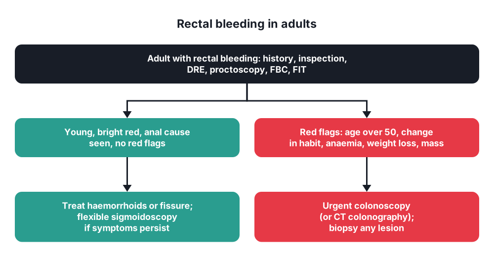
- Bright red on the paper or dripping into the pan, separate from the stool → anal source (haemorrhoids, fissure)
- Dark red, mixed with stool, with mucus → rectal or colonic source (cancer, polyps, proctitis)
- Painful bleeding → fissure; painless → haemorrhoids, cancer, polyp
- Tenesmus → feeling of incomplete evacuation from a mass or inflammation in the rectum (cancer, proctitis)
- Mucus discharge → proctitis, villous adenoma (large villous adenomas cause hypokalaemia), prolapse, fistula
- Pain → below the dentate line (fissure, thrombosed haemorrhoid, abscess, anal cancer); rectal cancer is usually painless until it invades the sphincter or sacral nerves
- Change in bowel habit → narrowing of the lumen (cancer), loss of reservoir (proctitis)
- Obstructed defaecation → pelvic floor dyssynergia, rectocele, intussusception, enterocele
- Incontinence → sphincter injury (obstetric, surgical), pudendal neuropathy, rectal prolapse, low reservoir (proctitis, after low anterior resection)
5. Classification
Symptoms of anorectal disease
- Bleeding, pain, lump or prolapse, discharge (pus, mucus), itching, change in bowel habit, tenesmus, incontinence, obstructed defaecation
Methods of examination
- Clinical → history, inspection, palpation (DRE), bimanual examination
- Endoscopy → proctoscopy (anoscopy), rigid sigmoidoscopy, flexible sigmoidoscopy, colonoscopy
- Imaging → MRI pelvis, endoanal and endorectal ultrasound, CT, CT colonography, defaecography (fluoroscopic or MR), fistulography
- Physiology → anorectal manometry, rectal sensation and compliance, balloon expulsion test, pudendal nerve terminal motor latency, EMG, colonic transit study
- Laboratory → FBC, iron studies, CEA, FIT (faecal immunochemical test), stool culture, calprotectin, STI tests
- Biopsy and histology; examination under anaesthesia (EUA)
Positions for examination
- Left lateral (Sims') → most common in clinic
- Lithotomy → theatre; position described by the clock face (12 o'clock anterior, 6 o'clock posterior)
- Knee-chest (genupectoral) and prone jack-knife → good exposure
6. Clinical picture
History
- Bleeding → colour, relation to stool, amount, pain
- Bowel habit → frequency, consistency, recent change, straining, incomplete evacuation, digitation (rectocele)
- Pain → during and after defaecation (fissure), constant throbbing (abscess)
- Lump → reduces on its own or by hand (haemorrhoids, prolapse)
- Discharge, itching, incontinence (flatus, liquid, solid; urgency; pads), obstetric history
- Red flags → age over 50, change in bowel habit, dark blood mixed with stool, weight loss, iron deficiency anaemia, abdominal or rectal mass, family history, nocturnal symptoms
- Systemic → weight loss, fever, joint or eye symptoms (IBD), sexual history (STIs)
Inspection
- Spread the buttocks → skin tags, fissure (posterior midline in 90%; sentinel tag), external haemorrhoids, prolapse, fistula openings (Goodsall's rule), abscess, warts, tumour, excoriation, scars
- Ask the patient to strain → prolapse of haemorrhoids or rectum, perineal descent
- Sensation and anal reflex ("anal wink" → S2–S4)
Digital rectal examination (DRE)
- Consent and chaperone; explain; left lateral position, knees to the chest
- Lubricated gloved index finger, pressed gently on the anal verge, then inserted
- Assess:
- Resting tone (internal sphincter) and squeeze (external sphincter)
- Anorectal ring, puborectalis
- Walls of the rectum all round (360°): mass, ulcer, stricture, tenderness, bulge (abscess)
- Mass → distance from the anal verge, size, position (clock face), fixity (mobile, tethered, fixed), relation to the sphincters and the prostate or vagina
- Prostate (men) → size, consistency, nodules, median groove; cervix (women)
- Pouch of Douglas → tenderness (pelvic abscess), nodules (Blumer's shelf: peritoneal metastases)
- Finger on withdrawal → blood, mucus, pus, stool colour (melaena)
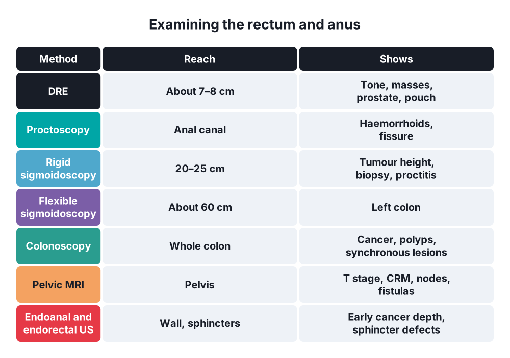
- DRE reaches about 7–8 cm; it finds most low rectal cancers
- Bimanual (with the other hand on the abdomen, or vaginal and rectal together) → pelvic masses, rectovaginal septum
Special situations
- Very painful anus (fissure, abscess) → examine under anaesthesia; do not force DRE
7. Diagnosis
Endoscopy
- Proctoscopy (anoscopy) → anal canal and lower rectum; haemorrhoids (seen when the patient strains as the scope is withdrawn), fissure, papillae, internal openings, banding and injection of haemorrhoids
- Rigid sigmoidoscopy → up to about 20–25 cm; quick, in clinic, no sedation; measures the exact height of a rectal tumour from the anal verge; biopsies; proctitis
- Flexible sigmoidoscopy → up to about 60 cm (splenic flexure); enema preparation only; bleeding in younger patients, screening, surveillance
- Colonoscopy → whole colon and terminal ileum; gold standard for colorectal cancer and polyps; biopsy, polypectomy, tattoo; synchronous tumours (3–5%) before rectal cancer surgery
- CT colonography → alternative when colonoscopy is incomplete or unsafe (frail)
Imaging
- MRI pelvis (high-resolution) → local staging of rectal cancer:
- T stage, depth of spread into mesorectal fat (mm), mesorectal fascia (CRM) involvement, nodes, EMVI (extramural venous invasion), tumour height, relation to the peritoneal reflection and sphincters, lateral nodes
- Fistula-in-ano → tracks, extensions, abscesses (gold standard for complex fistulas)
- MR defaecography → prolapse, rectocele, enterocele, pelvic floor descent
- Endorectal ultrasound (ERUS) → early rectal cancers (T1 vs T2; sm1–sm3 for local excision), rectal polyps
- Endoanal ultrasound (EAUS) → sphincter defects (incontinence after childbirth), fistulas, abscesses
- CT chest, abdomen and pelvis → distant metastases (liver, lung); abscesses
- PET-CT → recurrence, indeterminate metastases
- Fluoroscopic defaecography (evacuation proctography) → obstructed defaecation (intussusception, rectocele)
- Water-soluble contrast enema → anastomotic leak, strictures, before stoma reversal
Physiology tests
- Anorectal manometry → resting pressure (internal sphincter), squeeze pressure (external sphincter), rectoanal inhibitory reflex (absent in Hirschsprung's disease), dyssynergia
- Rectal sensation and compliance (balloon distension), balloon expulsion test (dyssynergia)
- Colonic transit study (radio-opaque markers) → slow transit constipation
- Pudendal nerve terminal motor latency, EMG
Laboratory
- FBC and ferritin (iron deficiency), CEA (cancer baseline), FIT (screening and triage of symptoms), faecal calprotectin (IBD), stool culture and C. difficile, STI swabs and serology, HIV
Biopsy
- Every rectal mass or ulcer → biopsy (histology); full-thickness or EUA for suspected deep lesions; swabs for infection
8. Differential diagnosis
Rectal bleeding
- Anal → haemorrhoids, fissure, anal cancer, prolapse
- Rectal and colonic → colorectal cancer, polyps, proctitis and colitis, diverticular disease, angiodysplasia, solitary rectal ulcer, radiation proctitis
- Upper GI (massive bleeding with high urea) → peptic ulcer
- Children → fissure, polyps, Meckel's, intussusception
Rectal mass
- Rectal cancer, villous adenoma, carcinoid, GIST, lymphoma
- Extrarectal → prostate cancer, cervical or ovarian cancer, Blumer's shelf, endometriosis, presacral tumours, faecal mass, abscess
Pain
- Fissure, thrombosed external haemorrhoid, abscess, anal cancer, proctalgia fugax (short, severe, at night), levator ani syndrome, coccydynia
9. Treatment
Conservative
- General measures for benign disease → high-fibre diet, fluids, stool softeners, avoid straining, sitz baths, topical treatments
- Proctitis → topical 5-ASA or steroids (UC), antibiotics (STIs), sucralfate or formalin (radiation)
- Pelvic floor disorders → biofeedback (dyssynergia, incontinence), pelvic floor exercises, loperamide (incontinence), laxatives and irrigation (obstructed defaecation)
- Office procedures → rubber band ligation and injection sclerotherapy of haemorrhoids, botulinum toxin and topical nitrates or diltiazem for fissure
Surgical
- Examination under anaesthesia (EUA) → when examination in clinic is too painful or incomplete; drain abscesses, lay open fistulas, biopsy
- Transanal procedures → polypectomy, TEM or TAMIS (local excision of polyps and early cancers), haemorrhoidectomy, sphincterotomy, fistula surgery
- Abdominal and pelvic procedures → anterior resection with total mesorectal excision, abdominoperineal resection, rectopexy (questions 100 and 102)
- Incontinence → sphincter repair (overlapping sphincteroplasty), sacral nerve stimulation, stoma as a last resort
- Specific diseases → see questions 100–106
10. Complications
Of the diseases
- Anaemia (chronic bleeding), strangulated haemorrhoids, sepsis (abscess), incontinence, stricture, malignant change (villous adenoma, chronic fistula, chronic UC)
Of examination
- Pain, bleeding after biopsy
- Perforation during sigmoidoscopy or colonoscopy (about 1 in 1000–3000 colonoscopies; higher with polypectomy)
- Vasovagal reactions, sedation complications
- Missed diagnosis → attributing bleeding to haemorrhoids without excluding cancer
Prognosis
- Depends on the disease; early diagnosis of rectal cancer gives the best results
Show as a list
Rectal disease: examination
- Anatomy
- 12-15 cm
- Mesorectum, TME plane
- Dentate line
- Sphincters, puborectalis
- Symptoms
- Bleeding
- Pain, lump
- Tenesmus, mucus
- Incontinence
- Clinical
- Inspection, strain
- DRE: tone, mass
- Bimanual
- Endoscopy
- Proctoscopy
- Rigid sigmoidoscopy
- Flexible sigmoidoscopy
- Colonoscopy
- Imaging
- MRI pelvis: CRM
- ERUS: early cancer
- EAUS: sphincters
- CT, defaecography
- Physiology
- Manometry
- Balloon expulsion
- Transit study


