
100. Rectal carcinoma
High yield
MRI-staged rectal cancer is treated with TME surgery, neoadjuvant therapy when advanced, and watch-and-wait after complete response.
Exam Prep · State examSurgery · General surgerySurgery · ColorectalSurgery · Surgical oncology
1. Definition
- Rectal carcinoma → malignant tumour of the rectum, over 90–95% adenocarcinoma
- Rectal cancer by height → lower edge within 15 cm of the anal verge on rigid sigmoidoscopy (UK and many European guidelines: within 15 cm; some use 12 cm or the sigmoid take-off on MRI)
- Low → 0–5 cm; middle → 5–10 cm; high → 10–15 cm
- Differs from colon cancer in anatomy (pelvis, sphincters, nerves), local recurrence risk and the role of radiotherapy
- Shared features with colon cancer (genetics, pathways, screening, chemotherapy) → question 86; anatomy and examination → question 99
2. Epidemiology
- About 30–35% of colorectal cancers
- Colorectal cancer → third most common cancer, second cause of cancer death
- Men more than women (about 1.5 : 1)
- Median age about 65–70; rising in under-50s (about 2% a year)
- Local recurrence → 20–40% in the era before TME; now under 5–10% with TME and neoadjuvant treatment
- 5-year survival → stage I about 90%, II about 75–80%, III about 60–70%, IV about 10–15%
3. Aetiology and risk factors
- Same as colon cancer (question 86):
- Age, male sex, family history
- Diet → red and processed meat, low fibre; obesity, inactivity, smoking, alcohol, type 2 diabetes
- Adenomas (adenoma–carcinoma sequence), especially large villous adenomas of the rectum
- Inflammatory bowel disease (UC, Crohn's colitis), PSC
- Hereditary → Lynch syndrome, FAP, MUTYH-associated polyposis
- Previous pelvic radiotherapy (prostate cancer)
- Molecular → mostly chromosomal instability (APC, KRAS, TP53); dMMR/MSI-high only about 2–5% of rectal cancers (much less than in the right colon)
4. Pathogenesis
Gross types
- Ulcerative (most common) → ulcer with raised everted edges
- Polypoid (cauliflower), annular (stenosing), diffusely infiltrating (linitis plastica-like, rare)
- Mucinous (over 50% mucin) and signet ring → worse prognosis
Spread
- Direct → through the wall into the mesorectum, then to adjacent organs (bladder, prostate, seminal vesicles, vagina, uterus, sacrum, pelvic side walls)
- Distal intramural spread is usually under 1 cm (basis of the 1 cm distal margin rule)
- Lymphatic:
- Upward along the superior rectal vessels to the inferior mesenteric nodes (main route; contained in the mesorectum)
- Lateral → internal iliac and obturator nodes for low tumours below the peritoneal reflection
- Downward to inguinal nodes → only if the tumour invades the anal canal below the dentate line
- Venous → superior rectal vein → portal vein → liver (most common); middle and inferior rectal veins → systemic → lungs (lung metastases more common than in colon cancer)
- Extramural venous invasion (EMVI) on MRI → high risk of distant spread
- Transcoelomic → peritoneum (upper rectum)
- Implantation → anastomosis, wounds
Circumferential resection margin (CRM)
- The distance from the tumour to the mesorectal fascia (the surgical plane)
- CRM involved (1 mm or less) → high local recurrence and worse survival → key target of MRI staging and surgery
5. Classification
TNM (AJCC/UICC 8th edition)
- Tis → intramucosal carcinoma
- T1 → submucosa; T2 → muscularis propria; T3 → through the muscle into the mesorectal fat (T3a under 1 mm, T3b 1–5 mm, T3c 5–15 mm, T3d over 15 mm on MRI); T4a → visceral peritoneum; T4b → other organs
- N1 → 1–3 nodes (N1c: tumour deposits); N2 → 4 or more
- M1a → one organ; M1b → more than one; M1c → peritoneum
Stage groups
- I → T1–2 N0; II → T3–4 N0; III → any T N1–2; IV → M1
- Dukes → A (wall), B (through the wall, N0), C (nodes), D (distant; Turnbull)
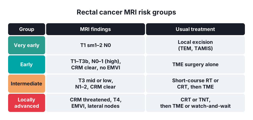
MRI risk groups (ESMO)
- Very early → T1 sm1–2 N0 → local excision
- Early ("good") → T1–T2, T3a–b, N0–N1 (high rectum), CRM clear, no EMVI → TME alone
- Intermediate ("bad") → T3 (mid or low), N1–N2, CRM clear, no EMVI → short-course RT or chemoradiotherapy, or TME alone in selected cases
- Locally advanced ("ugly") → CRM threatened or involved, T4, EMVI positive, lateral nodes, low tumours involving the levators → neoadjuvant chemoradiotherapy or total neoadjuvant therapy (TNT)
Pathology quality (Quirke)
- Mesorectal plane (complete, smooth) → best
- Intramesorectal plane (nearly complete)
- Muscularis propria plane (incomplete, defects to the muscle) → higher recurrence
Response to neoadjuvant treatment
- Clinical complete response (cCR) → no tumour on DRE, endoscopy (white scar) or MRI
- Pathological complete response (pCR) → no tumour in the specimen (ypT0N0); about 15–20% after CRT, up to about 28% after TNT
6. Clinical picture
Symptoms
- Rectal bleeding → most common; dark or bright red, mixed with stool or on the surface
- Change in bowel habit → frequent, loose or constipated stools, spurious diarrhoea
- Tenesmus (feeling of incomplete emptying), early morning bloody diarrhoea, mucus
- Pain → late (invasion of the sphincters or sacral nerves; perineal or sciatic pain)
- Obstruction → narrow stools, colic, distension (large or annular tumours)
- Urinary symptoms, pneumaturia (bladder invasion: fistula), vaginal discharge of faeces
- Weight loss, anorexia, fatigue (anaemia)
- Metastases → hepatomegaly, jaundice, cough
Signs
- DRE → hard, irregular, ulcerated mass with raised edges; note height from the anal verge, size, circumference involved, mobility or fixity, relation to the sphincters and prostate or vagina; blood on the glove
- Anaemia, abdominal distension, hepatomegaly, ascites
- Inguinal nodes (very low tumours)
- Blumer's shelf (pouch of Douglas deposits)
7. Diagnosis
Confirm
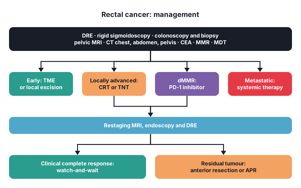
- DRE and rigid sigmoidoscopy (exact height)
- Colonoscopy with biopsy → histology; synchronous tumours (3–5%) and polyps; tattoo for surgery
- If colonoscopy is incomplete → CT colonography
Local staging
- High-resolution pelvic MRI → imaging of choice:
- T stage, depth of extramural spread (mm)
- CRM (distance to the mesorectal fascia; threatened if 1 mm or less, some use 2 mm)
- Nodes (mesorectal and lateral), tumour deposits
- EMVI
- Height, relation to the peritoneal reflection, levators and sphincters (low rectal tumours)
- Restaging after neoadjuvant treatment (tumour regression grade; diffusion-weighted imaging)
- Endorectal ultrasound (ERUS) → early tumours (T1 vs T2, submucosal depth) for local excision; less useful for advanced tumours
Distant staging
- CT chest, abdomen and pelvis with contrast → liver and lung metastases
- MRI liver (indeterminate lesions), PET-CT (selected: suspected recurrence, before major resection of metastases)
Bloods and markers
- CEA (baseline), FBC, LFT, U&E
- MMR or MSI testing of every tumour (Lynch screening; dMMR tumours respond to immunotherapy)
- RAS, BRAF (metastatic disease)
Function and fitness
- Baseline continence, urinary and sexual function, frailty, comorbidity, stoma counselling and siting
Multidisciplinary team (MDT)
- Every patient discussed before treatment and after restaging
8. Differential diagnosis
- Benign → large villous adenoma, solitary rectal ulcer syndrome, inflammatory polyp, endometriosis, stercoral ulcer
- Inflammatory → proctitis (UC, Crohn's, radiation), STI proctitis (LGV), amoebiasis
- Other tumours → anal squamous cell carcinoma (different treatment: chemoradiotherapy), rectal neuroendocrine tumour, GIST, lymphoma, melanoma, prostate cancer invading the rectum, presacral tumours
- Haemorrhoids and fissure (common cause of bleeding, but must not be assumed)
- Diverticular stricture (rectosigmoid)
9. Treatment
Conservative
- Neoadjuvant treatment → for intermediate and locally advanced tumours (mid and low rectum):
- Short-course radiotherapy (SCRT) → 5 × 5 Gy over 1 week, then surgery within 1 week, or delayed surgery after 4–8 weeks (Swedish, Dutch TME, Stockholm III trials)
- Long-course chemoradiotherapy (LCCRT) → 45–50.4 Gy in 25–28 fractions over 5 weeks with capecitabine or 5-FU; surgery at 8–12 weeks (allows downstaging; German CAO/ARO/AIO-94: lower local recurrence than postoperative CRT, 6% vs 13%)
- Total neoadjuvant therapy (TNT) → all chemotherapy and radiotherapy before surgery:
- RAPIDO → SCRT then 6 cycles of CAPOX or 9 of FOLFOX → fewer treatment failures (23.7% vs 30.4%), pCR 28% vs 14%
- PRODIGE-23 → FOLFIRINOX then CRT → better disease-free survival, pCR 27.8% vs 12.1%
- OPRA → CRT then consolidation chemotherapy → more organ preservation (about 53%)
- Now a standard option for locally advanced rectal cancer (NCCN, ESMO)
- Neoadjuvant chemotherapy alone (selected; PROSPECT trial: FOLFOX with selective CRT, non-inferior for intermediate tumours in the upper and mid rectum)
- Organ preservation ("watch-and-wait"):
- After clinical complete response to CRT or TNT (no mass on DRE, flat white scar with telangiectasia on endoscopy, no tumour on diffusion-weighted MRI)
- Habr-Gama: 5-year survival 100% and disease-free survival 92% in the first series
- Regrowth in about 25–30%, mostly in the first 2 years, usually salvageable by surgery
- Close follow-up → DRE, endoscopy and CEA every 3–6 months, MRI every 6 months, CT every 6–12 months for 2 years, then less often for 5 years
- dMMR/MSI-high rectal cancer → neoadjuvant immunotherapy (dostarlimab or other PD-1 inhibitors): 100% clinical complete response in the first trial (Cercek 2022); avoids radiotherapy and surgery in many (emerging standard)
- Adjuvant chemotherapy → after surgery for high-risk pathology (ypN+), if not given before (CAPOX or FOLFOX)
- Metastatic disease → systemic chemotherapy ± biologics (anti-EGFR in RAS wild-type, bevacizumab), immunotherapy for dMMR; resection of liver or lung metastases if possible; radiotherapy to the primary for symptoms
- Palliation → stent (upper rectum), defunctioning stoma, laser or argon plasma coagulation, radiotherapy for bleeding or pain, palliative care
Surgical
- Principles:
- Total mesorectal excision (TME, Heald 1982) → sharp dissection in the "holy plane" between the mesorectal fascia and the presacral fascia, removing the whole mesorectum intact (with its nodes) → local recurrence under 5–10%
- Partial mesorectal excision (PME) → upper rectal tumours; divide the mesorectum 5 cm below the tumour at right angles (no coning)
- Margins → distal 1 cm enough for low tumours after neoadjuvant treatment (2 cm without), CRM over 1 mm
- High or low tie of the inferior mesenteric artery; splenic flexure mobilisation for a tension-free anastomosis
- Preserve the hypogastric nerves and pelvic plexus (bladder and sexual function)
- Operations:
- Anterior resection (AR) → high AR (upper rectum, anastomosis above the peritoneal reflection) or low AR with TME (mid and low rectum); stapled colorectal or coloanal anastomosis; colonic J-pouch or side-to-end anastomosis for low ones (better function); defunctioning loop ileostomy for low anastomoses (closed at about 3 months)
- Ultra-low AR with intersphincteric resection (ISR) and hand-sewn coloanal anastomosis → very low tumours not invading the external sphincter (good continence needed)
- Abdominoperineal resection (APR, Miles 1908) → rectum, anal canal and sphincters removed with a permanent end colostomy; for tumours invading the sphincters or levators, poor continence, or where a clear distal margin cannot be achieved; extralevator APR (ELAPE) reduces positive margins and perforation
- Hartmann's procedure → frail, obstructed or perforated cases, or poor sphincter function
- Pelvic exenteration → T4b tumours invading the bladder, prostate, uterus or sacrum, and locally recurrent disease → en bloc multivisceral resection for R0 (specialist centres)
- Access → laparoscopic (COREAN and other trials: similar oncological results to open), robotic (ROLARR; better in narrow male pelvis, lower conversion), transanal TME (taTME) (bottom-up dissection; selected centres after concerns about urethral injury and multifocal recurrence)
- Local excision → TEM, TAMIS (full-thickness, en bloc):
- Early T1 (sm1, maybe sm2), under 3 cm, well differentiated, no lymphovascular invasion, under 30% of the circumference
- Also after a good response to neoadjuvant treatment in selected patients (organ preservation)
- Completion TME if pathology shows high-risk features
- Obstructing rectal cancer → defunctioning loop colostomy (or ileostomy), then staging and neoadjuvant treatment, then resection
- ERAS (enhanced recovery), prehabilitation, correction of anaemia, mechanical bowel preparation + oral antibiotics, stoma marking
Follow-up
- CEA every 3–6 months and CT chest, abdomen and pelvis at intervals for 3–5 years; colonoscopy at 1 year, then every 3–5 years; pelvic MRI in watch-and-wait
10. Complications
Of the disease
- Obstruction, perforation, bleeding, fistula (rectovesical, rectovaginal)
- Local recurrence → pelvic pain, bleeding, obstruction
- Liver and lung metastases
Of surgery
- Anastomotic leak → about 5–15% after low AR (higher in men, low anastomoses, after radiotherapy, smokers); diverting ileostomy reduces its severity
- Low anterior resection syndrome (LARS) → frequency, urgency, clustering, incontinence in up to 60–80% (worse after radiotherapy and low anastomoses); treat with diet, loperamide, transanal irrigation, sacral nerve stimulation, stoma
- Bladder dysfunction and sexual dysfunction (erectile and ejaculatory problems, dyspareunia) → pelvic nerve injury
- Perineal wound problems after APR (infection, breakdown, hernia; flap reconstruction after ELAPE)
- Stoma complications, high-output ileostomy (dehydration, AKI)
- Ureteric injury, presacral venous bleeding (difficult to control: pressure, thumbtacks, muscle welding)
- Urethral injury (taTME)
Of radiotherapy and chemotherapy
- Radiation → proctitis, enteritis, fistula, pelvic insufficiency fractures, infertility, ovarian failure, second cancers, worse bowel function
- Chemotherapy → oxaliplatin neuropathy, capecitabine hand-foot syndrome, DPD deficiency toxicity
Prognosis
- Depends on stage, CRM status, quality of the TME specimen, EMVI, response to neoadjuvant treatment
- Positive CRM → local recurrence over 20%; complete mesorectal specimen → under 5%
Show as a list
Rectal carcinoma
- Basics
- Within 15 cm of verge
- Low, mid, high
- Adenocarcinoma
- Spread
- Mesorectum
- Lateral nodes if low
- Portal + systemic veins
- CRM key
- Staging
- DRE, rigid scope
- Colonoscopy, biopsy
- Pelvic MRI: CRM, EMVI
- CT chest-abdomen-pelvis
- MMR testing
- Neoadjuvant
- SCRT 5x5 Gy
- CRT 50 Gy + capecitabine
- TNT: RAPIDO, PRODIGE
- dMMR: dostarlimab
- Surgery
- TME: holy plane
- Low AR + ileostomy
- APR if sphincters
- TEM/TAMIS: early T1
- Watch and wait
- cCR
- Regrowth 25-30%
- Close surveillance
- Complications
- Leak
- LARS
- Nerve injury


