
97. Rare forms of peritonitis and abdominal sepsis
High yield
Primary, tuberculous, tertiary, dialysis, chemical and rarer peritonitis; sepsis definitions, intra-abdominal abscesses and source control.
Exam Prep · State examSurgery · General surgerySurgery · Emergency and trauma
1. Definition
- Rare peritonitis → forms other than the usual secondary peritonitis from a perforation (question 96):
- Primary (spontaneous) peritonitis → infection without a gut breach (SBP in cirrhosis, children with nephrotic syndrome, pneumococcal and gonococcal peritonitis in young women)
- Peritoneal dialysis (PD) peritonitis
- Tuberculous peritonitis
- Tertiary peritonitis → persistent or recurrent infection over 48 hours after adequate source control
- Chemical (aseptic) peritonitis → bile, blood, urine, pancreatic juice, barium, chyle
- Granulomatous peritonitis → starch (glove powder), talc, sarcoid, fungi, parasites (hydatid, Ascaris)
- Encapsulating peritoneal sclerosis ("abdominal cocoon")
- Familial Mediterranean fever (periodic aseptic peritonitis)
- Actinomycosis, Chlamydia (Fitz-Hugh–Curtis syndrome)
- Sepsis (Sepsis-3, 2016) → life-threatening organ dysfunction caused by a dysregulated host response to infection; organ dysfunction = rise in SOFA score of 2 or more
- Septic shock → sepsis needing vasopressors to keep MAP 65 mmHg or more and lactate over 2 mmol/L despite adequate fluids
- Abdominal sepsis → sepsis from an intra-abdominal source (peritonitis, abscess, cholangitis, infected necrosis, ischaemic bowel)
2. Epidemiology
- SBP → about 10–30% of hospitalised patients with cirrhotic ascites; in-hospital mortality about 20%
- Primary peritonitis in children → under 2% of paediatric abdominal emergencies; girls 5–10 years, nephrotic syndrome
- PD peritonitis → about 1 episode every 2–3 patient-years; the main reason for stopping PD
- Abdominal TB → about 10% of extrapulmonary TB; common in India, Africa, Eastern Europe; rising with HIV and migration
- Tertiary peritonitis → about 5–10% of ICU patients with peritonitis; mortality 30–60%
- Abdominal sepsis → about 20–30% of sepsis in ICU; second source after the lungs
- Sepsis mortality → about 10–20%; septic shock 30–50%
3. Aetiology and risk factors
Primary peritonitis
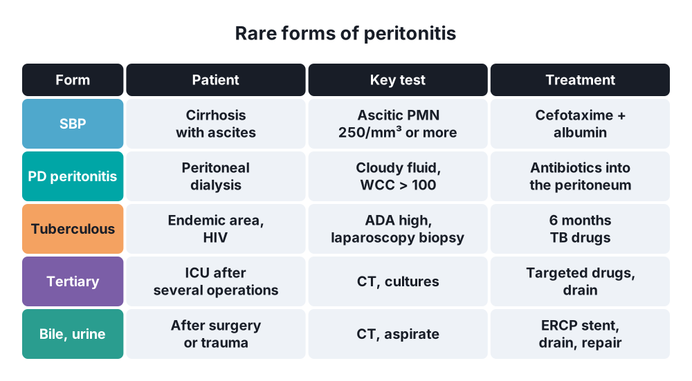
- SBP → cirrhosis with ascites (low ascitic protein under 15 g/L, previous SBP, GI bleeding, PPIs) → bacteria translocate from the gut; mostly monomicrobial: E. coli, Klebsiella, Streptococcus
- Children → nephrotic syndrome, SLE → Streptococcus pneumoniae, group A Streptococcus
- Young women → ascending infection from the genital tract (pneumococcus, gonococcus, Chlamydia)
PD peritonitis
- Touch contamination during exchanges, exit-site or tunnel infection
- Staphylococcus epidermidis and S. aureus (most), Streptococcus, Gram-negatives (Pseudomonas, Enterobacterales; think of a bowel source), fungi
Tuberculous peritonitis
- Mycobacterium tuberculosis (rarely M. bovis from milk)
- Spread → reactivation of latent peritoneal foci (from blood spread), ruptured mesenteric nodes, intestinal TB, fallopian tubes
- Risk factors → HIV, cirrhosis, dialysis, diabetes, steroids and anti-TNF drugs, malnutrition, alcohol, endemic area
Tertiary peritonitis
- Critically ill, immunosuppressed patients after several operations, long ICU stay, malnutrition, high APACHE II
- Organisms → low virulence, resistant: Enterococcus (including VRE), Candida, coagulase-negative Staphylococcus, Pseudomonas, Enterobacter, Acinetobacter
Chemical peritonitis
- Bile → bile leak after cholecystectomy, gangrenous gallbladder, liver trauma; becomes infected
- Blood → mild irritation; ruptured ectopic, spleen
- Urine → intraperitoneal bladder rupture (rise in creatinine from absorption)
- Pancreatic juice → pancreatitis, pancreatic trauma
- Barium → perforation during barium study → severe granulomatous reaction, mortality high (never use barium if perforation is possible)
- Chyle → chylous ascites (lymphoma, trauma, surgery)
- Meconium peritonitis → foetal bowel perforation (cystic fibrosis); calcification on X-ray in the newborn
Encapsulating peritoneal sclerosis
- Long-term PD (over 5 years), beta-blocker practolol (historical), cirrhosis, TB, idiopathic ("abdominal cocoon" in young women)
4. Pathogenesis
Primary peritonitis
- Bacteria translocate from the gut (cirrhosis: portal hypertension, gut oedema, bacterial overgrowth, poor immunity) or come through blood or the genital tract
- Ascites has low complement and opsonins → bacteria multiply
- Infection of the ascites without a perforation; one organism
Tuberculous peritonitis
- Mycobacteria seed the peritoneum → caseating granulomas
- Three forms develop → wet (ascitic, about 90%), fibrotic-fixed (matted loops, omental cake), dry (plastic: dense adhesions, doughy abdomen)
- Adhesions → obstruction, fistulas, cocoon
Tertiary peritonitis
- Initial secondary peritonitis treated by surgery and antibiotics
- Immune paralysis after SIRS (compensatory anti-inflammatory response) + antibiotic selection of resistant, low-virulence flora + gut barrier failure
- Diffuse, poorly localised infection with little pus; no single focus to drain
- Multiple organ dysfunction drives the high mortality
Sepsis
- Pathogen molecules (endotoxin, PAMPs) + tissue damage (DAMPs) → immune activation → cytokine release → vasodilatation, capillary leak, microvascular thrombosis (DIC), mitochondrial dysfunction → organ failure
- Immunosuppression phase follows → secondary infections
5. Classification
Peritonitis by origin
- Primary (no source), secondary (source in the abdomen), tertiary (persists after source control)
Specific forms
- Infective → SBP, PD, pneumococcal, gonococcal and chlamydial, TB, fungal, actinomycosis, parasitic
- Non-infective (aseptic) → bile, blood, urine, chyle, barium, starch or talc, familial Mediterranean fever, SLE serositis, sclerosing
TB peritonitis forms
- Wet (ascitic), fibrotic-fixed, dry (plastic)
Sepsis severity
- Infection → sepsis (SOFA rise of 2 or more) → septic shock (vasopressors + lactate over 2)
- qSOFA (bedside screen) → respiratory rate 22/min or more, altered mentation, systolic BP 100 mmHg or less; 2 or more → high risk (screen only, not for diagnosis)
- SIRS criteria → no longer used to define sepsis (too sensitive)
Intra-abdominal abscess by site
- Subphrenic (right more common), subhepatic, paracolic, interloop, pelvic, lesser sac, organ abscesses (liver, spleen, pancreas)
6. Clinical picture
SBP
- Fever, abdominal pain or tenderness (often mild), worsening ascites
- Often only encephalopathy, acute kidney injury, GI bleeding or "just unwell" in a cirrhotic patient
- Up to a third have no symptoms → tap every cirrhotic admitted with ascites
Primary peritonitis in children
- Sudden fever, vomiting, diffuse pain and tenderness → looks like perforated appendicitis
PD peritonitis
- Cloudy dialysis effluent (first sign), abdominal pain, fever
Tuberculous peritonitis
- Insidious over weeks to months → ascites, abdominal swelling, low-grade fever, night sweats, weight loss, anorexia, vague pain
- Doughy abdomen, abdominal mass (matted loops, omental cake), umbilical nodule
- Signs of obstruction (adhesions), fistulas
- Chest signs (active lung TB in about a third)
Tertiary peritonitis
- ICU patient after laparotomy for peritonitis → persistent fever, ongoing organ dysfunction, failure to wean, raised inflammatory markers, ileus, high output from drains, few local signs
Chemical peritonitis
- Bile → pain, ileus, jaundice (bile absorbed), fever later (infection)
- Urine → pain, distension, oliguria, haematuria, rising creatinine and potassium
Intra-abdominal abscess
- Swinging fever, rigors, sweats, malaise, raised WCC and CRP, ileus, local pain
- Subphrenic → shoulder tip pain, hiccups, pleural effusion, raised and fixed hemidiaphragm, basal crackles
- Pelvic → diarrhoea with mucus, tenesmus, urinary frequency, boggy tender mass on rectal examination
- "Pus somewhere, pus nowhere else, pus under the diaphragm" (classic saying)
Sepsis
- Fever or hypothermia, tachycardia, tachypnoea, confusion, hypotension, mottled skin, oliguria, low platelets, raised bilirubin
7. Diagnosis
Sepsis work-up
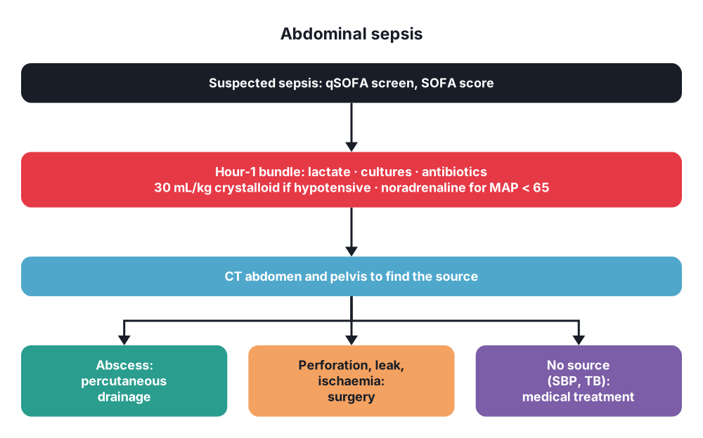
- Lactate, blood cultures (2 sets), FBC, CRP, procalcitonin, U&E, LFT, clotting, gas
- SOFA score → PaO2/FiO2, platelets, bilirubin, MAP and vasopressors, GCS, creatinine or urine output
- Find the source → CT abdomen and pelvis with IV contrast (most important), chest X-ray, urine
SBP
- Diagnostic paracentesis → ascitic neutrophils 250 cells/mm³ or more = SBP (diagnostic); send fluid in blood culture bottles at the bedside
- Secondary peritonitis suspected if → several organisms, protein over 10 g/L, glucose under 2.8 mmol/L, LDH above serum upper limit (Runyon's criteria), or no improvement in 48 hours → CT
PD peritonitis
- Effluent WCC over 100/mm³ with over 50% neutrophils (after a dwell of at least 2 hours); Gram stain and culture
Tuberculous peritonitis
- Ascitic fluid → exudate, lymphocytes predominate, protein over 25–30 g/L, SAAG under 11 g/L (unless cirrhosis), adenosine deaminase (ADA) over 30–40 U/L (sensitive and specific)
- AFB smear (positive in under 5%), culture (takes weeks, positive in 20–80%), GeneXpert MTB/RIF PCR
- Ultrasound or CT → ascites with septations, thickened smooth peritoneum, omental cake, mesenteric nodes with low-density (caseous) centres, matted loops, ileocaecal thickening
- Laparoscopy with peritoneal biopsy → gold standard: "millet seed" white nodules (miliary tubercles), adhesions, violin-string strands; histology shows caseating granulomas; diagnosis in over 90%
- Chest X-ray, IGRA, HIV test
- Differential → peritoneal carcinomatosis (CA-125 raised in both; cytology, biopsy)
Tertiary peritonitis
- CT (collections, leak), percutaneous aspiration of collections, peritoneal fluid culture at re-laparotomy (fungi, resistant organisms)
- Repeated cultures, fungal markers (beta-D-glucan)
Abscess
- CT with IV contrast → rim-enhancing fluid collection with gas; ultrasound (subphrenic, pelvic); MRI
- Chest X-ray → raised hemidiaphragm, effusion, gas-fluid level under the diaphragm
8. Differential diagnosis
- SBP vs secondary peritonitis in cirrhosis (Runyon's criteria, CT)
- TB peritonitis vs peritoneal carcinomatosis (ovarian, gastric, colonic), mesothelioma, pseudomyxoma peritonei, cirrhotic ascites, Crohn's disease, lymphoma
- Primary peritonitis in children vs appendicitis, mesenteric adenitis
- Tertiary peritonitis vs missed source (leak, abscess), other ICU infections (pneumonia, line, urine, C. difficile)
- Familial Mediterranean fever vs appendicitis (recurrent, self-limiting attacks; MEFV gene; colchicine)
9. Treatment
Conservative
- Sepsis bundle in the first hour → lactate, blood cultures, broad-spectrum antibiotics, 30 mL/kg crystalloid for hypotension or lactate 4 or more, noradrenaline for MAP under 65 mmHg; target urine output over 0.5 mL/kg/h
- ICU support → ventilation, renal replacement, glucose control, hydrocortisone in refractory shock, VTE and stress ulcer prophylaxis, early enteral nutrition
- SBP:
- Cefotaxime or ceftriaxone (or co-amoxiclav; piperacillin-tazobactam or a carbapenem if hospital-acquired) for 5–7 days
- IV albumin → 1.5 g/kg on day 1 and 1 g/kg on day 3 (prevents hepatorenal syndrome, lowers mortality)
- Prophylaxis after an episode → ciprofloxacin, norfloxacin or co-trimoxazole; consider liver transplant
- Primary peritonitis in children → antibiotics (ceftriaxone); laparoscopy if a perforation cannot be excluded
- PD peritonitis → intraperitoneal antibiotics (vancomycin + an aminoglycoside or ceftazidime), then by culture; remove the catheter if fungal, refractory (no response in 5 days) or relapsing
- Tuberculous peritonitis → antituberculous therapy for 6 months (2 months rifampicin, isoniazid, pyrazinamide, ethambutol, then 4 months rifampicin and isoniazid); steroids (to prevent adhesions) are debated and not routine
- Tertiary peritonitis → antibiotics against resistant organisms (vancomycin or linezolid for Enterococcus, echinocandin for Candida, a carbapenem or newer agents for resistant Gram-negatives) guided by cultures; organ support; nutrition; avoid overuse of antibiotics
- Chemical peritonitis:
- Bile leak → ERCP with sphincterotomy and stent + percutaneous drainage of the biloma
- Urine → catheter for extraperitoneal bladder injury
- Chylous ascites → low-fat diet with medium-chain triglycerides, somatostatin analogues, paracentesis, treat the cause
- Familial Mediterranean fever → colchicine
- Actinomycosis → long course of penicillin (6–12 months)
Surgical
- Source control → the core of treatment in abdominal sepsis:
- Percutaneous drainage (CT or ultrasound guided) → first choice for most abscesses (unilocular, accessible)
- Surgical drainage → multiloculated, thick pus, failed percutaneous drainage, interloop abscesses, or an ongoing source (leak)
- Pelvic abscess → transrectal or transvaginal drainage, or percutaneous
- Subphrenic abscess → percutaneous; open drainage (extraperitoneal, posterior through the 12th rib bed, or anterior subcostal) if it fails
- Tertiary peritonitis → re-laparotomy only if a drainable source is found on CT; repeated "blind" laparotomies do not help; open abdomen if ACS
- TB peritonitis → surgery only for complications: obstruction (adhesiolysis, stricturoplasty, limited resection), perforation, fistula, abscess, or diagnostic biopsy
- Bile, urine → laparoscopic washout and drain if diffuse; repair of an intraperitoneal bladder rupture
- PD peritonitis → catheter removal; laparotomy if a bowel perforation is suspected (several Gram-negative organisms or anaerobes)
- Encapsulating peritoneal sclerosis → stop PD, tamoxifen and steroids; enterolysis (peritonectomy of the cocoon) in specialist centres
- Familial Mediterranean fever → avoid unnecessary laparotomies (often an appendicectomy has been done before)
10. Complications
- Septic shock, multiple organ dysfunction, death
- SBP → hepatorenal syndrome, encephalopathy, recurrence (about 70% at 1 year without prophylaxis)
- TB → bowel obstruction, perforation, fistula, cocoon, infertility (genital TB), drug toxicity (hepatitis, optic neuritis with ethambutol, peripheral neuropathy with isoniazid: give pyridoxine)
- Tertiary peritonitis → enterocutaneous fistula, open abdomen complications, long ICU stay, death
- Abscess → rupture, empyema (subphrenic), recurrence, fistula
- Barium peritonitis → dense adhesions, granulomas, high mortality
- Post-sepsis syndrome → muscle weakness, cognitive problems, depression, readmissions
Prognosis
- SBP → about 20% in-hospital mortality; poor long-term survival without transplant
- TB peritonitis → good with treatment; worse with HIV and late diagnosis
- Tertiary peritonitis and septic shock → 30–60% mortality
Show as a list
Rare peritonitis and sepsis
- Primary
- SBP: cirrhosis
- Children: pneumococcus
- Tap: PMN >=250
- Cefotaxime + albumin
- Tuberculous
- Wet, fibrotic, dry
- Lymphocytes, ADA high
- Laparoscopy: millet seeds
- 6 months drugs
- Tertiary
- After source control
- Enterococcus, Candida
- ICU, high mortality
- Other
- PD: cloudy fluid
- Bile, urine, barium
- Sclerosing cocoon
- FMF: colchicine
- Sepsis
- SOFA rise >=2
- Shock: pressor + lactate >2
- 1-hour bundle
- Source control
- Abscess
- Subphrenic, pelvic
- Swinging fever
- Percutaneous drain


