73. Portal hypertension and oesophageal varices
High yield
Raised portal pressure, mostly from cirrhosis, opens portosystemic collaterals; bleeding varices need resuscitation, terlipressin, antibiotics and banding.
Exam Prep · State examSurgery · HPBSurgery · Upper GISurgery · Emergency and trauma
1. Definition
- Portal hypertension → raised pressure in the portal venous system; hepatic venous pressure gradient (HVPG) over 5 mmHg (normal 1–5)
- Clinically significant portal hypertension → HVPG ≥ 10 mmHg (varices form, decompensation)
- Variceal bleeding risk → HVPG ≥ 12 mmHg
- Oesophageal varices → dilated submucosal veins in the lower oesophagus, a portosystemic collateral
- Gastric varices → in the cardia and fundus
2. Epidemiology
- Cirrhosis → cause in about 90% in the West; schistosomiasis is the main non-cirrhotic cause worldwide
- Varices → present in about 50% of cirrhotic patients at diagnosis; new varices form at about 7–8% a year
- Variceal bleeding → 10–15% a year in patients with varices; about 70% of upper GI bleeds in cirrhosis
- Each bleed → mortality 15–20% at 6 weeks; rebleeding in 60% within a year without prevention
- Children → extrahepatic portal vein obstruction (umbilical sepsis, catheters) most common
3. Aetiology and risk factors
Anatomy (revision)
- Portal vein → formed behind the neck of the pancreas by the superior mesenteric and splenic veins (inferior mesenteric vein joins the splenic vein); runs in the hepatoduodenal ligament; carries 75% of liver blood flow
- No valves → pressure passes back to all tributaries
- Portosystemic anastomoses (open up in portal hypertension):
- Oesophagus → left gastric (portal) to oesophageal and azygos veins (systemic) → oesophageal varices
- Rectum → superior rectal (portal) to middle and inferior rectal (systemic) → anorectal varices (not haemorrhoids)
- Umbilicus → paraumbilical veins in the falciform ligament (portal) to epigastric veins (systemic) → caput medusae
- Retroperitoneum → colonic and splenic veins to renal and lumbar veins (veins of Retzius); spontaneous splenorenal shunts
Causes by level
- Prehepatic:
- Portal vein thrombosis (neonatal sepsis, pancreatitis, cancer, thrombophilia, myeloproliferative disease)
- Splenic vein thrombosis → sinistral (left-sided) portal hypertension → isolated gastric varices (pancreatitis, pancreatic cancer)
- Arteriovenous fistula (rare; raised flow)
- Hepatic:
- Presinusoidal → schistosomiasis, primary biliary cholangitis (early), sarcoidosis, non-cirrhotic portal hypertension
- Sinusoidal → cirrhosis (alcohol, hepatitis B and C, MASLD), alcoholic hepatitis
- Postsinusoidal → veno-occlusive disease (sinusoidal obstruction syndrome: chemotherapy, bone marrow transplant)
- Posthepatic:
- Budd-Chiari syndrome (hepatic vein thrombosis)
- IVC obstruction (web)
- Right heart failure, constrictive pericarditis, tricuspid regurgitation
4. Pathogenesis
- Portal pressure = flow × resistance:
- Increased resistance → fibrosis and nodules distort the sinusoids (fixed); stellate cell contraction and low nitric oxide (dynamic)
- Increased portal inflow → splanchnic vasodilatation (nitric oxide, glucagon) → hyperdynamic circulation
- Collaterals open → blood bypasses the liver → varices, encephalopathy (toxins skip the liver)
- Varices bleed when wall tension rises (size, thin wall, red signs, pressure) → Laplace's law
- Ascites → portal hypertension + low albumin + sodium and water retention (renin-angiotensin-aldosterone from low effective arterial volume)
- Splenomegaly → congestion → hypersplenism (low platelets, white cells)
- Hepatorenal syndrome → renal vasoconstriction in advanced disease
- Portal hypertensive gastropathy → congested gastric mucosa, slow bleeding
5. Classification
- By level → prehepatic, hepatic (pre-, sinusoidal, post-), posthepatic
Varices (endoscopic size)
- Small (grade 1) → flatten with insufflation
- Medium (grade 2) → do not flatten, under one-third of the lumen
- Large (grade 3) → over one-third of the lumen
- Red signs (red wale marks, cherry red spots) → high bleeding risk
Gastric varices (Sarin)
- GOV1 → along the lesser curve (continuation of oesophageal varices); most common
- GOV2 → into the fundus
- IGV1 → isolated fundal varices (think splenic vein thrombosis)
- IGV2 → elsewhere in the stomach
Liver function
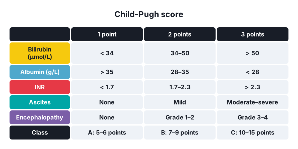
- Child-Pugh (bilirubin, albumin, INR, ascites, encephalopathy) → A (5–6 points), B (7–9), C (10–15)
- MELD (bilirubin, INR, creatinine, ± sodium) → transplant priority and TIPS risk
6. Clinical picture
Symptoms
- Haematemesis (often large, bright red) and melaena → variceal bleeding
- Abdominal swelling (ascites)
- Confusion, drowsiness, day-night reversal (hepatic encephalopathy)
- Jaundice, itch, bruising
- History → alcohol, hepatitis, blood transfusion, IV drugs, previous bleeds
Signs
- Shock in an active bleed → tachycardia, hypotension
- Splenomegaly (key sign of portal hypertension)
- Ascites → shifting dullness, fluid thrill
- Caput medusae (flow away from the umbilicus)
- Signs of chronic liver disease → spider naevi, palmar erythema, gynaecomastia, testicular atrophy, Dupuytren's contracture, leuconychia, clubbing
- Asterixis (flapping tremor), fetor hepaticus
- Cruveilhier-Baumgarten murmur (venous hum at the umbilicus; rare)
7. Diagnosis
- Bloods → FBC (low platelets from hypersplenism), LFT, INR, albumin, U&E, glucose, group and crossmatch
- Child-Pugh and MELD scores
- Upper GI endoscopy → varices, size, red signs; gastric varices; portal hypertensive gastropathy
- Screening endoscopy in every new cirrhosis, unless liver stiffness under 20 kPa and platelets over 150 × 10⁹/L (Baveno VII) → varices needing treatment unlikely
- Doppler ultrasound → liver texture, splenomegaly, ascites, portal vein patency and flow direction (hepatofugal flow in severe disease), HCC screening
- Transient elastography (FibroScan) → liver stiffness ≥ 25 kPa → clinically significant portal hypertension
- CT or MR angiography → collaterals, portal vein thrombosis, planning for TIPS or shunts
- HVPG measurement → wedged minus free hepatic venous pressure (gold standard; trials and specialist centres)
- Diagnostic paracentesis in ascites → serum-ascites albumin gradient ≥ 11 g/L = portal hypertension; neutrophils ≥ 250/mm³ = spontaneous bacterial peritonitis
- Cause → viral serology, autoantibodies, ferritin, caeruloplasmin, alpha-1 antitrypsin
8. Differential diagnosis
- Upper GI bleeding in cirrhosis → peptic ulcer (about 30%), portal hypertensive gastropathy, Mallory-Weiss tear, oesophagitis, gastric antral vascular ectasia, cancer
- Splenomegaly → haematological disease, infection (malaria, leishmaniasis), storage disorders
- Ascites → malignancy (peritoneal), heart failure, nephrotic syndrome, tuberculosis, pancreatitis
9. Treatment
Conservative: primary prevention (never bled)
- Medium or large varices, or small with red signs or Child-Pugh C → non-selective beta-blocker:
- Carvedilol (preferred; 6.25–12.5 mg daily) or propranolol
- Lowers portal pressure and the risk of decompensation (PREDESCI trial)
- Or endoscopic variceal band ligation (EVL) if beta-blockers are not tolerated
Conservative: acute variceal bleeding
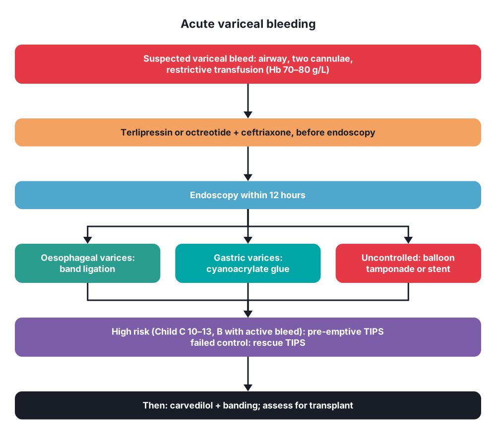
- Resuscitation → airway (intubate if encephalopathic or massive haematemesis), two large-bore cannulae
- Restrictive transfusion → target haemoglobin 70–80 g/L (over-transfusion raises portal pressure and rebleeding)
- Vasoactive drug at once, before endoscopy → terlipressin (2 mg IV every 4 hours) or octreotide or somatostatin, for 2–5 days
- Antibiotic prophylaxis → ceftriaxone 1 g daily for up to 7 days (lowers infection, rebleeding and death)
- Correct coagulation selectively; avoid routine FFP; platelets if under 50 × 10⁹/L
- Prevent encephalopathy → lactulose, rifaximin
- Stop beta-blockers in the acute bleed and shock
- Endoscopy within 12 hours (after resuscitation):
- Band ligation → treatment of choice for oesophageal varices
- Gastric varices (GOV2, IGV1) → cyanoacrylate glue injection; or EUS-guided coil and glue
- Sclerotherapy → second line
- Uncontrolled bleeding → balloon tamponade (Sengstaken-Blakemore or Minnesota tube; temporary, up to 24 hours) or self-expanding oesophageal stent (Danis) → bridge to TIPS
- TIPS (transjugular intrahepatic portosystemic shunt):
- Rescue TIPS → failed endoscopic and drug therapy
- Pre-emptive (early) TIPS within 72 hours → high-risk patients: Child-Pugh C 10–13 or Child-Pugh B with active bleeding at endoscopy → lowers rebleeding and death
Conservative: secondary prevention (after a bleed)
- Non-selective beta-blocker plus band ligation every 2–4 weeks until varices are gone, then surveillance endoscopy
- TIPS if rebleeding despite this
- Assess for liver transplantation (any decompensation, MELD ≥ 15)
Conservative: other complications
- Ascites → salt restriction, spironolactone ± furosemide; large-volume paracentesis with albumin (8 g per litre removed, over 5 L); TIPS for refractory ascites
- SBP → cefotaxime or ceftriaxone plus albumin; prophylaxis with ciprofloxacin or co-trimoxazole after an episode
- Hepatic encephalopathy → lactulose, rifaximin, find the trigger (bleeding, infection, constipation, drugs)
- Treat the cause → stop alcohol, antivirals; anticoagulation for portal vein thrombosis
Surgical
- Liver transplantation → definitive treatment for end-stage cirrhosis with portal hypertension (cures both)
- Surgical portosystemic shunts → rarely now (replaced by TIPS); for Child-Pugh A with good liver function, non-cirrhotic portal hypertension, or where TIPS is not possible:
- Total (non-selective) → portocaval shunt (end-to-side or side-to-side) → good control of bleeding but encephalopathy and liver failure
- Partial → small-diameter H-graft interposition shunt (8 mm; portocaval or mesocaval)
- Selective → distal splenorenal (Warren) shunt → decompresses the varices through the spleen while keeping portal flow to the liver
- Devascularisation → Sugiura procedure (oesophageal transection, gastric devascularisation, splenectomy), or Hassab operation (gastro-oesophageal devascularisation with splenectomy) → when shunts are not possible (diffuse thrombosis); schistosomiasis
- Emergency oesophageal transection with a stapler → historical last resort
- Splenectomy → cures sinistral portal hypertension (splenic vein thrombosis)
- Meso-Rex bypass → children with extrahepatic portal vein obstruction (SMV to left portal vein; restores portal flow)
- Budd-Chiari → anticoagulation, angioplasty or stent, TIPS, transplant
10. Complications
- Variceal haemorrhage → death in 15–20%
- Ascites, spontaneous bacterial peritonitis, hepatic hydrothorax
- Hepatic encephalopathy
- Hepatorenal syndrome
- Hepatopulmonary syndrome, portopulmonary hypertension
- Hypersplenism, portal vein thrombosis
- Hepatocellular carcinoma
- Treatment:
- Band ligation → ulcers after banding, stricture
- Balloon tamponade → oesophageal rupture, aspiration, airway obstruction
- TIPS → encephalopathy (about 30%), stent stenosis or occlusion, liver failure, heart failure
- Shunt surgery → encephalopathy, liver failure, shunt thrombosis
- Beta-blockers → hypotension, fatigue; stop in refractory ascites with low blood pressure
Show as a list
Portal hypertension
- Definition
- HVPG > 5 mmHg
- CSPH >= 10
- Bleed risk >= 12
- Causes
- Prehepatic: portal vein thrombosis
- Hepatic: cirrhosis, schistosomiasis
- Posthepatic: Budd-Chiari
- Collaterals
- Oesophageal varices
- Rectal varices
- Caput medusae
- Assess
- Child-Pugh, MELD
- Endoscopy
- Doppler, FibroScan
- Acute bleed
- Hb 70-80
- Terlipressin
- Ceftriaxone
- Band ligation
- Glue for gastric
- Sengstaken
- TIPS
- Prevention
- Carvedilol
- Banding
- Surgery
- Transplant
- Warren shunt
- Sugiura


