40. Spontaneous pneumothorax, pleural empyema and pleural metastases
High yield
Bleb rupture in tall young smokers; infected pleural fluid in three stages; malignant effusion treated by pleurodesis or catheter.
Exam Prep · State examSurgery · General surgerySurgery · Emergency and traumaAnatomy · Thorax
1. Definition
- Spontaneous pneumothorax → air in the pleural space without trauma, with collapse of the lung
- Primary → no known lung disease
- Secondary → underlying lung disease
- Pleural empyema → pus in the pleural space, or fluid with organisms on Gram stain or culture
- Pleural metastases → secondary tumour deposits on the pleura, usually with a malignant pleural effusion (MPE)
2. Epidemiology
- Primary spontaneous pneumothorax → tall, thin young men, 15–35 years; smokers; right side more often; bilateral in < 10%
- Secondary → age > 50 years; COPD most common cause
- Recurrence after a first primary pneumothorax → about 30%, mostly within 1 year
- Empyema → most follow pneumonia; 1 in 5 die or need surgery
- MPE → about 15% of patients with cancer; median survival about 5 months
3. Aetiology and risk factors
Spontaneous pneumothorax
- Primary → rupture of apical subpleural blebs (< 2 cm) or bullae; smoking (risk × 10–20), tall stature, Marfan's syndrome
- Secondary:
- COPD, bullous emphysema
- Cystic fibrosis (10% develop one)
- Asthma, interstitial lung disease
- Infection → Pneumocystis (HIV), tuberculosis, necrotising pneumonia
- Lung cancer, sarcoma metastases
- Catamenial → endometriosis; women 20–30 years, 2–3 days after menstruation starts, right side
- Lymphangioleiomyomatosis, Ehlers-Danlos syndrome
Empyema
- Parapneumonic → about 60%
- Postoperative → lung resection, oesophagectomy, bronchopleural fistula
- Trauma → haemothorax, penetrating injury
- Oesophageal perforation
- Subphrenic or liver abscess
- Ruptured lung abscess
- Organisms → Streptococcus pneumoniae, Streptococcus anginosus (milleri), S. aureus, anaerobes, Gram-negatives; tuberculosis
Pleural metastases
- Lung and breast cancer → about 75% of MPEs
- Lymphoma, ovary, stomach, unknown primary
- Primary pleural tumour → malignant mesothelioma (asbestos, latency 15–40 years)
4. Pathogenesis
Pneumothorax
- Bleb ruptures
- Air enters the pleural space → lung collapses
- One-way valve → tension pneumothorax → mediastinal shift, reduced venous return, cardiac arrest
Empyema stages
- Exudative (stage I) → 2–5 days after pneumonia; thin fluid, low cell count, normal pH and glucose; antibiotics alone may cure
- Fibrinopurulent (stage II) → 5–14 days; turbid fluid, fibrin, loculations; pH < 7.2, low glucose, high lactate dehydrogenase (LDH); needs drainage
- Organising (stage III) → after 2–6 weeks; thick fibrous peel → trapped lung; needs decortication
MPE
- Tumour cells seed the pleura
- Increased capillary permeability, blocked lymphatic drainage
- Exudate accumulates → recurs after drainage
5. Classification
Pneumothorax
- By cause → primary spontaneous, secondary spontaneous, traumatic, iatrogenic (central line, lung biopsy, ventilation)
- By size (British Thoracic Society, BTS) → large if rim ≥ 2 cm at the hilum level on chest X-ray (≥ 2 cm ≈ 50% volume)
- American → large if ≥ 3 cm at the apex
- By physiology → simple, tension
Pleural effusion (Light's criteria)
- Exudate if any one:
- Pleural fluid to serum protein ratio > 0.5
- Pleural fluid to serum LDH ratio > 0.6
- Pleural fluid LDH > 2/3 of the upper limit of normal serum LDH
- Transudate → heart failure, cirrhosis, nephrotic syndrome, hypothyroidism
- Exudate → infection, cancer, pulmonary embolism, pancreatitis, rheumatoid arthritis, tuberculosis
6. Clinical picture
Symptoms
- Pneumothorax → sudden pleuritic chest pain and dyspnoea, often at rest; secondary → severe dyspnoea with small pneumothorax
- Empyema → persistent fever after pneumonia, rigors, weight loss, pleuritic pain, malaise
- MPE → progressive dyspnoea, cough, chest heaviness, weight loss
Signs
- Pneumothorax:
- Reduced expansion
- Hyper-resonant percussion
- Absent breath sounds
- Subcutaneous emphysema
- Tension → tachycardia, hypotension, tracheal deviation away, distended neck veins, hypoxia
- Effusion or empyema → stony dull percussion, absent breath sounds, reduced vocal resonance
- Chronic empyema → clubbing, chest wall sinus (empyema necessitans)
- Mesothelioma → chest wall mass, contracted hemithorax
7. Diagnosis
Imaging
- Erect posteroanterior chest X-ray → visible pleural line, no lung markings beyond it
- CT → small or uncertain pneumothorax, bullous disease, blebs before surgery
- Lung ultrasound → no "lung sliding", lung point
- Effusion → blunted costophrenic angle, meniscus
- Ultrasound → effusion size, septations, guides aspiration; all pleural procedures under ultrasound
- Contrast CT → empyema: "split pleura" sign (enhancing thickened pleura); pleural nodules in metastases; underlying tumour
Laboratory
- Diagnostic pleural aspiration:
- Appearance → pus, blood, milky (chyle)
- pH → < 7.2 in a parapneumonic effusion → chest drain
- Glucose low, LDH high → infection, rheumatoid, cancer
- Protein and LDH → Light's criteria
- Gram stain, culture, acid-fast bacilli
- Cytology → positive in about 60% of MPE
- Pleural biopsy → thoracoscopic (VATS) or local anaesthetic thoracoscopy; CT-guided biopsy; mesothelioma needs histology
8. Differential diagnosis
- Pneumothorax → large bulla (CT shows lung around it), myocardial infarction, pulmonary embolism, pericarditis
- Empyema → lung abscess, simple parapneumonic effusion, subphrenic abscess
- MPE → heart failure, tuberculous effusion, pulmonary embolism, mesothelioma
9. Treatment
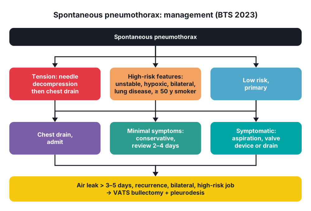
Conservative: pneumothorax (BTS 2023)
- Tension → immediate needle decompression (2nd intercostal space mid-clavicular line, or 4th–5th space in the mid-axillary line), then chest drain
- High-risk features → chest drain:
- Haemodynamic instability
- Significant hypoxia
- Bilateral pneumothorax
- Underlying lung disease
- Age ≥ 50 years with significant smoking history
- Haemothorax
- Primary, minimal symptoms → conservative, regardless of size; review in 2–4 days
- Primary, symptomatic → needle aspiration, ambulatory device (one-way valve), or chest drain
- Secondary → chest drain usually; admit
- Oxygen → speeds reabsorption (four times faster)
- No flying until resolved on chest X-ray; no diving for life unless pleurectomy
- Stop smoking
Surgical: pneumothorax
- Indications:
- Second ipsilateral pneumothorax
- First contralateral pneumothorax
- Persistent air leak > 3–5 days or failure to re-expand
- Bilateral simultaneous
- Haemopneumothorax
- High-risk occupations → pilots, divers
- Previous tension pneumothorax
- Operation → VATS bullectomy/blebectomy + pleurodesis (parietal pleurectomy or pleural abrasion)
- Recurrence → about 1–2% after open pleurectomy, about 5% after VATS, 5–10% after chemical pleurodesis alone
- Unfit → talc slurry or autologous blood patch via the drain
Conservative: empyema
- IV antibiotics → cover anaerobes (co-amoxiclav, or cefuroxime + metronidazole); 2–6 weeks
- Chest drain under ultrasound for pus, pH < 7.2, positive Gram stain or culture
- Loculated → intrapleural tissue plasminogen activator (tPA) + DNase (MIST2)
- Nutrition, physiotherapy, thromboprophylaxis
Surgical: empyema
- Failure to improve after 5–7 days of drain and antibiotics
- VATS debridement and decortication → stage II, early stage III
- Open decortication → stage III, thick peel, trapped lung
- Unfit → open drainage, rib resection, Eloesser flap (open window)
- Bronchopleural fistula → closure, muscle flap
Pleural metastases and MPE
- Aim → relief of dyspnoea; not curative
- Therapeutic aspiration → symptom relief; ≤ 1.5 L at a time (re-expansion pulmonary oedema)
- Recurrent MPE, lung expands → talc pleurodesis (slurry via drain, or poudrage at thoracoscopy)
- Trapped lung or failed pleurodesis → indwelling pleural catheter (IPC), drained at home
- Treat the primary → chemotherapy, endocrine, targeted therapy
- Mesothelioma → chemotherapy, immunotherapy; radical surgery (extended pleurectomy and decortication) only in selected epithelioid disease
10. Complications
- Pneumothorax:
- Tension pneumothorax
- Recurrence
- Persistent air leak, bronchopleural fistula
- Haemopneumothorax
- Re-expansion pulmonary oedema → large, long-standing collapse drained fast
- Surgical emphysema, pneumomediastinum
- Empyema:
- Trapped lung, fibrothorax, restrictive lung function
- Bronchopleural fistula
- Empyema necessitans (through the chest wall)
- Sepsis, death
- MPE → recurrence, loculation, catheter infection
Show as a list
Pleural disease
- Spontaneous pneumothorax
- Primary: blebs
- Secondary: COPD
- Tension
- Conservative, aspiration
- Chest drain
- VATS pleurectomy
- Empyema
- Parapneumonic
- Exudative
- Fibrinopurulent
- Organising
- pH below 7.2
- Drain, antibiotics
- Decortication
- Pleural metastases
- Lung, breast
- Mesothelioma
- Cytology, thoracoscopy
- Talc pleurodesis
- Indwelling catheter


