
60. Perforated peptic ulcer
High yield
Gastric or duodenal ulcer perforating into the peritoneum; resuscitation, omental patch repair and H. pylori eradication.
Exam Prep · State examSurgery · Upper GISurgery · Emergency and trauma
1. Definition
- Perforated peptic ulcer → full-thickness breach of a gastric or duodenal ulcer with leakage of contents into the peritoneal cavity
- Free perforation → into the general peritoneal cavity
- Covered (sealed) perforation → plugged by omentum, liver or fibrin
- Penetration → ulcer erodes into an adjacent organ (pancreas, liver) without free leak
2. Epidemiology
- Lifetime risk in peptic ulcer disease → 2–10%
- Mortality → 5–15% despite surgery
3. Aetiology and risk factors
- Helicobacter pylori (H. pylori) → 70% of perforated duodenal ulcers (DU); 80% excluding NSAID users
- Non-steroidal anti-inflammatory drugs (NSAIDs) → especially gastric ulcer (GU) and the elderly
- Steroids
- Smoking
- Gastric cancer, lymphoma, gastrointestinal stromal tumour (GIST) → perforated GU in a minority
4. Pathogenesis
- Site → anterior wall of the first part of the duodenum (most common), prepyloric, lesser curvature
- Spillage of gastroduodenal contents:
- Chemical peritonitis (acid, bile, enzymes)
- Bacterial superinfection after 6–12 h
- Systemic inflammatory response syndrome (SIRS)
- Multi-organ dysfunction
- Fluid tracks down the right paracolic gutter → right iliac fossa pain
- 50% have sealed spontaneously by the time of admission
- Classical clinical phases:
- Shock phase → first 6 h, severe pain, rigidity
- Phase of false calm → 6–12 h, pain eases, rigidity persists
- Phase of diffuse peritonitis → after 12 h, sepsis
5. Classification
- By site → duodenal, pyloric, gastric
- By size:
- Small → < 1 cm (most DU perforations)
- Giant → > 2 cm
- By course → free, covered, atypical (into the lesser sac or retroperitoneum)
- Boey score (risk factors):
- Major medical illness
- Preoperative shock (systolic blood pressure (SBP) < 100 mmHg)
- Perforation > 24 h
- Boey score 0, 1, 2, 3 → mortality 0%, 10%, 45.5%, 100%
- Peptic Ulcer Perforation (PULP) score → 8 variables; predicts 30-day mortality better than Boey
6. Clinical picture
Symptoms
- Sudden, severe epigastric pain ("dagger-like"), rapidly generalised
- Shoulder-tip pain → diaphragmatic irritation
- Vomiting
- History of dyspepsia → absent in 1/3
- Elderly, steroid users, inpatients → subtle presentation
Signs
- Board-like rigidity of the abdomen
- Patient lies still; shallow breathing
- Tachycardia, hypotension → shock
- Loss of liver dullness on percussion → free gas
- Right iliac fossa tenderness → fluid in the right paracolic gutter
- Absent bowel sounds → later
7. Diagnosis
Laboratory
- Full blood count → leucocytosis
- Serum amylase → mildly raised; a very high level suggests pancreatitis
- Urea, creatinine, electrolytes
- Arterial blood gas, lactate
- Group and save, clotting
Imaging
- Erect chest X-ray → free subdiaphragmatic gas in up to 70%
- Left lateral decubitus X-ray → if the patient cannot sit up
- Computed tomography (CT) → most sensitive; free gas, fluid, site; less useful < 6 h from onset
- Water-soluble contrast study → confirms sealing before non-operative treatment
- Ultrasound → free fluid
8. Differential diagnosis
- Acute pancreatitis
- Perforated appendicitis
- Acute cholecystitis
- Mesenteric ischaemia
- Ruptured abdominal aortic aneurysm
- Perforated diverticulitis
- Inferior myocardial infarction, basal pneumonia
9. Treatment
Conservative
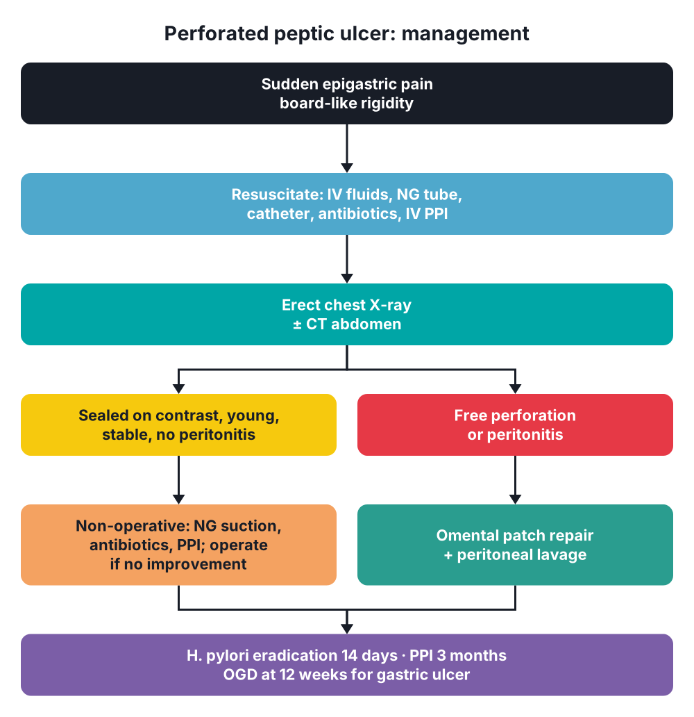
- Resuscitation in all patients:
- Intravenous fluids
- Nasogastric tube
- Urinary catheter
- Intravenous antibiotics
- Intravenous PPI (proton pump inhibitor)
- Analgesia
- Non-operative treatment (Taylor method) → selected patients only:
- Sealed perforation on water-soluble contrast
- Young, stable, no generalised peritonitis
- Nasogastric suction, intravenous antibiotics, PPI
- 28% fail and need surgery within 12 h
- Poor results > 70 years
Surgical
- Operation of choice → simple closure with omental patch and peritoneal lavage
- Approach → laparoscopic or upper midline laparotomy
- Laparoscopy → less pain, shorter stay, fewer wound infections; longer operation, more suture-site leaks
- Open surgery preferred → Boey score 3, age > 70 years, symptoms > 24 h
- Steps:
- Aspirate fluid; send for culture
- Identify the perforation; check the posterior stomach through the lesser sac if none found
- Place 3 interrupted sutures (3-0 absorbable), 5 mm from each edge, left untied
- Lay a viable pedicle of omentum over the perforation
- Tie the sutures over the omentum, side sutures first
- Lavage with 5–10 L warm saline
- Close the abdomen; drain optional
- Graham patch → free omental plug; replaced by pedicle omentopexy (Cellan-Jones)
- Perforated GU → biopsy the edge or excise the ulcer; resection only if neither is possible
- Giant perforation (> 2 cm) → options:
- Seromuscular sutures with omental plug
- Controlled tube duodenostomy
- Distal gastrectomy with Roux-en-Y reconstruction
- Definitive acid-reducing surgery → no longer routine; emergency resection raises mortality
- After surgery:
- H. pylori eradication (triple therapy, 14 days) + PPI for 3 months
- Ulcer recurrence after eradication → about 5% at 1 year (35% without)
- Repeat endoscopy at 12 weeks → mandatory after perforated GU
10. Complications
- Subphrenic, subhepatic or pelvic abscess
- Leak from the repair, reperforation
- Duodenal fistula
- Duodenal obstruction → persistent vomiting
- Wound infection, burst abdomen
- Pneumonia
- Multi-organ failure
11. Prognosis
- Mortality → 5–15%
- Worse with age > 65 years, shock, comorbidity, delay > 24 h, perforation during hospital stay
Show as a list
Perforated peptic ulcer
- Causes
- H. pylori
- NSAIDs
- Steroids
- Clinical
- Sudden epigastric pain
- Board-like rigidity
- Loss of liver dullness
- Shock
- Investigations
- Erect chest X-ray
- CT abdomen
- Amylase
- Risk scores
- Boey score
- PULP score
- Treatment
- Resuscitation
- Omental patch repair
- Peritoneal lavage
- H. pylori eradication
- Special cases
- Sealed: non-operative
- Gastric: biopsy edge
- Giant: resection options
- Complications
- Abscess
- Leak
- Fistula