58. Peptic ulcer: modern surgical treatment
High yield
Mucosal defect beyond the muscularis mucosae from acid-pepsin; H. pylori and NSAIDs cause most; surgery mainly for complications.
Exam Prep · State examSurgery · Upper GI
1. Definition
- Peptic ulcer → defect of the gastric or duodenal mucosa extending through the muscularis mucosae
- Erosion → defect confined to the mucosa
- Duodenal ulcer (DU) → 90% in the first part of the duodenum
- Gastric ulcer (GU) → most on the lesser curvature, at the incisura
2. Epidemiology
- DU → more common than GU, younger patients
- GU → older patients, more often linked to NSAIDs
3. Aetiology and risk factors
- Helicobacter pylori infection → 70–80% of DU
- Non-steroidal anti-inflammatory drugs (NSAIDs) and aspirin
- Smoking
- Zollinger–Ellison syndrome → gastrinoma, multiple or atypical ulcers
- Stress ulcers:
- Curling's ulcer → after major burns
- Cushing's ulcer → after head injury
- Hyperparathyroidism → hypercalcaemia, raised gastrin
4. Pathogenesis
- Imbalance → aggressive factors exceed mucosal defence
- Aggressive factors:
- Acid and pepsin
- H. pylori → antral gastritis → raised gastrin → raised acid (DU)
- NSAIDs → loss of prostaglandins → reduced mucus and bicarbonate
- Defensive factors:
- Mucus-bicarbonate layer
- Mucosal blood flow
- Epithelial renewal
- DU → high acid output
- GU types I and IV → normal or low acid; mucosal defence fails
5. Classification
Modified Johnson classification of gastric ulcer
- Type I → lesser curvature at the incisura; normal or low acid
- Type II → gastric body ulcer with a DU; high acid
- Type III → prepyloric; high acid
- Type IV → high lesser curvature near the gastro-oesophageal junction; low acid
- Type V → anywhere in the stomach; NSAID-induced
By course
- Acute → stress, NSAIDs, burns, head injury
- Chronic → fibrotic base, may penetrate neighbouring organs
6. Clinical picture
Symptoms
- Epigastric pain, burning or gnawing
- DU:
- Pain 2–3 h after meals and at night
- Relieved by food or antacids
- GU:
- Pain soon after meals
- Food fear, weight loss
- Periodicity → episodes over weeks with pain-free months
- Nausea, vomiting, bloating
- Back pain → posterior ulcer penetrating the pancreas
- Complications as first presentation → bleeding, perforation, obstruction
Signs
- Epigastric tenderness
- Pallor → chronic blood loss
- Succussion splash → gastric outlet obstruction
7. Diagnosis
Laboratory
- Full blood count → iron-deficiency anaemia
- H. pylori tests:
- Urea breath test
- Stool antigen test
- Rapid urease test (CLO test) on antral biopsy
- Histology
- Fasting serum gastrin → suspected Zollinger–Ellison syndrome; stop proton pump inhibitor (PPI) first
Imaging
- Oesophagogastroduodenoscopy (OGD) → investigation of choice
- Every GU → multiple biopsies from the edge and base
- Repeat OGD after 8–12 weeks → confirm healing of GU
- Barium meal → only if OGD not possible
- Computed tomography (CT) → complications, penetration
8. Differential diagnosis
- Gastro-oesophageal reflux disease
- Functional dyspepsia
- Gastric carcinoma
- Biliary colic, cholecystitis
- Pancreatitis
- Myocardial infarction
9. Treatment
Conservative
- H. pylori eradication → triple therapy (PPI + 2 antibiotics) for 14 days
- PPI for 4–8 weeks (DU) and 8–12 weeks (GU)
- Stop NSAIDs; if essential → PPI cover or a cyclo-oxygenase-2 (COX-2) inhibitor with PPI
- Stop smoking
- Confirm eradication after treatment
- Medical treatment heals most ulcers → elective ulcer surgery now rare
Surgical
- Indications:
- Perforation
- Bleeding not controlled by endoscopy
- Gastric outlet obstruction
- Intractable ulcer despite full medical therapy
- Suspected malignancy, GU that does not heal
- Zollinger–Ellison syndrome not controlled medically
- Operations for DU:
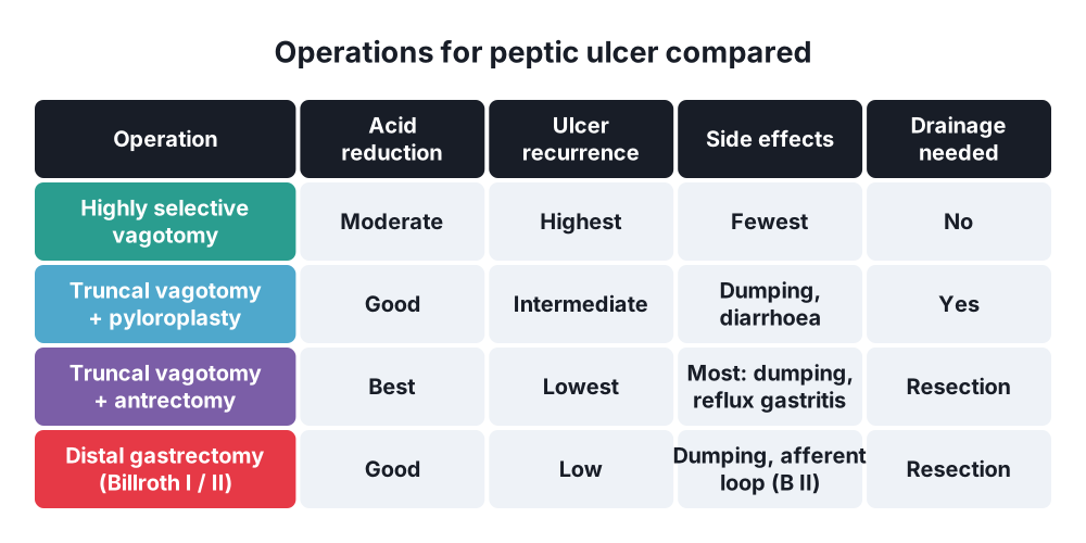
- Highly selective vagotomy (HSV) → nerves of Latarjet preserved, no drainage, fewest side effects, highest recurrence
- Truncal vagotomy + drainage (pyloroplasty or gastrojejunostomy)
- Truncal vagotomy + antrectomy → lowest recurrence, most side effects
- Operations for GU:
- Type I → distal gastrectomy with Billroth I gastroduodenostomy
- Types II and III → antrectomy + truncal vagotomy
- Type IV → resection including the ulcer (Pauchet or Csendes) with Roux-en-Y
- Any GU → resection or excision must include the ulcer for histology
- Reconstruction after distal gastrectomy:
- Billroth I → gastroduodenostomy
- Billroth II (Polya) → gastrojejunostomy, duodenal stump closed
- Roux-en-Y → less bile reflux and dumping
10. Complications
Of the ulcer
- Bleeding
- Perforation
- Penetration → pancreas, liver, colon
- Gastric outlet obstruction
- Malignant change → GU only
Of ulcer surgery
- Early:
- Duodenal stump blow-out (Billroth II)
- Anastomotic leak
- Stomal obstruction
- Gastric remnant necrosis → left gastric artery ligated at origin with splenectomy
- Late:
- Alkaline reflux gastritis → commonest late problem; treated by Roux-en-Y conversion
- Early dumping → 30–60 min after meals; osmotic fluid shift; 10%
- Late dumping → 2–3 h after meals; reactive hypoglycaemia; 5%
- Post-vagotomy diarrhoea
- Afferent loop syndrome → long afferent loop after Billroth II
- Recurrent ulcer → exclude Zollinger–Ellison syndrome
- Small stomach syndrome
- Iron and vitamin B12 deficiency, bone disease
- Gastric stump carcinoma
Show as a list
Peptic ulcer
- Causes
- H. pylori
- NSAIDs
- Zollinger–Ellison
- Stress ulcers
- Johnson types
- I incisura
- II body + DU
- III prepyloric
- IV high lesser curve
- V NSAID
- Diagnosis
- OGD + biopsy
- H. pylori tests
- Fasting gastrin
- Medical treatment
- Eradication 14 days
- PPI
- Stop NSAIDs
- Surgery indications
- Perforation
- Bleeding
- Obstruction
- Intractability
- Suspected cancer
- Operations
- Highly selective vagotomy
- Vagotomy + drainage
- Vagotomy + antrectomy
- Distal gastrectomy
- Post-gastrectomy problems
- Dumping
- Reflux gastritis
- Afferent loop
- Recurrent ulcer