
104. Paraproctitis and anorectal abscesses
High yield
Anal gland infection spreads into the spaces around the anus; drain it promptly and keep the sphincters safe.
Exam Prep · State examSurgery · General surgerySurgery · ColorectalSurgery · Emergency and trauma
1. Definition
- Paraproctitis → inflammation of the tissues around the rectum and anal canal (term used in Bulgarian and Russian textbooks)
- Acute paraproctitis → an anorectal (perianal) abscess: a collection of pus in the spaces around the anus and rectum
- Chronic paraproctitis → the fistula-in-ano that often follows the abscess (see question 105)
- Cryptoglandular abscess → begins in an infected anal gland at the dentate line (about 90%)
2. Epidemiology
- Common → about 1 in 5000 people a year need an operation
- Men 2–3 times more often than women
- Peak at 20–40 years
- About 30–50% of patients later develop a fistula-in-ano
- Children → mostly boys under 2 years
3. Aetiology and risk factors
Primary (cryptoglandular, about 90%)
- Anal glands (6–12) open into the crypts at the dentate line and reach into the intersphincteric space
- Blockage of the gland duct (stool debris, oedema) → stasis → infection
- Organisms → gut flora (E. coli, Bacteroides, enterococci); skin organisms (Staphylococcus aureus) when the abscess is a skin infection
Secondary (about 10%)
- Crohn's disease, ulcerative colitis
- Tuberculosis, actinomycosis, lymphogranuloma venereum
- Malignancy → rectal or anal cancer, leukaemia, lymphoma
- Trauma → foreign body, impalement, episiotomy, haemorrhoidectomy, prostatectomy
- Radiotherapy
- Immunosuppression → diabetes, HIV, chemotherapy, steroids
- Anal fissure, infected haemorrhoids
4. Pathogenesis
- Infection starts in the intersphincteric space and spreads along the paths of least resistance:
- Downwards → to the anal margin → perianal abscess
- Outwards through the external sphincter → ischiorectal (ischioanal) abscess
- Upwards in the intersphincteric plane → high intersphincteric or supralevator abscess
- Inwards under the mucosa → submucosal abscess
- Around the anus (horseshoe) → through the deep postanal space to one or both ischiorectal fossae
- If the abscess opens (by itself or by drainage) and the crypt remains infected, the track persists as a fistula-in-ano
- Necrotising spread along fascial planes → Fournier's gangrene (necrotising fasciitis of the perineum)
5. Classification
By site (relative frequency)
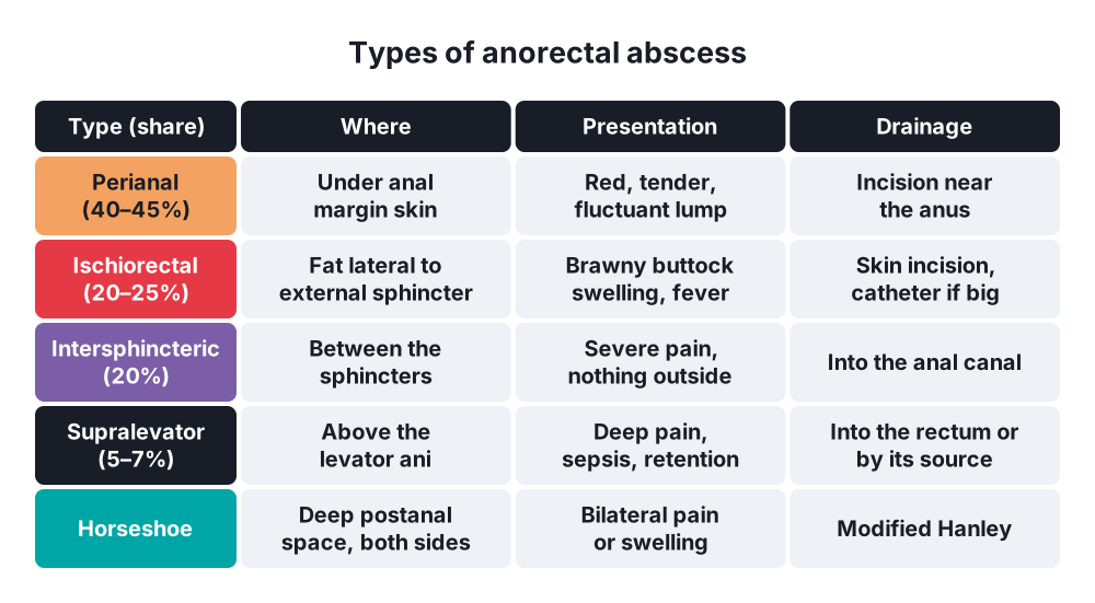
- Perianal (about 40–45%) → just under the skin of the anal margin
- Ischiorectal (ischioanal) (about 20–25%) → in the fat of the ischiorectal fossa, lateral to the external sphincter
- Intersphincteric (about 20%) → between the internal and external sphincters
- Supralevator (pelvirectal) (about 5–7%) → above the levator ani; may come from the intersphincteric space or from the pelvis (diverticulitis, appendicitis, Crohn's)
- Submucosal (about 5%) → under the rectal mucosa
- Horseshoe → spreads around the anus through the deep postanal space
By course
- Acute (abscess) and chronic (fistula)
Older Russian and Bulgarian terms
- Subcutaneous, ischiorectal, submucosal, pelviorectal, retrorectal paraproctitis
6. Clinical picture
- Constant, throbbing anal pain, worse on sitting, walking and defecation, building over a few days
- Fever, malaise, chills
- Spontaneous discharge of pus brings sudden relief
By type
- Perianal → red, hot, tender, fluctuant swelling at the anal margin; few systemic signs
- Ischiorectal → larger, diffuse, brawny induration of the buttock; fluctuance may be late; fever and sepsis common
- Intersphincteric → severe pain with nothing to see outside; tender bulge on DRE
- Supralevator → deep pelvic or rectal pain, fever, urinary retention, sepsis; tender mass high on DRE; little or nothing outside
- Submucosal → pain and fullness in the rectum; smooth swelling on DRE
- Horseshoe → bilateral swelling or pain, posterior midline tenderness
- Fournier's gangrene → pain out of proportion, rapidly spreading redness, dusky skin, crepitus, foul discharge, septic shock
- Diabetics and the immunosuppressed may have few local signs but severe sepsis
7. Diagnosis
- Clinical in most patients (inspection and gentle palpation)
- DRE → often too painful awake; do it under anaesthesia
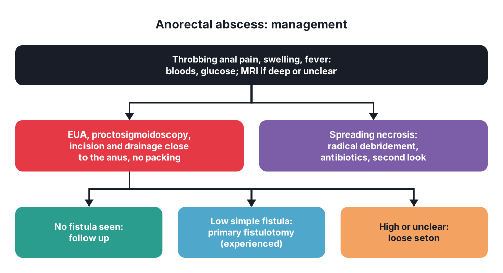
- Examination under anaesthesia (EUA) with proctoscopy and sigmoidoscopy → confirms the abscess, looks for an internal opening, excludes other disease
- Imaging when the diagnosis is unclear (deep, recurrent, Crohn's, no visible swelling):
- MRI pelvis → best for deep abscesses and fistula tracks
- Endoanal ultrasound → in experienced hands, as good as MRI
- CT pelvis → quick in the emergency department; supralevator and pelvic sources
- Bloods → FBC, CRP, glucose (undiagnosed diabetes), lactate if septic
- Pus culture → gut organisms suggest a fistula; skin organisms suggest a simple skin abscess
- Suspected necrotising infection → LRINEC score may help, but clinical suspicion and early surgery matter most
8. Differential diagnosis
- Thrombosed external haemorrhoid → blue, tense lump, sudden onset, no fever
- Anal fissure → pain with defecation, visible tear
- Pilonidal abscess → natal cleft, higher, midline pits
- Bartholin's abscess → labial swelling
- Hidradenitis suppurativa, infected sebaceous cyst, furuncle
- Proctalgia fugax, levator ani syndrome
- Prostatitis, rectal or anal cancer, presacral tumours
9. Treatment
Conservative
- Antibiotics alone are not enough → pus needs drainage
- Antibiotics are added only for:
- Cellulitis, systemic sepsis
- Diabetes, immunosuppression, prosthetic heart valves
- Necrotising infection
- Analgesia, stool softeners, sitz baths after drainage
- Children over 2 years with a small abscess → sometimes aspiration and antibiotics
Surgical
- Prompt incision and drainage is the rule; do not wait for fluctuance
- General anaesthesia allows a full EUA, proper drainage and fewer re-operations (local anaesthetic is used for small superficial abscesses in some countries)
- Principles:
- Incise over the most fluctuant point, as close to the anus as possible → shortens any future fistula
- Radial or cruciate incision, or excise a small disc of skin to stop early closure
- Gently break down loculi; avoid forceful finger dissection near the sphincters
- Do not pack the cavity routinely → painful and does not reduce recurrence
- Large cavity → a de Pezzer or Malecot catheter through a small incision, or two incisions bridged by a loose drain
- By type:
- Perianal → incision over the abscess
- Ischiorectal → incision through the skin near the anus; counter-incisions or a catheter for big cavities
- Intersphincteric → drain into the anal canal by dividing the internal sphincter over the abscess
- Supralevator → drain according to the source:
- From the intersphincteric space → into the rectum (draining through the ischiorectal fossa creates a high extrasphincteric fistula)
- From an ischiorectal abscess → through the perineal skin
- From a pelvic source → percutaneous (CT-guided) drainage or laparoscopy, and treat the cause
- Submucosal → incise through the mucosa into the rectum
- Horseshoe → modified Hanley procedure: open the deep postanal space in the posterior midline, place a loose seton through the internal opening, and add counter-incisions over each ischiorectal fossa
- Fistula found at drainage:
- Low, simple tract with an obvious internal opening → primary fistulotomy can be done by an experienced surgeon (fewer recurrences)
- High, anterior in women, Crohn's, uncertain anatomy → loose seton only; definitive treatment later
- Do not search hard for an internal opening in oedematous tissue → false passages
- Fournier's gangrene → resuscitation, broad-spectrum IV antibiotics, urgent radical debridement, repeat look at 24–48 hours, sometimes a defunctioning stoma, later NPWT and grafts
- Follow-up → clinic review after healing; EUA or MRI if it recurs or keeps discharging (suggests a fistula)
10. Complications
- Fistula-in-ano (30–50%)
- Recurrent abscess (about 10%) → incomplete drainage, missed horseshoe or deep collection, undrained fistula
- Incontinence from sphincter damage during drainage
- Spreading sepsis → Fournier's gangrene, pelvic sepsis, septic shock
- Bleeding, urinary retention
- Rare → portal pyaemia, liver abscess
Prognosis
- Excellent with prompt drainage; Fournier's gangrene still carries about 20–40% mortality
Show as a list
Paraproctitis and anorectal abscess
- Cause
- Anal gland at dentate line
- Secondary: Crohn's, cancer, TB
- Sites
- Perianal
- Ischiorectal
- Intersphincteric
- Supralevator
- Horseshoe
- Signs
- Throbbing pain, fever
- Deep: nothing outside
- Tests
- EUA
- MRI or endoanal US
- Treatment
- Prompt drainage
- Close to the anus
- No packing
- Seton if unclear
- Complications
- Fistula 30-50%
- Fournier's gangrene


