17. Osteomyelitis
High yield
Bone infection by haematogenous, contiguous or post-traumatic spread; acute disease evolves to sequestrum and chronic infection.
Exam Prep · State examSurgery · General surgery
1. Definition
- Osteomyelitis → infection and inflammation of bone, including marrow, cortex and periosteum
- Acute → < 2 weeks
- Subacute → 2–6 weeks
- Chronic → > 6 weeks, with dead bone
2. Aetiology
Organisms
- Staphylococcus aureus → most common at all ages
- Streptococci, Haemophilus influenzae → children
- Group B streptococcus, Escherichia coli → neonates
- Salmonella → sickle cell disease
- Pseudomonas → puncture wounds of the foot, drug users
- Mycobacterium tuberculosis → spine (Pott's disease)
Routes of entry
- Haematogenous → from a distant focus (skin, tonsils, teeth); mostly children
- Contiguous → from adjacent soft tissue infection (diabetic foot, pressure ulcer)
- Exogenous (post-traumatic) → open fractures, gunshot wounds, surgery, implants
3. Pathogenesis
Acute haematogenous osteomyelitis
- Site → metaphysis of long bones; distal femur, proximal tibia
- Slow flow in metaphyseal hairpin capillary loops → bacteria settle
- Few phagocytes in the metaphysis
- Evolution of the purulent process:
- Inflammation, oedema in the rigid bone → raised pressure
- Pus spreads through Haversian and Volkmann canals to the cortex
- Subperiosteal abscess → periosteum lifted
- Cortex loses blood supply from both sides → necrosis
- Sequestrum → dead bone separated from living bone
- Involucrum → new subperiosteal bone around the sequestrum
- Cloacae → openings in the involucrum through which pus drains
- Sinus → track to the skin
- Infants → transphyseal vessels allow spread into the joint (septic arthritis)
- Joints with intra-articular metaphysis → hip, shoulder, elbow, ankle → septic arthritis
Chronic osteomyelitis
- Sequestrum, dead space, biofilm → protect bacteria from antibiotics and immune cells
- Sclerotic, poorly perfused bone
- Brodie's abscess → localised subacute abscess in the metaphysis with sclerotic wall
4. Classification
- By route → haematogenous, contiguous, post-traumatic
- By course → acute, subacute, chronic
- Primary chronic forms:
- Brodie's abscess
- Garré's sclerosing osteomyelitis → dense sclerosis, no pus
- Ollier's albuminous osteomyelitis
- Cierny-Mader (adults):
- Medullary
- Superficial
- Localised
- Diffuse
- Host type → A (healthy), B (compromised), C (treatment worse than disease)
5. Clinical picture
Acute haematogenous osteomyelitis
- Children, often boys
- Sudden high fever, rigors, toxaemia
- Severe bone pain, refusal to move or bear weight (pseudoparalysis)
- Local tenderness over the metaphysis
- Later → swelling, redness, heat (subperiosteal or soft tissue abscess)
- Sympathetic joint effusion
- Toxic form → septic shock
Chronic osteomyelitis
- History of acute infection, fracture or surgery
- Discharging sinus, periods of remission and flare
- Thickened, irregular bone, scars
- Pain, low-grade fever during flares
- Long-term → Marjolin's ulcer (squamous cell carcinoma in the sinus), amyloidosis
6. Diagnosis
Laboratory
- Leucocytosis, raised erythrocyte sedimentation rate (ESR) and CRP
- Blood cultures → positive in 50% of haematogenous cases
- Bone aspirate or biopsy culture → definitive
- Sinus swabs → unreliable
Imaging
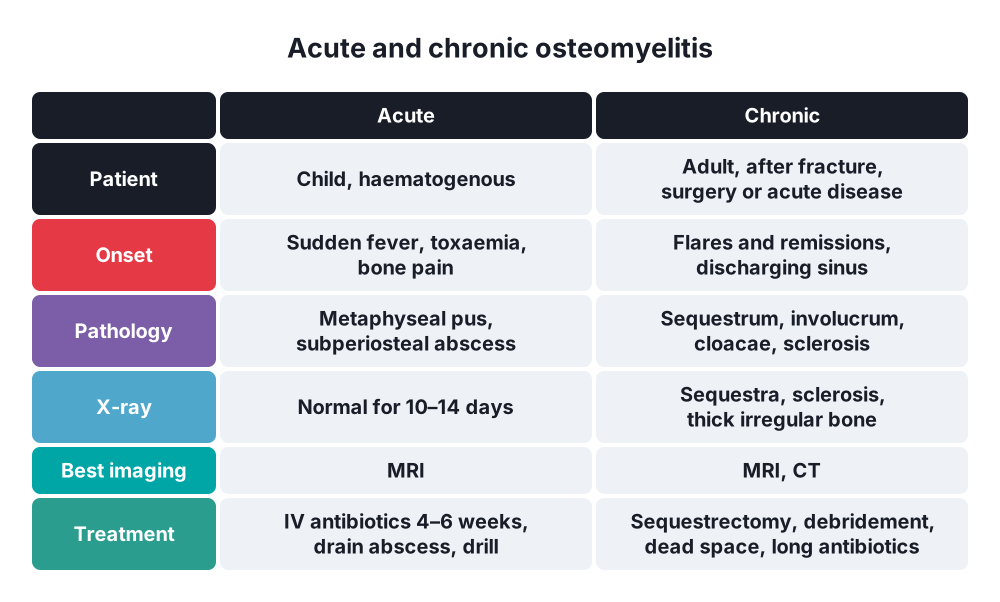
- X-ray → normal for the first 10–14 days; then periosteal reaction, lytic areas, sequestrum, involucrum
- MRI → investigation of choice; marrow oedema within days
- Ultrasound → subperiosteal abscess, joint effusion
- Bone scan → early, multifocal disease
- CT → sequestra in chronic disease
- Fistulography → sinus track
7. Differential diagnosis
- Septic arthritis
- Cellulitis
- Ewing's sarcoma → fever, pain, onion-skin periosteal reaction
- Fracture, trauma
- Rheumatic fever, juvenile arthritis
- Bone infarction in sickle cell disease
8. Treatment
Acute osteomyelitis: conservative
- Blood cultures and aspirate before antibiotics
- Intravenous antibiotics → anti-staphylococcal (flucloxacillin), adjusted by culture
- Duration → 4–6 weeks total; switch to oral after clinical and CRP response
- Immobilisation → splint, rest
- Analgesia, fluids
Acute osteomyelitis: surgical
- Indications:
- Abscess (subperiosteal, soft tissue)
- No response to antibiotics in 48–72 h
- Drain the subperiosteal abscess
- Drill holes or a cortical window to decompress the medulla
- Drain coexistent septic arthritis
Chronic osteomyelitis
- Combined surgical and antibiotic treatment
- Sequestrectomy and radical debridement (saucerisation) of dead bone and sinus
- Dead space management → antibiotic-loaded beads or cement spacer, muscle flaps, bone graft
- Stabilise bone → external fixator (Ilizarov)
- Soft tissue cover → flaps
- Prolonged antibiotics, 6 weeks or more
- Remove infected implants
- Amputation → last resort
9. Complications
- Septic arthritis
- Sepsis, metastatic abscesses
- Pathological fracture
- Growth disturbance → limb shortening, deformity
- Chronic osteomyelitis
- Marjolin's ulcer
- Amyloidosis
Show as a list
Osteomyelitis
- Routes
- Haematogenous
- Contiguous
- Post-traumatic
- Pathology
- Metaphysis
- Subperiosteal abscess
- Sequestrum
- Involucrum
- Cloaca, sinus
- Forms
- Acute
- Chronic
- Brodie's abscess
- Garré's
- Diagnosis
- Blood cultures
- MRI
- X-ray after 10–14 days
- Treatment
- IV antibiotics
- Drainage, drilling
- Sequestrectomy