
43. Oesophagus: congenital diseases, diverticula and strictures
High yield
Oesophageal atresia with fistula, Zenker's and epiphrenic diverticula, and benign strictures treated by dilatation or surgery.
Exam Prep · State examSurgery · Upper GISurgery · General surgeryAnatomy · Thorax
1. Definition
- Oesophageal atresia (OA) → congenital break in the continuity of the oesophagus, usually with a tracheo-oesophageal fistula (TOF)
- Oesophageal diverticulum → outpouching of the oesophageal wall:
- True → all layers of the wall
- False (pseudodiverticulum) → mucosa and submucosa only, through a gap in the muscle
- Oesophageal stricture → fixed narrowing of the lumen that causes dysphagia
2. Epidemiology
- OA → 1 in 3,000–4,500 live births; about half have other anomalies
- Zenker's (pharyngeal pouch) → most common diverticulum; elderly, age > 70 years, men
- Epiphrenic → rare; middle-aged adults
- Peptic stricture → most common benign stricture; older patients with long-standing reflux
- Caustic injury → children (accidental), adults (deliberate)
3. Aetiology and risk factors
Anatomy (revision)
- Length about 25 cm; from C6 (cricopharyngeus) to T11
- Three narrowings → cricopharyngeus (15 cm from incisors), aortic arch and left main bronchus (25 cm), diaphragm (40 cm)
- Upper third → striated muscle; middle third → mixed; lower third → smooth muscle
- No serosa → leaks spread easily; anastomoses heal less well
Congenital
- Failed separation of the foregut into trachea and oesophagus (week 4–6)
- VACTERL association → vertebral, anorectal, cardiac, tracheo-oesophageal, renal, limb anomalies
- Other → congenital stenosis, webs, duplication cysts, vascular rings (dysphagia lusoria)
Diverticula
- Pulsion → high pressure behind a poorly relaxing sphincter:
- Zenker's → upper oesophageal sphincter (cricopharyngeus) does not relax in time
- Epiphrenic → achalasia, diffuse oesophageal spasm in about 70%
- Traction → pulled out by inflamed, scarred mediastinal nodes (tuberculosis, histoplasmosis)
Strictures
- Peptic → gastro-oesophageal reflux disease (GORD)
- Caustic → alkali (liquefactive necrosis, deep) or acid (coagulative necrosis)
- Pill-induced → doxycycline, NSAIDs, potassium chloride, iron, alendronate
- Post-surgical (anastomotic), post-radiotherapy, post-sclerotherapy
- Eosinophilic oesophagitis → rings, young men with food bolus
- Schatzki ring, scleroderma, Crohn's disease
- Malignant → always excluded
4. Pathogenesis
OA with TOF
- Upper pouch → saliva cannot pass → drooling, aspiration
- Distal fistula → air enters the stomach; gastric acid enters the lungs → pneumonitis
Zenker's diverticulum
- Cricopharyngeus fails to relax or is fibrosed
- Raised pressure in the hypopharynx on swallowing
- Mucosa herniates through Killian's dehiscence → weak triangle between thyropharyngeus and cricopharyngeus (parts of the inferior constrictor)
- Pouch at C5–C6, usually on the left; it fills, swings down and compresses the oesophagus
Strictures
- Injury → inflammation → collagen and fibrosis → narrowing over weeks to months
- Caustic → stricture forms 3–8 weeks after injury
5. Classification
Oesophageal atresia (Gross)
- Type A → isolated OA, no fistula (about 8%); gasless abdomen
- Type B → proximal fistula (about 1%)
- Type C → proximal atresia + distal TOF (about 85%)
- Type D → both fistulas (about 1%)
- Type E → H-type fistula without atresia (about 4%); late presentation with chest infections
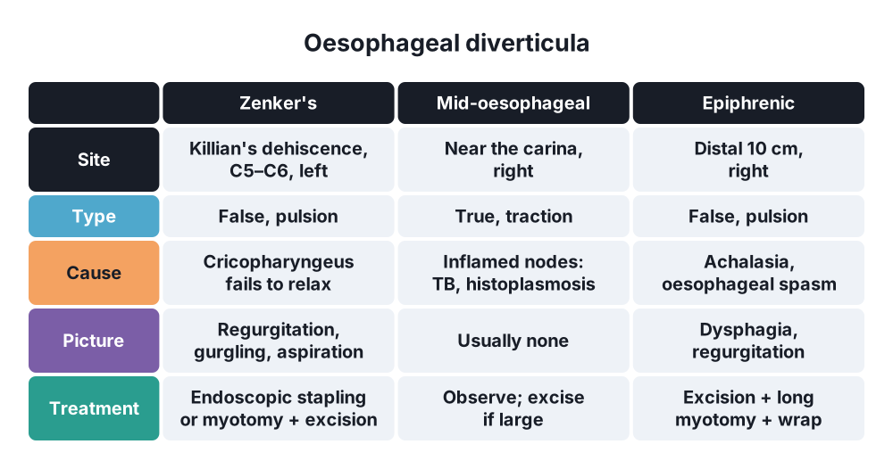
Diverticula by site
- Pharyngo-oesophageal (Zenker's) → false, pulsion
- Mid-oesophageal → true, traction; near the carina
- Epiphrenic → false, pulsion; distal 10 cm, usually right side
Strictures
- Simple → short (< 2 cm), straight, wide enough for an endoscope
- Complex → long, angulated, narrow; need more dilatations
- By cause → benign (peptic, caustic, anastomotic) or malignant
6. Clinical picture
Symptoms
- OA in a newborn:
- Polyhydramnios in pregnancy
- Frothy saliva, drooling
- Choking, coughing and cyanosis with the first feed
- H-type fistula → coughing with feeds, recurrent pneumonia
- Zenker's:
- Cervical dysphagia
- Regurgitation of undigested food, hours after eating
- Gurgling in the neck, halitosis
- Nocturnal cough, aspiration pneumonia
- Weight loss
- Epiphrenic → dysphagia, regurgitation, chest pain from the motility disorder
- Mid-oesophageal → usually none
- Stricture → slowly progressive dysphagia to solids, food bolus obstruction; long reflux history (peptic)
Signs
- OA → nasogastric tube (10 Fr) will not pass beyond 10 cm from the lips
- Zenker's → soft swelling in the left neck that gurgles on pressure (Boyce's sign)
- Stricture → weight loss, signs of aspiration
7. Diagnosis
Imaging
- OA → chest and abdominal X-ray with the tube in place:
- Tube coiled in the upper pouch
- Air in the stomach → distal fistula
- Gasless abdomen → no distal fistula (type A or B)
- Look for VACTERL → echocardiography (right aortic arch changes the side of thoracotomy), renal ultrasound, spine X-ray
- H-type → prone tube oesophagogram, bronchoscopy
- Barium swallow → test of choice for diverticula and strictures; shows size, neck, site
- Stricture → barium shows length and shape before endoscopy
Endoscopy
- Stricture → endoscopy with biopsy in every stricture to exclude cancer
- Zenker's → avoid blind endoscopy (perforation risk); done with care to exclude cancer in the pouch
- Caustic injury → endoscopy in 12–48 h to grade the burn (Zargar grade)
Physiology
- Manometry → epiphrenic and mid-oesophageal diverticula (look for achalasia or spasm)
- pH monitoring → reflux in peptic stricture
8. Differential diagnosis
- Dysphagia → oesophageal cancer, achalasia, external compression, neurological disease
- Zenker's → neck mass, goitre, laryngocoele
- Diverticulum versus hiatus hernia, ulcer crater
- Neonate with choking → laryngeal cleft, neurological swallowing disorder
9. Treatment
Surgical: atresia
- Before surgery → upright position, Replogle tube on continuous suction in the upper pouch, no feeds, antibiotics
- Risk stratification → birth weight and cardiac disease (Waterston, Spitz)
- Operation → right extrapleural thoracotomy (or thoracoscopy): divide and close the fistula, end-to-end oesophageal anastomosis
- Long gap (type A) → delayed primary anastomosis, traction (Foker), or replacement with stomach or colon
Surgical: diverticula
- Zenker's:
- Endoscopic stapled diverticulotomy (or laser, flexible endoscopy) → divides the wall between pouch and oesophagus, including cricopharyngeus; first choice in the elderly
- Open (left neck) → cricopharyngeal myotomy plus diverticulectomy (> 5 cm) or diverticulopexy
- Z-POEM → newer submucosal tunnel myotomy
- Epiphrenic (symptomatic only) → laparoscopic or thoracoscopic diverticulectomy + long myotomy on the opposite side + partial fundoplication
- Mid-oesophageal, large and symptomatic → diverticulectomy by VATS, plus myotomy if motility is abnormal
Conservative: strictures
- Endoscopic dilatation:
- Bougies (Savary-Gilliard over a guide wire, Maloney) or through-the-scope balloon
- "Rule of 3" → no more than three sizes up (3 mm) per session after resistance is felt
- Perforation risk about 0.1–0.4%; higher in caustic and radiation strictures
- Peptic → high-dose PPI for life, then antireflux surgery if needed
- Refractory → intralesional steroids, temporary removable stent, incisional therapy
- Caustic injury → no vomiting or neutralising agents; nil by mouth, PPI, early endoscopy; dilatation from about 3 weeks
- Eosinophilic oesophagitis → PPI, topical steroids, diet
Surgical: strictures
- Failure of dilatation, perforation, or suspected cancer → resection (oesophagectomy)
- Severe caustic damage → oesophageal replacement (colon or stomach); caustic oesophagus carries a lifelong cancer risk
10. Complications
- OA → anastomotic leak, stricture (30–40%), recurrent fistula, tracheomalacia, GORD
- Zenker's → aspiration pneumonia, perforation at endoscopy, cancer in the pouch (rare)
- Diverticulectomy → leak, recurrent laryngeal nerve injury (neck)
- Dilatation → perforation, bleeding, restenosis
- Caustic injury → perforation, mediastinitis, stricture, squamous cell carcinoma decades later
Show as a list
Oesophagus
- Congenital
- Atresia with TOF
- Gross type C 85%
- VACTERL
- Thoracotomy, anastomosis
- Diverticula
- Zenker's: pulsion, false
- Mid-oesophageal: traction, true
- Epiphrenic: motility disorder
- Myotomy
- Strictures
- Peptic
- Caustic
- Pill, anastomotic
- Biopsy all
- Dilatation
- PPI


