45. Oesophageal carcinoma
High yield
Squamous cell and adenocarcinoma; progressive dysphagia; staged by CT, EUS and PET; neoadjuvant therapy then oesophagectomy.
Exam Prep · State examSurgery · Upper GISurgery · Surgical oncologyAnatomy · Thorax
1. Definition
- Oesophageal carcinoma → malignant epithelial tumour of the oesophagus
- Two main types → squamous cell carcinoma (SCC) and adenocarcinoma
- Oesophagogastric junction (OGJ) tumour → centre within 2 cm of the junction; staged as oesophageal cancer
2. Epidemiology
- About the 7th most common cancer and 6th cause of cancer death worldwide
- SCC → most common type worldwide; "oesophageal cancer belt" from Iran to China
- Adenocarcinoma → most common type in the UK, USA and Western Europe; rising fast
- Men more than women → adenocarcinoma 5:1, SCC 3:1
- Age 60–70 years
- Most present late; 5-year survival about 20%
3. Aetiology and risk factors
Squamous cell carcinoma
- Smoking and alcohol → synergistic
- Hot drinks, nitrosamines, poor diet (low fruit and vegetables)
- Achalasia, caustic stricture
- Plummer-Vinson syndrome (iron deficiency, web)
- Tylosis (inherited palmar hyperkeratosis)
- Head and neck cancer, previous radiotherapy
Adenocarcinoma
- GORD → Barrett's oesophagus
- Obesity (central)
- Smoking
- Male sex, white ethnicity
- Helicobacter pylori → may protect
4. Pathogenesis
Adenocarcinoma
- Chronic reflux injures the squamous lining
- Intestinal metaplasia → Barrett's oesophagus (columnar lining with goblet cells)
- Low-grade dysplasia → high-grade dysplasia (HGD) → invasive adenocarcinoma
- Risk about 0.3% a year in non-dysplastic Barrett's; much higher with HGD
SCC
- Chronic irritation → squamous dysplasia → carcinoma; often multifocal ("field change")
Site
- SCC → upper and middle thirds
- Adenocarcinoma → lower third and OGJ
Spread
- Local → no serosa, so early spread to trachea, bronchus, aorta, recurrent laryngeal nerve, pericardium
- Lymphatic → rich submucosal lymphatics; skip lesions; nodes in neck, chest and abdomen (coeliac)
- Blood → liver, lung, bone, brain
- Peritoneum → OGJ tumours
5. Classification
Histology
- SCC
- Adenocarcinoma
- Rare → small cell carcinoma, melanoma, gastrointestinal stromal tumour (GIST), leiomyosarcoma
- Benign tumours → leiomyoma (most common), lipoma, polyps, granular cell tumour
TNM (AJCC 8th edition)
- Tis → HGD
- T1a → lamina propria or muscularis mucosae; T1b → submucosa
- T2 → muscularis propria
- T3 → adventitia
- T4a → pleura, pericardium, azygos vein, diaphragm, peritoneum (resectable); T4b → aorta, vertebra, trachea (unresectable)
- N1 1–2 nodes, N2 3–6, N3 ≥ 7
- M1 → distant metastases
Siewert (OGJ adenocarcinoma)
- Type I → centre 1–5 cm above the OGJ (distal oesophagus)
- Type II → 1 cm above to 2 cm below (true cardia)
- Type III → 2–5 cm below (subcardial stomach)
6. Clinical picture
Symptoms
- Progressive dysphagia → solids, then soft food, then liquids
- Weight loss, anorexia
- Odynophagia, retrosternal pain
- Regurgitation, cough on swallowing (tracheo-oesophageal fistula)
- Hoarseness → recurrent laryngeal nerve invasion
- Haematemesis, anaemia
- Early cancer → dyspepsia only, or found on Barrett's surveillance
- Red flag → new dysphagia at any age, or dyspepsia with weight loss over 55, needs urgent endoscopy (2-week wait)
Signs
- Often none
- Cachexia
- Supraclavicular node (Virchow's node, left)
- Hepatomegaly, ascites
- Horner's syndrome (rare)
7. Diagnosis
Endoscopy
- Upper GI endoscopy with multiple biopsies → test of choice
- Distance from incisors, length, relation to OGJ, Barrett's segment
- HER2 and PD-L1 testing on tissue (adenocarcinoma)
Staging
- CT chest, abdomen and pelvis with contrast → metastases, local invasion
- PET-CT → if curative treatment is planned; finds hidden metastases
- Endoscopic ultrasound (EUS) → T and N stage; fine-needle aspiration of nodes
- Staging laparoscopy → OGJ and Siewert II–III tumours; peritoneal spread
- Bronchoscopy → upper and middle third tumours near the airway
- Barium swallow → "apple core" or shouldered stricture; less used now
Fitness
- Performance status, cardiopulmonary exercise testing, spirometry, nutrition
- Decision by the upper GI MDT
8. Differential diagnosis
- Benign peptic stricture
- Achalasia and pseudoachalasia
- Leiomyoma, GIST
- External compression → lung cancer, nodes, aortic aneurysm
- Gastric cancer of the cardia
9. Treatment
Surgical
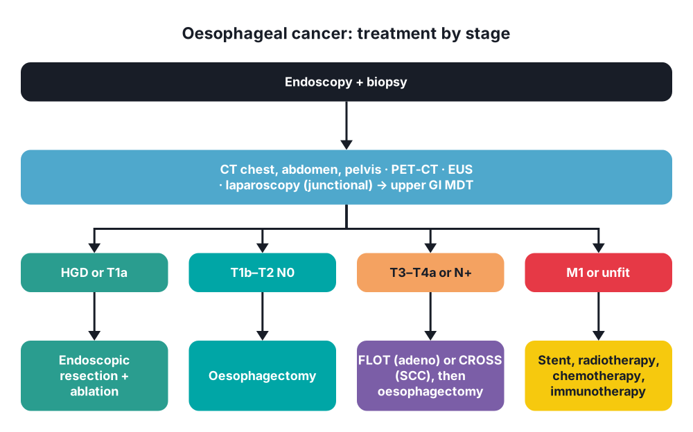
- Early disease (HGD, T1a) → endoscopic resection (EMR or ESD) + radiofrequency ablation of remaining Barrett's; cure rate > 90%
- T1b–T2 N0 → oesophagectomy (some T1b by endoscopy if low risk)
- Locally advanced (T3–T4a or N+) → neoadjuvant therapy, then oesophagectomy:
- Adenocarcinoma → perioperative FLOT chemotherapy (FLOT4; ESOPEC 2024 favoured it over CROSS)
- SCC → neoadjuvant chemoradiotherapy (CROSS); complete response in about half of SCC
- Residual disease after CROSS → adjuvant nivolumab (CheckMate 577)
- Oesophagectomy → main types:
- Ivor Lewis → laparotomy (or laparoscopy) + right thoracotomy; anastomosis in the upper chest; lower and middle third tumours
- McKeown (three-stage) → right thoracotomy + abdomen + neck anastomosis; upper and middle third tumours
- Transhiatal → abdomen + neck, no thoracotomy; fewer lung problems, less node clearance
- Left thoracoabdominal → OGJ tumours
- Minimally invasive or robotic → fewer lung complications
- Principles:
- Margins → 5–10 cm proximal, 5 cm distal (longitudinal submucosal spread)
- Two-field lymphadenectomy (abdominal and thoracic)
- Conduit → stomach (gastric tube) on the right gastroepiploic artery; colon or jejunum if the stomach cannot be used
- Pyloric drainage, feeding jejunostomy
- Enhanced recovery, high-volume centres
- Operative mortality → 2–5% in high-volume centres
Conservative
- Definitive chemoradiotherapy → SCC of the cervical oesophagus, or patients unfit for or refusing surgery
- Palliation (most patients):
- Self-expanding metal stent → fast relief of dysphagia; covered stent seals a TOF
- Palliative radiotherapy, brachytherapy
- Chemotherapy ± trastuzumab (HER2+) or immunotherapy (nivolumab, pembrolizumab)
- Nutrition, dietitian, analgesia
- Laser, argon plasma or photodynamic therapy for unfit patients
- Barrett's surveillance → endoscopy every 2–5 years; HGD treated by endoscopy
10. Complications
Of disease
- Malnutrition, aspiration pneumonia
- Tracheo-oesophageal fistula
- Bleeding, aorto-oesophageal fistula
- Recurrent laryngeal nerve palsy
Of oesophagectomy
- Pneumonia, respiratory failure → most common
- Anastomotic leak (5–15%) → neck leaks are easier to treat than chest leaks
- Conduit necrosis
- Atrial fibrillation
- Chylothorax (thoracic duct injury)
- Recurrent laryngeal nerve injury (neck anastomosis)
- Anastomotic stricture, reflux, delayed gastric emptying, dumping
11. Prognosis
- 5-year survival about 20% for all patients
- After neoadjuvant therapy and resection → about 45–50%
- T1a after endoscopic resection → > 90%
- Stage IV → median survival about 1 year with modern systemic therapy
Show as a list
Oesophageal carcinoma
- Types
- SCC: upper, middle
- Adenocarcinoma: lower
- Leiomyoma (benign)
- Risks
- Smoking, alcohol
- Achalasia, caustic
- Barrett's, GORD
- Obesity
- Clinical
- Progressive dysphagia
- Weight loss
- Hoarseness
- Staging
- Endoscopy, biopsy
- CT, PET-CT
- EUS
- Laparoscopy
- Treatment
- Endoscopic resection
- FLOT or CROSS
- Ivor Lewis
- McKeown
- Stent, palliation


