
95. Non-occlusive mesenteric ischaemia
High yield
Bowel ischaemia from low flow and vasospasm with open vessels, in critically ill patients; treat the cause, intra-arterial vasodilators.
Exam Prep · State examSurgery · General surgerySurgery · Emergency and trauma
1. Definition
- Non-occlusive mesenteric ischaemia (NOMI) → acute intestinal ischaemia without an occlusion of the mesenteric arteries or veins
- Caused by splanchnic vasoconstriction and low blood flow (low cardiac output, shock, vasopressors), sometimes on top of existing atherosclerotic narrowing
- The bowel is starved of blood while the vessels are still patent
- Related conditions → ischaemic colitis (low-flow ischaemia of the colon, usually milder), occlusive AMI (question 93), MVT (question 94)
2. Epidemiology
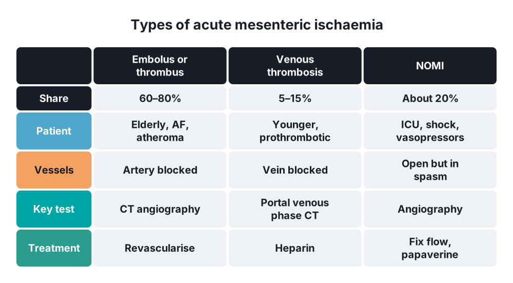
- About 20% of acute mesenteric ischaemia (5–30% in different series)
- Almost always in critically ill, elderly patients in ICU or after major surgery
- Highest risk → after cardiac surgery (especially with cardiopulmonary bypass and low output), haemodialysis patients, septic shock
- After cardiac surgery → incidence about 0.5–1%, but mortality very high
- Highest mortality of all forms of AMI → about 50–80% (partly due to the underlying illness)
- Diagnosis is often late (patients are sedated and ventilated)
3. Aetiology and risk factors
Low flow states
- Cardiogenic shock, severe heart failure, arrhythmias, after cardiac surgery (low cardiac output, intra-aortic balloon pump)
- Hypovolaemia → haemorrhage, burns, dehydration, pancreatitis
- Septic shock
- Haemodialysis (fluid removal, hypotension during dialysis)
Vasoconstriction
- Vasopressors → noradrenaline, adrenaline, vasopressin, high-dose dopamine
- Digoxin (splanchnic vasoconstrictor), alpha-agonists, ergotamine, cocaine, amphetamines
- High intra-abdominal pressure (abdominal compartment syndrome)
- Strenuous endurance exercise (marathon runners)
Patient factors
- Age over 50–70, atherosclerosis (existing SMA stenosis), chronic kidney disease, diabetes, previous mesenteric ischaemia
- Major cardiovascular or abdominal surgery (aortic repair: IMA ligation → colonic ischaemia)
4. Pathogenesis
- Low cardiac output or shock → the body protects the brain and heart by sympathetic splanchnic vasoconstriction (renin-angiotensin, vasopressin)
- Mesenteric arteries spasm (mainly SMA branches); vasopressors worsen it
- Vasospasm persists even after cardiac output is restored
- Mucosa is hit first (countercurrent exchange in the villi makes the tips hypoxic) → barrier loss, bacterial translocation, SIRS
- Patchy then transmural necrosis → perforation, peritonitis, sepsis, multiple organ failure
- Reperfusion injury worsens the damage when flow returns
- Ischaemia is often patchy and segmental, sometimes extensive; can affect the small bowel, colon or both
- Watershed areas of the colon (splenic flexure, rectosigmoid) are at particular risk
5. Classification
By site
- Small bowel NOMI (SMA territory)
- Colonic NOMI → overlaps with ischaemic colitis
By depth of injury
- Mucosal (reversible) → mural → transmural (gangrene)
Ischaemic colitis (Brandt) for comparison
- Non-gangrenous (transient or chronic, most) → conservative
- Gangrenous (about 15–20%) → surgery
Typical angiographic signs of NOMI (Siegelman)
- Narrowing of the origins of SMA branches
- Irregular "string of sausages" pattern (alternating spasm and dilatation)
- Spasm of the mesenteric arcades
- Poor filling of intramural vessels ("pruned tree")
6. Clinical picture
- The patient is often sedated, ventilated, on vasopressors → typical pain may be absent
Symptoms (if awake)
- Abdominal pain → may be absent in about 20–25%; severe and out of proportion when present
- Nausea, vomiting, diarrhoea, bloody stools
Signs
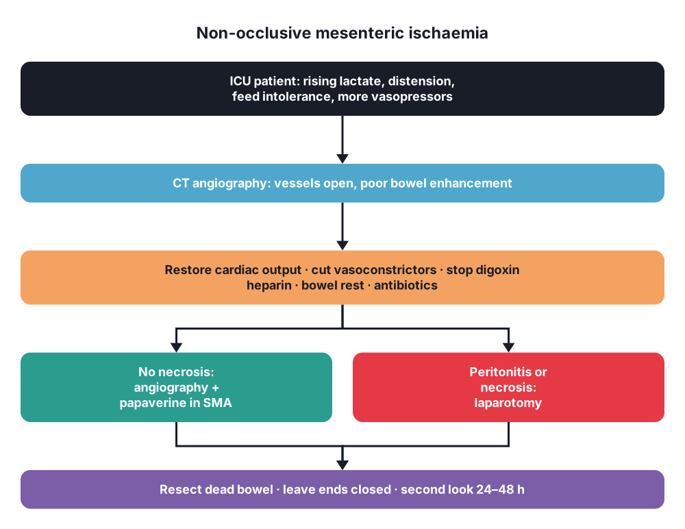
- Unexplained deterioration in an ICU or post-cardiac surgery patient:
- Rising lactate and metabolic acidosis without another cause
- Increasing vasopressor need, new organ failure, fever, rising WCC
- Abdominal distension, ileus, high NG aspirates, feed intolerance
- GI bleeding (melaena, bloody stool)
- Late → peritonitis, septic shock
- Raised intra-abdominal pressure
7. Diagnosis
- High index of suspicion → think of NOMI in any critically ill patient with unexplained acidosis, distension or feed intolerance
Bloods
- Lactate (rising), base deficit, WCC, CRP, D-dimer, amylase, LFT, CK
- Daily monitoring in high-risk ICU patients
Imaging
- CT angiography (biphasic) → first test:
- Vessels patent (no occlusion)
- Narrow SMA and branches, poor peripheral filling
- Bowel signs → reduced wall enhancement, wall thickening, dilated fluid-filled loops, pneumatosis, portal venous gas
- CTA is less reliable for NOMI than for occlusive AMI
- Selective mesenteric angiography → gold standard for NOMI:
- Shows the Siegelman signs (branch narrowing, string of sausages, arcade spasm, pruned tree)
- Allows treatment at the same time (intra-arterial papaverine)
- Needs a stable enough patient and transfer to the angiography suite
- Endoscopy → flexible sigmoidoscopy or colonoscopy at the bedside for colonic ischaemia (pale or dusky mucosa, ulcers, pseudomembranes)
- Intra-abdominal pressure (bladder pressure) → exclude abdominal compartment syndrome
- Laparotomy or laparoscopy (bedside in ICU in some centres) → when peritonitis or necrosis is suspected; the bowel may look patchy, with pulses present
8. Differential diagnosis
- Occlusive AMI (embolus or thrombus) → CTA shows the occlusion
- Mesenteric venous thrombosis
- Abdominal compartment syndrome (also causes and is caused by gut ischaemia)
- Acalculous cholecystitis, pancreatitis, perforated stress ulcer
- C. difficile colitis, toxic megacolon
- Ogilvie's syndrome (pseudo-obstruction)
- Other causes of lactic acidosis in ICU → sepsis, liver failure, metformin, adrenaline
9. Treatment
Conservative
- Treat the cause (most important):
- Restore circulating volume and cardiac output (fluids guided by monitoring, inotropes such as dobutamine or milrinone that dilate splanchnic vessels)
- Reduce or stop vasoconstrictors where possible (vasopressin, high-dose noradrenaline); stop digoxin
- Treat sepsis, arrhythmia and heart failure; adjust dialysis to avoid hypotension
- Supportive care → bowel rest (stop enteral feeding), NG decompression, broad-spectrum antibiotics, correct acidosis and electrolytes
- Systemic anticoagulation (heparin) → to prevent secondary thrombosis in low-flow vessels (if no contraindication)
- Intra-arterial vasodilators through a catheter left in the SMA at angiography:
- Papaverine → about 30–60 mg/h continuous infusion; repeat angiography after 24 hours; continue until the spasm resolves (often 24–72 hours)
- Alternatives → prostaglandin E1 (alprostadil), nitroglycerin, prostacyclin
- Do not run heparin through the same catheter as papaverine (they precipitate)
- Evidence comes from case series; no randomised trials
- Monitor intra-abdominal pressure
Surgical
- Indications → peritonitis, perforation, signs of transmural necrosis on CT, failure to improve despite treatment
- Laparotomy:
- Resect necrotic bowel only (ischaemia is often patchy)
- Ends left closed (no anastomosis in unstable patients), temporary abdominal closure
- Second-look laparotomy at 24–48 hours (ischaemia often progresses)
- Continue the papaverine infusion during and after surgery
- Colonic gangrene (ischaemic colitis) → resection (often subtotal colectomy) with end stoma, no anastomosis
- Extensive ischaemia in a patient with multiple organ failure → may be non-survivable; consider palliation with the family
10. Complications
Of the disease
- Bowel necrosis, perforation, peritonitis
- Septic shock, multiple organ failure, death (50–80%)
- Ischaemic stricture (late obstruction)
- GI haemorrhage
Of treatment
- Short bowel syndrome after extensive resection
- Hypotension from systemic spill of vasodilator (papaverine)
- Catheter complications (thrombosis, dislodgement, bleeding)
- Anastomotic leak, wound complications, open abdomen problems
Prognosis
- Worst of all mesenteric ischaemia types; depends on the underlying disease and time to diagnosis
- Prevention → avoid prolonged hypotension, use the lowest effective vasopressor dose, early enteral feeding when perfusion is adequate, monitor lactate in high-risk patients
Show as a list
NOMI
- Who
- ICU, elderly
- After cardiac surgery
- Dialysis, shock
- Vasopressors, digoxin
- Mechanism
- Low flow
- Splanchnic spasm
- Vessels patent
- Patchy ischaemia
- Clinical
- Often no pain
- Rising lactate
- Distension, ileus
- GI bleeding
- Tests
- CT angiography
- Angiography: gold standard
- String of sausages
- Treatment
- Fix cardiac output
- Cut vasopressors
- Papaverine in SMA
- Resect dead bowel
- Second look


