28. Congenital, traumatic and inflammatory diseases of the neck
High yield
Thyroglossal and branchial cysts, lymphatic malformations, penetrating neck trauma by zones, lymphadenitis and deep neck abscesses.
Exam Prep · State examSurgery · General surgeryAnatomy · Head and neck
1. Congenital diseases
A. Thyroglossal duct cyst
- Remnant of the thyroglossal duct (descent of the thyroid from the foramen caecum)
- Most common congenital neck mass in children
- Midline, at or below the hyoid bone
- Moves up on swallowing and on tongue protrusion
- May become infected → abscess, fistula
- Ultrasound → confirms cyst and a normal thyroid gland
- Treatment → Sistrunk operation: excision of the cyst, the tract and the central part of the hyoid bone
- Simple excision → recurrence up to 50%
- Ectopic (lingual) thyroid → may be the only thyroid tissue
B. Branchial (cleft) cyst
- Remnant of the second branchial cleft in 95%
- Young adults, 20–30 years
- Anterior border of sternocleidomastoid, junction of upper and middle thirds
- Smooth, fluctuant, transilluminates poorly; contains cholesterol crystals
- Branchial fistula → opening at the lower third of the anterior border of sternocleidomastoid; tract passes between internal and external carotid arteries to the tonsillar fossa
- Differential diagnosis in an adult → cystic metastasis of HPV-related oropharyngeal or papillary thyroid cancer
- Treatment → complete surgical excision, after infection has settled
C. Lymphatic malformation (cystic hygroma)
- Malformation of lymphatic channels
- Infants, posterior triangle of the neck
- Soft, compressible, brilliantly transilluminant
- Complications → airway compression, infection, haemorrhage
- Treatment → sclerotherapy (OK-432, doxycycline, bleomycin), surgical excision, sirolimus
D. Other congenital lesions
- Dermoid cyst → midline, does not move with tongue protrusion
- Congenital muscular torticollis → fibrosis of sternocleidomastoid; head tilted to the affected side, chin to the other side
- Cervical rib → thoracic outlet syndrome
- Haemangioma
- Laryngocele
2. Traumatic diseases
Mechanism
- Penetrating → stab, gunshot
- Blunt → strangulation, hanging, road traffic accidents, seat belt
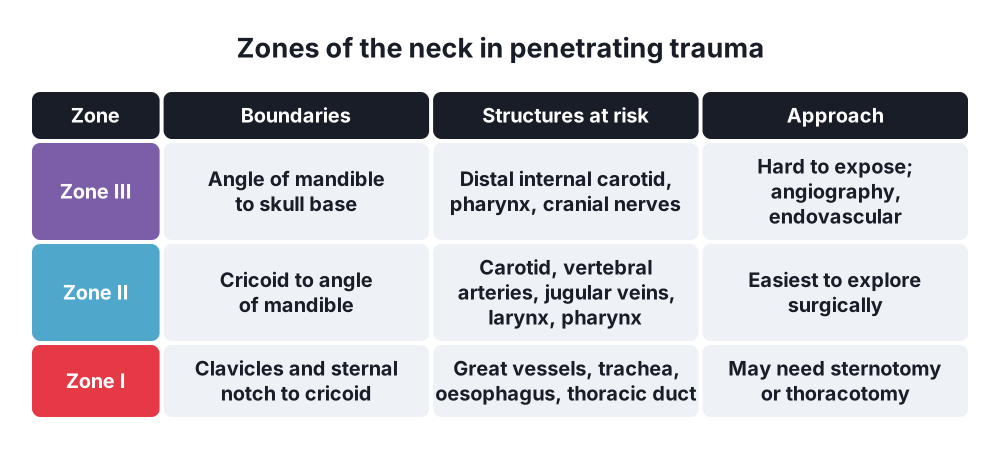
Penetrating injuries breach platysma → zones of the neck
- Zone I → clavicles and sternal notch to cricoid cartilage; great vessels, trachea, oesophagus, thoracic duct
- Zone II → cricoid to angle of the mandible; carotid and vertebral arteries, jugular veins, larynx, pharynx
- Zone III → angle of the mandible to skull base; distal carotid, pharynx
Hard signs (immediate surgery)
- Airway compromise
- Expanding or pulsatile haematoma
- Active bleeding
- Shock
- Bruit or thrill
- Air bubbling from the wound
- Massive haemoptysis or haematemesis
Soft signs (CT angiography first)
- Dysphagia, dysphonia
- Subcutaneous emphysema
- Non-expanding haematoma
Management
- ATLS → airway first; intubate early before swelling
- Do not probe a neck wound; do not remove an impaled object outside theatre
- Hard signs → neck exploration
- Stable with soft signs → CT angiography, endoscopy, contrast swallow
- Blunt injury → laryngotracheal fracture, carotid dissection (stroke)
3. Inflammatory diseases
A. Acute lymphadenitis
- Secondary to infection of the tonsils, teeth, scalp, skin
- Tender, enlarged nodes; may suppurate → abscess
- Treatment → treat the source, antibiotics; drain the abscess
B. Tuberculous lymphadenitis (scrofula)
- Most common extrapulmonary tuberculosis
- Matted, painless nodes in the posterior triangle or jugular chain
- Cold abscess → collar-stud abscess, discharging sinus
- Diagnosis → FNA, culture, polymerase chain reaction (PCR)
- Treatment → anti-tuberculous drugs for 6 months; aspirate cold abscess through healthy skin; excise residual nodes
C. Deep neck space infections
- Source → teeth, tonsils, pharynx, intravenous drug use
- Peritonsillar abscess (quinsy) → trismus, uvula pushed to the other side, hot potato voice; needle aspiration or incision
- Parapharyngeal abscess → swelling behind the angle of the mandible, trismus; risk to carotid sheath
- Retropharyngeal abscess → children; neck stiffness, dysphagia, stridor; widened prevertebral space on lateral X-ray; spreads to the mediastinum (danger space)
- Ludwig's angina → floor of the mouth
- Diagnosis → contrast CT of the neck and chest
- Treatment → airway, intravenous antibiotics, surgical drainage
- Complications → mediastinitis, internal jugular vein thrombosis (Lemierre's syndrome, Fusobacterium necrophorum), carotid artery erosion, sepsis
D. Other
- Sialadenitis, thyroiditis
- Actinomycosis → woody hard swelling, multiple sinuses, sulphur granules
- Cat-scratch disease, toxoplasmosis, HIV
Show as a list
Neck diseases
- Congenital
- Thyroglossal cyst
- Branchial cyst
- Cystic hygroma
- Dermoid
- Torticollis
- Traumatic
- Zones I, II, III
- Hard signs
- Soft signs
- Inflammatory
- Lymphadenitis
- Tuberculous nodes
- Quinsy
- Parapharyngeal abscess
- Retropharyngeal abscess
- Treatment
- Sistrunk operation
- Neck exploration
- Airway and drainage