88. Multiple and combined injuries
High yield
Polytrauma, associated and combined injuries; ATLS primary survey, damage control resuscitation and surgery, and injury scoring.
Exam Prep · State examSurgery · General surgerySurgery · Emergency and trauma
1. Definition
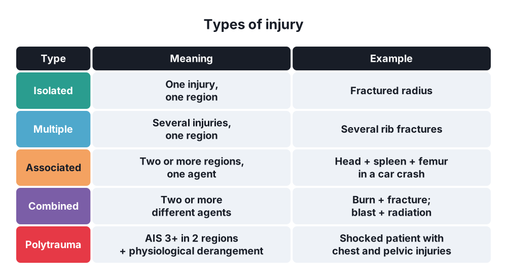
- Isolated injury → one injury in one body region
- Multiple injury → several injuries in one body region (several rib fractures, several bowel perforations)
- Associated (concomitant) injury → injuries in two or more body regions from one agent (head + abdomen + femur in a car crash)
- Combined injury → injury from two or more different agents (mechanical + thermal, mechanical + radiation, burn + chemical, blast + burn)
- Polytrauma → associated injuries where at least one, or their combination, threatens life
- Berlin definition (2014) → AIS 3 or more in at least two body regions plus one or more physiological variables: systolic BP 90 mmHg or less, GCS 8 or less, base deficit 6 or more, INR 1.4 or more (or PTT 40 s or more), age 70 or more
- Major trauma → Injury Severity Score (ISS) over 15
- Basic terms → question 1; abdominal injuries → question 87; chest → question 33; shock → question 13
2. Epidemiology
- Trauma is the leading cause of death under 45 years; road traffic collisions cause most polytrauma
- Trimodal distribution of trauma deaths (Trunkey):
- Immediate (seconds to minutes, about 50%) → brain stem, high spinal cord, heart, aortic rupture; only prevention helps
- Early (minutes to hours, about 30%) → haemorrhage, tension pneumothorax, airway loss, intracranial haematoma; the target of ATLS and the "golden hour"
- Late (days to weeks, about 20%) → sepsis, multiple organ failure, pulmonary embolism
- With modern trauma systems the curve is now closer to bimodal (fewer late deaths)
- Haemorrhage → the leading cause of preventable death after injury
- Combined injuries → war, terror attacks, industrial explosions, house fires, nuclear accidents
3. Aetiology and risk factors
High-energy mechanisms (trauma team activation)
- Road traffic collision at high speed, ejection, rollover, death in the same vehicle, pedestrian or cyclist struck
- Fall from over 3 m (or over twice body height)
- Crush, entrapment, explosion, gunshot wounds
Typical injury patterns
- Frontal impact (driver) → head, cervical spine, sternum, heart, aorta, liver or spleen, knee–femur–hip (dashboard)
- Side impact → head, chest wall, spleen or liver, pelvis, femur
- Pedestrian (adult) → tibia and knee (bumper), then head and chest (bonnet, windscreen), then head (ground)
- Fall from height onto the feet → calcaneus, tibial plateau, hip, lumbar spine, aortic or renal pedicle shear
Combined injury agents
- Mechanical + thermal → burns with fractures or blunt trauma (car fires, explosions)
- Blast → primary (pressure wave: ears, lungs, bowel), secondary (fragments), tertiary (body thrown), quaternary (burns, crush, toxic gases)
- Radiation + trauma or burns → nuclear accidents
- Chemical + trauma or burns
Risk factors for poor outcome
- Age over 70, anticoagulants, comorbidity
- Severe head injury, shock on arrival, delay to definitive care
4. Pathogenesis
Mutual aggravation syndrome
- In associated and combined injuries each injury makes the others worse → the effect is greater than their sum
- Examples:
- Blood loss from fractures + abdominal bleeding → shock earlier
- Hypotension and hypoxia double the mortality of head injury (secondary brain injury)
- Burns + trauma → more fluid loss, more infection, slower healing
- Radiation → bone marrow suppression → bleeding, infection, poor wound healing
Lethal diamond (formerly the lethal triad)
- Hypothermia (under 35 °C) → slows clotting enzymes, platelet dysfunction
- Acidosis (pH under 7.2) → poor perfusion, lactate; impairs clotting
- Coagulopathy → trauma-induced coagulopathy, consumption, dilution by crystalloid
- Hypocalcaemia → citrate in transfused blood; calcium is needed for clotting and heart contraction
- Each worsens the others → a vicious cycle that surgery alone cannot stop
Acute traumatic coagulopathy
- Present in about 25% of severely injured patients on arrival, before any fluids
- Tissue injury + shock → activated protein C, hyperfibrinolysis, fibrinogen depletion
- Doubles to quadruples mortality
Systemic response
- Two hit model → first hit (injury, shock) primes the immune system; second hit (surgery, infection, transfusion) → SIRS, ARDS, multiple organ failure
- Fat embolism after long bone and pelvic fractures
5. Classification
By number and location
- Isolated, multiple (one region), associated (two or more regions), combined (two or more agents)
By leading injury
- The dominant injury decides the priority and the outcome (head, chest, abdomen, pelvis, limbs)
Injury scores
- Abbreviated Injury Scale (AIS) → each injury scored 1 (minor) to 6 (unsurvivable)
- Injury Severity Score (ISS) → sum of the squares of the highest AIS in the three most injured of six body regions; range 1–75; ISS over 15 = major trauma; any AIS 6 → ISS 75
- New ISS (NISS) → three highest AIS anywhere (even in the same region)
- Revised Trauma Score (RTS) → GCS, systolic BP, respiratory rate (physiology)
- TRISS → combines ISS, RTS, age and mechanism → probability of survival
- Glasgow Coma Scale → 3–15; 8 or less → intubate
- Shock index → heart rate / systolic BP; over 1 suggests major bleeding
Patient condition (damage control orthopaedics)
- Stable, borderline, unstable, in extremis → guides early total care vs damage control
6. Clinical picture
- Life-threatening problems first, then a full head-to-toe search
- One obvious injury (an open fracture) often distracts from a hidden, deadly one (splenic bleed)
Signs of the main threats (ATLS order)
- Catastrophic external haemorrhage → pulsatile or pouring bleeding from a limb or junctional wound
- Airway → stridor, gurgling, facial or neck injury, burns to the face (soot, singed hair: inhalation injury)
- Breathing → tension pneumothorax (absent breath sounds, hyper-resonance, tracheal shift late), open chest wound, flail chest, massive haemothorax
- Circulation → shock: tachycardia, low BP, cold clammy skin, capillary refill over 2 s, low urine output, falling consciousness
- Five places for blood loss → "on the floor and four more": chest, abdomen, pelvis and retroperitoneum, long bones (femur up to 1.5 L each)
- Disability → GCS, pupils, lateralising signs, blood glucose
- Exposure → full examination, log-roll, back and perineum; prevent hypothermia
Combined injuries
- Burns + trauma → the burn is obvious, but early shock (in the first hour) is from bleeding, not from the burn; look for the injury
- Blast lung → hypoxia, haemoptysis, "butterfly" shadowing; ruptured eardrums are a marker of primary blast
- Radiation → nausea and vomiting within hours (time to vomiting predicts dose), then a latent phase, then marrow, gut and skin effects
- Crush syndrome → limb trapped for hours; on release: hyperkalaemia, myoglobinuria, acute kidney injury
7. Diagnosis
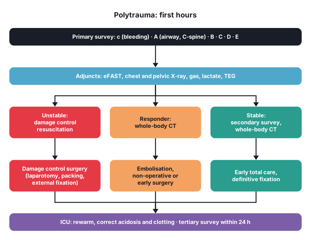
Primary survey with resuscitation (ATLS, cABCDE)
- c → Catastrophic haemorrhage → direct pressure, tourniquet, haemostatic dressings, pelvic binder
- A → Airway with cervical spine protection → jaw thrust, suction, airway adjuncts, rapid sequence intubation (GCS 8 or less, airway burns, unprotected airway), surgical airway if needed; manual in-line stabilisation
- B → Breathing → oxygen, needle or finger thoracostomy for tension pneumothorax, chest drain, occlusive dressing for an open wound
- C → Circulation → two large-bore IV or intraosseous lines, bloods (crossmatch, FBC, coagulation, gas with lactate), blood not crystalloid, find the source
- D → Disability → GCS, pupils, glucose
- E → Exposure and environment → undress, log-roll, keep warm
Adjuncts to the primary survey
- eFAST → blood in the abdomen, pericardium, pleura; pneumothorax
- Chest X-ray and pelvic X-ray
- ECG, pulse oximetry, end-tidal CO2, urinary catheter (after urethral injury is excluded), NG tube (orogastric if base of skull fracture)
- Arterial blood gas → lactate and base deficit (shock severity)
- Viscoelastic tests (TEG, ROTEM) → guide blood products
Secondary survey (once stable)
- AMPLE history → Allergies, Medications, Past history and pregnancy, Last meal, Events and environment
- Head-to-toe examination, including ears, eyes, back, perineum, rectum, all limbs and pulses
Definitive imaging
- Whole-body CT (head, cervical spine, chest, abdomen, pelvis) → fast, finds hidden injuries; for stable or responding patients; some centres scan carefully selected unstable patients if the CT is next to the resuscitation room
- Angiography (bleeding pelvis, solid organs); X-rays of limbs
Tertiary survey
- Repeat full examination within 24 hours (and when the patient wakes) → finds about 10% missed injuries (hand, foot, spine fractures)
Combined injuries
- Burns → percentage of body surface (rule of nines, Lund and Browder), carboxyhaemoglobin, bronchoscopy for inhalation injury
- Radiation → dosimetry, lymphocyte count at 24–48 hours (falling count = higher dose), decontamination before surgery
- Crush → CK, potassium, myoglobin, renal function
8. Differential diagnosis
- Causes of shock in polytrauma:
- Haemorrhagic (most common) → chest, abdomen, pelvis, long bones, external
- Obstructive → tension pneumothorax, cardiac tamponade
- Neurogenic → spinal cord injury (low BP, low heart rate, warm skin)
- Cardiogenic → blunt cardiac injury, myocardial infarction that caused the crash
- Low GCS → head injury vs hypoxia, hypotension, hypoglycaemia, alcohol and drugs
- A medical event that caused the injury → stroke, seizure, arrhythmia, hypoglycaemia
9. Treatment
Conservative
- Pre-hospital → control catastrophic bleeding, airway, splint, pelvic binder, tranexamic acid, rapid transfer to a major trauma centre ("scoop and run" for penetrating trauma)
- Damage control resuscitation (DCR):
- Permissive hypotension → systolic about 80–90 mmHg (or a palpable radial pulse) until bleeding is controlled; not with traumatic brain injury (keep systolic over 100–110 mmHg)
- Haemostatic resuscitation → early red cells, plasma and platelets in a 1:1:1 ratio (massive transfusion protocol), or whole blood; fibrinogen (cryoprecipitate or concentrate)
- Minimise crystalloid (worsens dilution, acidosis, oedema)
- Tranexamic acid → 1 g over 10 minutes then 1 g over 8 hours, within 3 hours of injury (CRASH-2)
- Calcium replacement, warming (warm fluids, blankets, warm room)
- Reverse anticoagulants (prothrombin complex concentrate for warfarin; specific reversal agents for DOACs)
- Non-operative management of selected injuries in stable patients → solid organs with angioembolisation, many chest injuries with a chest drain alone
- ICU → ventilation, rewarming, correction of coagulopathy, monitoring for abdominal compartment syndrome, nutrition, VTE prophylaxis once bleeding stops
- Combined injuries:
- Burns → fluid resuscitation (Parkland formula), adjusted for blood loss; early excision once stable
- Radiation → decontaminate first; surgery within 48 hours (before marrow suppression), then delay until recovery
- Crush → IV fluids before release if possible, urine output over 200–300 mL/h, treat hyperkalaemia, dialysis if needed
Surgical
- Priorities → life before limb; bleeding before contamination; the most threatening region first
- Order when several cavities bleed → the patient's physiology and the FAST, chest drain and pelvic X-ray findings decide; often chest drain → laparotomy → pelvic packing → limb
- Damage control surgery (DCS) → for patients in shock with the lethal diamond (pH under 7.2, temperature under 34–35 °C, coagulopathy, over 10 units of blood):
- Stage 1: abbreviated surgery (under 60–90 minutes) → stop bleeding (ligation, shunts, packing), control contamination (staple off bowel, no anastomosis), temporary abdominal closure (vacuum dressing)
- Stage 2: ICU resuscitation (24–48 hours) → rewarm, correct acidosis and clotting
- Stage 3: planned re-operation → remove packs, definitive repair, anastomoses, close the abdomen
- Damage control orthopaedics → external fixation of long bone and pelvic fractures in unstable or borderline patients; definitive fixation (nailing, plating) at days 5–10 once stable
- Early total care → definitive fixation in the first 24 hours for stable patients (fewer lung complications, shorter stay)
- Pelvic bleeding → pelvic binder, preperitoneal pelvic packing, angioembolisation, external fixation, REBOA
- Head injury → craniotomy for an expanding haematoma; prevent secondary injury (oxygen, BP, CO2, glucose)
- Resuscitative thoracotomy → penetrating chest injury with recent loss of output
- Hybrid theatres → surgery and angiography together
10. Complications
Early
- Haemorrhagic shock, death
- Trauma-induced coagulopathy, transfusion reactions, TRALI, TACO, hyperkalaemia, hypocalcaemia
- Missed injuries
- Abdominal compartment syndrome (see question 98)
- Fat embolism syndrome (petechiae, hypoxia, confusion 24–72 hours after fractures)
- Crush syndrome and acute kidney injury
Late
- ARDS, pneumonia
- Sepsis and multiple organ dysfunction syndrome (MODS)
- Venous thromboembolism (very high risk) → mechanical prophylaxis early, low molecular weight heparin when bleeding is controlled
- Pressure sores, malnutrition, muscle loss
- Post-traumatic stress disorder, long-term disability
Prognosis
- Mortality rises with ISS, age, head injury, shock on arrival and coagulopathy
- Combined injuries (especially radiation and burns) have a much worse outcome than either injury alone
Show as a list
Multiple and combined injuries
- Terms
- Multiple: 1 region
- Associated: 2+ regions
- Combined: 2+ agents
- Polytrauma: life threat
- Scores
- AIS 1-6
- ISS >15 major
- RTS, TRISS
- GCS
- Deaths
- Immediate
- Early: golden hour
- Late: sepsis, MOF
- Assessment
- cABCDE
- eFAST, CXR, pelvis XR
- Whole-body CT
- Tertiary survey
- Treatment
- Permissive hypotension
- 1:1:1, TXA
- Damage control surgery
- Damage control orthopaedics
- Lethal diamond
- Hypothermia
- Acidosis
- Coagulopathy
- Hypocalcaemia


