
94. Mesenteric venous thrombosis
High yield
Clot in the superior mesenteric vein causes congested, swollen bowel; most respond to heparin, surgery if bowel dies.
Exam Prep · State examSurgery · General surgerySurgery · Emergency and trauma
1. Definition
- Mesenteric venous thrombosis (MVT) → thrombosis of the mesenteric veins (mostly the superior mesenteric vein, SMV), which blocks venous return from the bowel and can cause venous infarction
- Primary (idiopathic) → no cause found (now under 10% with modern testing)
- Secondary → a known prothrombotic, local or inflammatory cause (over 90%)
- Forms → acute, subacute (weeks to months of vague pain), chronic (found by chance or with portal hypertension)
- Arterial AMI → question 93; NOMI → question 95
2. Epidemiology
- About 5–15% of all mesenteric ischaemia
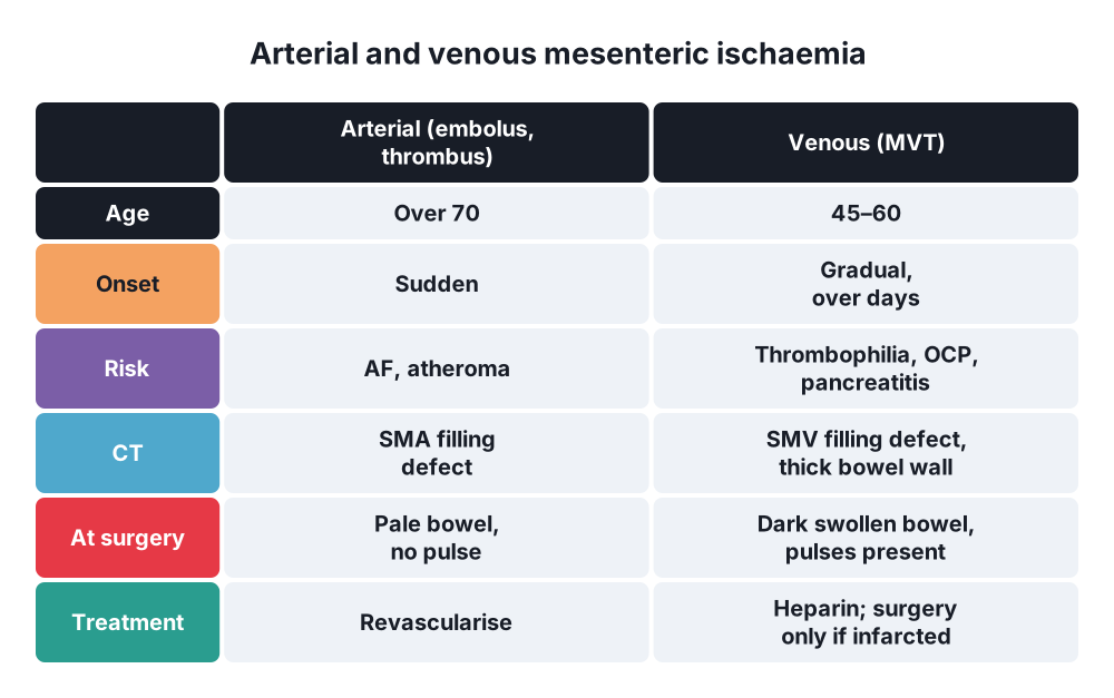
- Younger patients than arterial AMI → mean age about 45–60
- Men and women about equal
- SMV involved in over 90%; inferior mesenteric vein in only 5–10%
- Often extends into the portal and splenic veins
- 30-day mortality about 10–30% (much lower than arterial AMI); about 70% present acutely
- Recurrence up to 25% (mostly in the first 30 days) without anticoagulation; about 3% with it
3. Aetiology and risk factors
- Virchow's triad → stasis, vessel wall injury, hypercoagulability
Thrombophilia (found in 60–70%)
- Inherited → factor V Leiden, prothrombin G20210A mutation, protein C, protein S or antithrombin deficiency
- Acquired → myeloproliferative neoplasms (polycythaemia vera, essential thrombocythaemia; JAK2 V617F mutation), antiphospholipid syndrome, malignancy, oral contraceptive pill, pregnancy, hyperhomocysteinaemia, paroxysmal nocturnal haemoglobinuria, heparin-induced thrombocytopenia
Local causes
- Intra-abdominal inflammation → pancreatitis, appendicitis, diverticulitis, inflammatory bowel disease, abscess, cholangitis
- Surgery and trauma → splenectomy (most typical), bariatric surgery (sleeve gastrectomy), portocaval procedures, abdominal trauma
- Venous stasis → portal hypertension and cirrhosis, heart failure, obesity, abdominal compartment syndrome
- Compression → tumours (pancreas), nodes
Other
- Dehydration, infection (CMV, COVID-19), nephrotic syndrome
4. Pathogenesis
- Thrombus forms in small venous arcades (peripheral, e.g. in inflammation) or in the main SMV (central, e.g. in thrombophilia) and spreads
- Venous outflow blocked → bowel and mesentery become congested, oedematous and haemorrhagic (dark red to purple, thick wall)
- Fluid and blood leak into the wall, lumen and peritoneum → bloody ascites, hypovolaemia
- Rising pressure in the wall → arterial inflow falls (secondary arterial spasm) → haemorrhagic infarction
- Necrosis → perforation, peritonitis, sepsis
- Usually affects the jejunum and ileum; the duodenum and colon are usually spared
- The change from congestion to infarction is gradual and the border between healthy and diseased bowel is less sharp than in arterial ischaemia
- Collaterals may develop in chronic MVT → cavernous transformation, varices
5. Classification
By cause
- Primary (idiopathic), secondary (thrombophilia, local causes, stasis)
By course
- Acute → sudden pain, risk of infarction
- Subacute → weeks of vague pain, no infarction
- Chronic → asymptomatic or portal hypertension (varices, splenomegaly, ascites)
By extent
- Central (SMV trunk, portal vein, splenic vein) vs peripheral (small mesenteric veins and arcades)
- Extension into the portal vein → portomesenteric thrombosis
6. Clinical picture
Symptoms
- Abdominal pain → central, colicky, then constant; more gradual onset than arterial AMI (days, sometimes 1–2 weeks); out of proportion to signs early
- Anorexia, nausea, vomiting, diarrhoea or constipation
- GI bleeding (haematemesis, melaena, haematochezia) in about 15–50%
- History → previous DVT or PE (about half), family history, OCP, recent surgery, pancreatitis, cirrhosis
Signs
- Early → mild tenderness, soft abdomen, distension
- Fever, tachycardia
- Ascites
- Late → peritonism, shock (infarction)
- Chronic → splenomegaly, varices, ascites (portal hypertension)
7. Diagnosis
Bloods
- FBC (high haematocrit from dehydration; high WCC), lactate (late), U&E, LFT, amylase or lipase (pancreatitis), CRP, D-dimer (sensitive)
- Clotting, blood cultures
- Thrombophilia screen → ideally before or soon after starting heparin, but do not delay treatment (protein C, S and antithrombin fall during acute thrombosis and with anticoagulants; test after treatment ends if needed)
- JAK2 V617F mutation, antiphospholipid antibodies, PNH flow cytometry
- Look for an occult cancer (age-appropriate screening)
Imaging
- CT with IV contrast in the portal venous phase (with arterial phase: biphasic) → investigation of choice (sensitivity over 90%):
- Filling defect in the SMV or portal vein, enlarged vein with an enhancing rim (target sign)
- Bowel wall thickening (often circumferential, "target" appearance), mesenteric oedema and fat stranding, dilated loops, ascites
- Late signs → reduced wall enhancement, pneumatosis, portal venous gas, free gas
- Collaterals and cavernous transformation (chronic)
- Duplex ultrasound → portal and SMV thrombosis; less sensitive for the mesenteric veins
- MR venography → alternative (pregnancy, contrast allergy)
- Plain X-ray → non-specific (ileus, thumbprinting)
- Diagnostic paracentesis → bloody ascites (not routine)
- Angiography → only for interventional treatment (catheter-directed thrombolysis)
- Upper GI endoscopy → varices (chronic)
8. Differential diagnosis
- Arterial AMI (embolism or thrombosis), NOMI
- Acute pancreatitis (may cause MVT)
- Small bowel obstruction, strangulation
- Inflammatory bowel disease, infectious enteritis
- Perforated peptic ulcer, cholecystitis, appendicitis
- Portal vein thrombosis from other causes (cirrhosis, cancer)
- Budd-Chiari syndrome (hepatic veins)
9. Treatment
Conservative
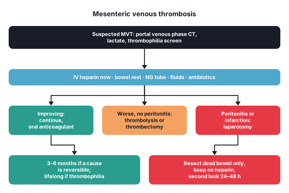
- Anticoagulation is the mainstay → start IV unfractionated heparin (or LMWH) as soon as the diagnosis is made, even with GI bleeding (bleeding is rarely a contraindication)
- Unfractionated heparin preferred if surgery is likely (short half-life, reversible)
- Prevents spread, allows recanalisation; avoids surgery in most (about 80–90% need no bowel resection)
- Supportive care → bowel rest (NBM), NG tube, IV fluids, electrolytes, analgesia, broad-spectrum antibiotics (translocation), catheter
- Serial clinical examination and lactate; repeat CT if not improving
- Treat the cause → pancreatitis, sepsis, stop the OCP, haematology referral (myeloproliferative disease)
- Long-term anticoagulation (warfarin or a DOAC):
- 3–6 months for a reversible cause (surgery, pancreatitis, OCP)
- Lifelong for thrombophilia, myeloproliferative neoplasms, recurrent or unexplained (idiopathic) MVT
- DOACs are increasingly used (not in antiphospholipid syndrome or advanced cirrhosis)
- Endovascular treatment → for patients without peritonitis who worsen despite anticoagulation:
- Catheter-directed thrombolysis (via the SMA or transhepatic or transjugular access to the portal vein)
- Mechanical thrombectomy, TIPS with thrombectomy
- Higher bleeding risk; specialist centres
- Chronic MVT → anticoagulation if a prothrombotic state is found; manage varices like portal hypertension (beta-blockers, banding, TIPS for refractory bleeding)
Surgical
- Indications → peritonitis, perforation, signs of bowel infarction on CT or clinically, deterioration despite anticoagulation
- Laparoscopy or laparotomy:
- Continue heparin during and after surgery
- Findings → dark, swollen, congested bowel; bloody ascites; pulsating arteries (unlike arterial ischaemia) but thrombosed veins
- Resect only clearly necrotic bowel, with margins (the border is often indistinct; the thrombus can extend into the margins)
- Primary anastomosis if stable, or ends left closed
- Second-look laparotomy at 24–48 hours (planned at the first operation) → maximises bowel preserved
- Venous thrombectomy of the SMV → rarely; for acute central thrombosis, with heparin, in selected centres
- Temporary abdominal closure if the bowel is very oedematous (risk of abdominal compartment syndrome)
10. Complications
Of the disease
- Bowel infarction, perforation, peritonitis, sepsis, death
- GI bleeding
- Stricture of the bowel after healing (late obstruction)
- Portal hypertension → oesophageal and gastric varices, splenomegaly, ascites (chronic or portal extension)
- Recurrent thrombosis (other sites too: DVT, PE)
Of treatment
- Bleeding on anticoagulation or after thrombolysis
- Short bowel syndrome after large resection
- Anastomotic leak, heparin-induced thrombocytopenia
Prognosis
- 30-day mortality 10–30%; better than arterial AMI
- Long-term outcome depends on the cause (cancer, cirrhosis, myeloproliferative disease); 5-year survival in chronic MVT about 80%
Show as a list
Mesenteric venous thrombosis
- Causes
- Thrombophilia
- JAK2, OCP, cancer
- Pancreatitis
- Splenectomy, cirrhosis
- Features
- Younger patients
- Gradual pain
- Bleeding 15-50%
- Ascites
- Tests
- Portal venous CT
- SMV filling defect
- Thick bowel wall
- Thrombophilia screen
- Treatment
- Heparin first
- Bowel rest, antibiotics
- Thrombolysis if worse
- Surgery if infarcted
- Long term
- 3-6 months if reversible
- Lifelong if thrombophilia
- Varices, recurrence


