
82. Mesenteric lymphadenitis
High yield
Inflamed mesenteric nodes in children after a viral illness mimic appendicitis; ultrasound helps, and most settle without surgery.
Exam Prep · State examSurgery · General surgerySurgery · Emergency and trauma
1. Definition
- Mesenteric lymphadenitis (mesenteric adenitis) → inflammation of the lymph nodes in the small bowel mesentery, mainly in the ileocaecal region, causing right lower abdominal pain
- Primary → enlarged nodes without another cause found (most cases; usually viral)
- Secondary → nodes enlarged because of another disease (appendicitis, terminal ileitis, Crohn's disease, tuberculosis, lymphoma)
- Pseudoappendicitis → mesenteric adenitis that looks like appendicitis
2. Epidemiology
- Mainly children and adolescents (5–15 years); uncommon over 20
- Boys and girls about equal
- Second most common cause of acute RIF pain in children after appendicitis; found in about 10–20% of children admitted with suspected appendicitis
- More in winter and spring (viral infections)
- In negative appendicectomies in children, mesenteric adenitis is the most common finding
3. Aetiology and risk factors
Anatomy (revision)
- Mesenteric lymph nodes (100–200) lie along the superior mesenteric vessels, densest near the ileocaecal junction
- Drain the small bowel and right colon → superior mesenteric nodes → cisterna chyli
- Lymphoid tissue is largest in childhood → strong reaction to infection
Causes
- Viral (most common):
- Adenovirus, Coxsackie, Epstein-Barr virus, rotavirus, norovirus, CMV, HIV
- Often after an upper respiratory tract infection or sore throat
- Bacterial:
- Yersinia enterocolitica and Y. pseudotuberculosis → classic cause; terminal ileitis and nodes; undercooked pork, unpasteurised milk
- Beta-haemolytic streptococcus (after tonsillitis)
- Salmonella, Campylobacter, Shigella, E. coli, Staphylococcus
- Tuberculous mesenteric lymphadenitis → Mycobacterium tuberculosis or M. bovis (unpasteurised milk); endemic areas; chronic
- Others → Bartonella (cat-scratch), toxoplasmosis, Giardia
- Secondary → appendicitis, Crohn's disease, coeliac disease, lymphoma, metastases, sarcoidosis, SLE
4. Pathogenesis
- Infection in the gut or upper airway (swallowed organisms)
- Organisms or antigens reach the ileocaecal mesenteric nodes through lymphatics
- Nodes become enlarged, red and tender (reactive hyperplasia)
- Stretching of the mesentery and irritation of the peritoneum → pain in the RIF and around the umbilicus
- Usually self-limiting within days to 2 weeks
- Yersinia → invades Peyer's patches of the terminal ileum → ileitis + suppurative nodes
- Tuberculosis → caseation, matted nodes, calcification, abscess; nodes may adhere to bowel → obstruction
- Rarely nodes suppurate → abscess, peritonitis
5. Classification
- Acute non-specific (primary) → viral; most common
- Acute specific → Yersinia, streptococcal, Salmonella
- Chronic specific → tuberculous
- Suppurative (abscess forms)
- Secondary (reactive) → to appendicitis, Crohn's, lymphoma
6. Clinical picture
Symptoms
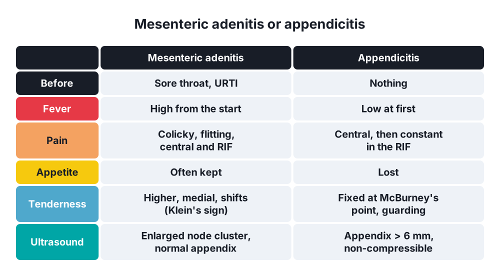
- Abdominal pain → around the umbilicus and in the RIF; often colicky, comes and goes, less constant than appendicitis
- Pain may begin at the same time as or after an upper respiratory infection, sore throat or fever
- Fever often high (over 38.5 °C) from the start (appendicitis usually has low fever at first)
- Nausea, vomiting (less than in appendicitis)
- Appetite often kept (anorexia is typical of appendicitis)
- Diarrhoea (Yersinia, gastroenteritis)
- Tuberculous → weeks of pain, weight loss, night sweats, fever, diarrhoea or constipation
Signs
- Child often looks well between bouts; flushed with fever
- Tenderness in the RIF, but:
- Less localised and shifts (moves with the mesentery)
- Higher and more medial than McBurney's point, along the root of the mesentery
- Guarding and rebound often absent
- Klein's sign → tender point moves to the left when the patient turns from supine onto the left side (the mesentery shifts); in appendicitis the tenderness stays in the RIF
- Upper respiratory infection → red throat, enlarged tonsils, cervical lymphadenopathy (generalised lymph node enlargement)
- Rectal examination → may be tender on the right
- TB → doughy abdomen, ascites, mass of matted nodes
7. Diagnosis
- Mesenteric adenitis is mainly a diagnosis of exclusion; the main aim is to not miss appendicitis
Blood tests
- WCC → normal or raised; lymphocytosis (viral) or neutrophilia (bacterial)
- CRP → raised; can be high
- Throat swab, stool culture (Yersinia, Salmonella), Yersinia serology
- Urinalysis (exclude urinary infection); pregnancy test in girls
- TB → interferon gamma release assay (IGRA), Mantoux test, chest X-ray
Imaging
- Ultrasound (graded compression) → test of choice:
- Cluster of three or more enlarged mesenteric nodes in the RIF, short-axis diameter over 5–8 mm
- Normal compressible appendix (under 6 mm) → key finding to exclude appendicitis
- Thickened terminal ileum wall (Yersinia ileitis)
- CT (adults, uncertain cases) → enlarged clustered nodes, normal appendix, ileal thickening; finds secondary causes
- MRI → alternative in children or pregnancy
- TB → CT: nodes with low-density (caseating) centres and peripheral rim enhancement, calcification, ascites, ileocaecal thickening
Laparoscopy
- When appendicitis cannot be excluded → shows enlarged, soft, reddish nodes in the ileocaecal mesentery, clear peritoneal fluid, a normal appendix
- Take a node biopsy if chronic disease (TB, lymphoma) is suspected
8. Differential diagnosis
- Acute appendicitis (main differential)
- Meckel's diverticulitis
- Intussusception (younger children; nodes may act as a lead point)
- Terminal ileitis (Crohn's disease, Yersinia)
- Gastroenteritis, constipation
- Urinary tract infection, pyelonephritis
- Right lower lobe pneumonia
- Ovarian cyst or torsion (girls)
- Lymphoma (Burkitt's in children), tuberculosis
- Henoch-Schönlein purpura
9. Treatment
Conservative
- Primary mesenteric adenitis is self-limiting → no surgery
- Supportive care:
- Rest, oral fluids, normal diet as tolerated
- Analgesia and antipyretics → paracetamol, ibuprofen
- Treat the upper respiratory infection (antibiotics only for proven streptococcal infection)
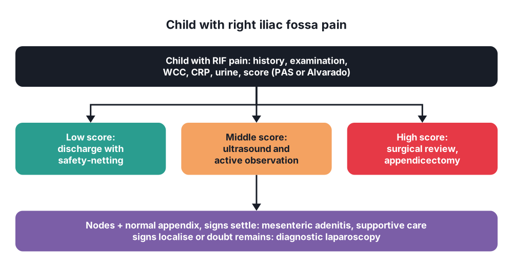
- Active observation when the diagnosis is uncertain:
- Admit, repeat examination every few hours by the same senior doctor
- Repeat WCC and CRP; repeat ultrasound
- If signs settle → mesenteric adenitis likely → discharge with safety-netting advice
- If signs localise or worsen → treat as appendicitis
- Yersinia → usually self-limiting; antibiotics (co-trimoxazole, ciprofloxacin in adults, doxycycline) for severe disease, sepsis or immunosuppression
- Salmonella and other bacteria → antibiotics if systemically unwell
- Tuberculous mesenteric lymphadenitis → antituberculous drugs for 6 months (rifampicin, isoniazid, pyrazinamide, ethambutol for 2 months; then rifampicin and isoniazid for 4 months)
- Follow-up → recurrent attacks occur in some children
Surgical
- No role in uncomplicated mesenteric adenitis
- Diagnostic laparoscopy → when appendicitis cannot be excluded:
- If nodes are enlarged and the appendix is normal → appendicectomy is usually done (prevents future confusion; low morbidity), and biopsy a node if the picture is atypical
- Inspect the terminal ileum (Crohn's), look for Meckel's diverticulum (60–100 cm from the ileocaecal valve)
- Suppurative node or abscess → percutaneous or surgical drainage
- TB complications → surgery for obstruction (adhesions, strictures: stricturoplasty or limited ileocaecal resection), perforation, abscess, or diagnostic biopsy
- Lymphoma → biopsy for diagnosis; treatment is chemotherapy
10. Complications
- Usually none; resolves in days to 2 weeks
- Suppuration → node abscess, peritonitis (rare)
- Intussusception (enlarged node as a lead point)
- Negative appendicectomy → wound infection, adhesions
- Missed appendicitis → perforation
- Tuberculous → intestinal obstruction, abscess, fistula, perforation, chylous ascites, calcified nodes
- Yersinia → reactive arthritis, erythema nodosum
Prognosis
- Excellent for primary mesenteric adenitis
Show as a list
Mesenteric adenitis
- Who
- Children 5-15
- After viral URTI
- Causes
- Adenovirus, EBV
- Yersinia: ileitis
- Streptococcus
- TB: chronic
- Clinical
- High fever early
- Colicky RIF pain
- Appetite kept
- Klein's sign
- Neck nodes
- Tests
- WCC: lymphocytes
- Ultrasound: nodes > 5-8 mm
- Normal appendix
- Treatment
- Self-limiting
- Analgesia, fluids
- Active observation
- Laparoscopy if doubt
- TB: 6 months drugs
- Complications
- Abscess
- Intussusception
- Missed appendicitis


