
93. Acute mesenteric ischaemia from arterial embolism and thrombosis
High yield
Sudden SMA blockage causes pain out of proportion to signs; CT angiography, then urgent revascularisation and bowel resection.
Exam Prep · State examSurgery · General surgerySurgery · Emergency and trauma
1. Definition
- Acute mesenteric ischaemia (AMI) → sudden reduction of blood flow to the bowel, enough to cause ischaemia and, if untreated, infarction (necrosis) of the bowel
- Four causes:
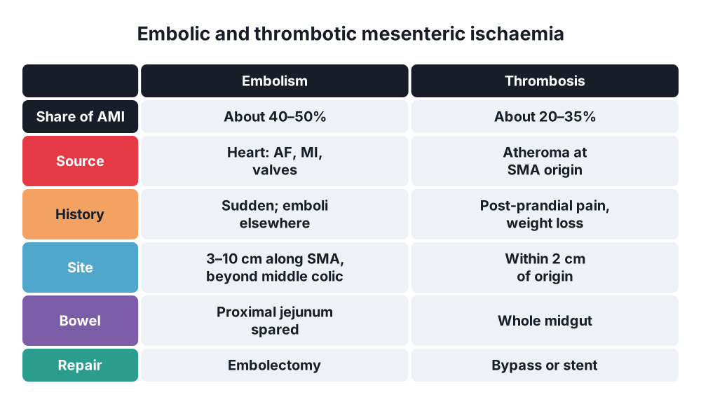
- Arterial embolism → about 40–50%
- Arterial thrombosis → about 20–35%
- Non-occlusive mesenteric ischaemia (NOMI) → about 20% (question 95)
- Mesenteric venous thrombosis → about 5–15% (question 94)
- This article covers occlusive arterial AMI, mostly of the superior mesenteric artery (SMA)
- Chronic mesenteric ischaemia → "intestinal angina" from gradual narrowing (pain after meals, weight loss, fear of eating)
2. Epidemiology
- Rare → under 1 per 1000 hospital admissions; about 1 in 100 patients with an acute abdomen (more in the elderly)
- Incidence doubles with every 5 years over 70
- Women more than men in some series
- Mortality 50–80% in older series; about 30–50% with modern CT and revascularisation
- Embolus without treatment → mortality about 54%; thrombosis → about 77% (more bowel involved)
- Delay → survival falls from about 50% to 30% when the diagnosis of SMA embolism is made more than 24 hours after onset
- About 68% of embolic patients have emboli elsewhere too (brain, limbs, kidneys, spleen)
3. Aetiology and risk factors
Anatomy (revision)
- Coeliac trunk (T12) → foregut; SMA (L1) → midgut (from the second part of the duodenum to two thirds of the transverse colon); IMA (L3) → hindgut
- The SMA leaves the aorta at an acute (oblique) angle and is wide → emboli enter it easily
- Collaterals → pancreaticoduodenal arcades (coeliac–SMA), arc of Bühler (coeliac–SMA, rare), arc of Riolan and marginal artery of Drummond (SMA–IMA)
- Watershed areas → splenic flexure (Griffiths' point), rectosigmoid (Sudeck's point)
Embolism
- Heart source in about 90%:
- Atrial fibrillation (most common), recent myocardial infarction (mural thrombus), low ejection fraction, ventricular aneurysm
- Valve disease, endocarditis (septic emboli), prosthetic valves, atrial myxoma
- Aorta → aneurysm thrombus, atheroma, after catheterisation (cholesterol emboli)
- Paradoxical embolus (patent foramen ovale)
- Embolus lodges 3–10 cm beyond the SMA origin, often just distal to the middle colic artery (about 50%); about 15% at the origin
Thrombosis
- Atherosclerosis of the SMA origin (within 2 cm of the aorta) → a narrowed artery finally clots (acute on chronic)
- Often previous chronic mesenteric ischaemia (post-prandial pain, weight loss)
- Triggers → dehydration, low cardiac output, hypercoagulable states
- Other → aortic dissection extending into the SMA, vasculitis, thrombophilia (factor V Leiden, antiphospholipid syndrome), trauma, stent or graft thrombosis, isolated SMA dissection
Risk factors
- Age over 70, atrial fibrillation, heart failure, recent MI
- Atherosclerosis (smoking, diabetes, hypertension, peripheral arterial disease), previous embolic events
- No anticoagulation in AF
4. Pathogenesis
- Occlusion → mucosa is the most sensitive layer (highest metabolic rate)
- Mucosal ischaemia within minutes to hours → loss of the barrier, bacterial translocation, mucosal sloughing, bleeding into the lumen
- Spasm of the vessels around the block worsens the ischaemia (can persist after revascularisation)
- Transmural infarction within about 6–12 hours → wall necrosis, fluid and blood loss into the lumen and peritoneum
- Perforation, peritonitis, septic shock, multiple organ failure
- Reperfusion injury after revascularisation → oxygen free radicals, oedema, systemic inflammation, hyperkalaemia, acidosis
- Embolus → sudden; no collaterals; ischaemia spares the proximal jejunum and transverse colon (supplied by branches above the clot)
- Thrombosis → at the origin; collaterals already formed; slower onset; infarction of the whole midgut (from the duodenojejunal flexure to the mid-transverse colon) when collaterals fail
- Pain comes from ischaemic visceral fibres → severe; the parietal peritoneum is not involved until transmural necrosis → pain out of proportion to the signs
5. Classification
By cause
- Occlusive arterial → embolism, thrombosis
- Non-occlusive (NOMI)
- Venous (mesenteric venous thrombosis)
By stage
- Early (reversible) ischaemia → pain, few signs (first 6 hours)
- Infarction → transmural necrosis, peritonism (6–12 hours)
- Peritonitis and sepsis → perforation, shock, multiple organ failure (over 12–24 hours)
Embolus vs thrombosis
- Embolus → sudden, often AF, proximal jejunum spared, emboli elsewhere, distal SMA on CT (meniscus sign)
- Thrombosis → history of post-prandial pain and weight loss, atherosclerosis elsewhere, calcified SMA origin, slower onset, longer segment of bowel
6. Clinical picture
Symptoms
- Sudden, severe, central or diffuse abdominal pain → out of proportion to the examination (the classic sign)
- Gut emptying → sudden vomiting and diarrhoea (forceful evacuation)
- Later → bloody diarrhoea, melaena or haematemesis (mucosal sloughing)
- Thrombosis → previous post-prandial pain ("intestinal angina", 15–30 minutes after meals), fear of eating (sitophobia), weight loss
- Elderly or ventilated patients may only show confusion, unexplained acidosis or distension
Signs
- Early → soft, mildly tender abdomen; bowel sounds present or increased; patient writhing
- Atrial fibrillation (irregular pulse), signs of emboli elsewhere (cold limb, stroke)
- Late → distension, absent bowel sounds, peritonism (rigidity, rebound), fever
- Shock → tachycardia, hypotension, oliguria, mottled skin
- Rectal examination → blood
Classic triad (Bergan)
- Severe pain without signs + gut emptying + a source of emboli (heart disease)
7. Diagnosis
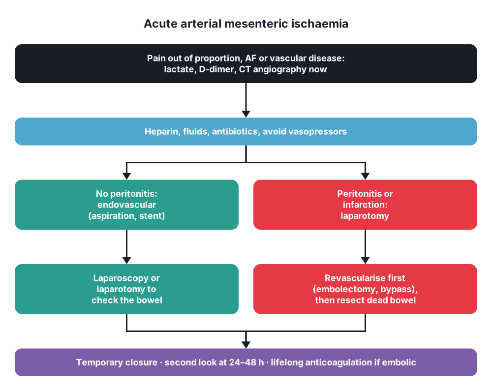
- High suspicion in any older patient with AF and severe abdominal pain → go straight to CT angiography
Bloods
- Lactate → raised (late; a normal lactate does not exclude AMI)
- WCC often over 15–20 × 10⁹/L, metabolic acidosis, raised base deficit
- D-dimer → sensitive (a normal D-dimer makes AMI unlikely) but not specific
- Amylase, LDH, CK, phosphate, ALP may rise (non-specific); raised urea, haematocrit (fluid loss)
- ECG (AF, MI), troponin, clotting, group and crossmatch
- Intestinal fatty acid-binding protein (I-FABP) → research marker
Imaging
- CT angiography (CTA, biphasic: arterial and portal venous phases) → investigation of choice (sensitivity about 93%, specificity about 96%):
- Filling defect in the SMA → embolus (round cut-off, distal; "meniscus") or thrombus (at the origin on calcified plaque)
- Bowel signs → reduced or absent wall enhancement, thin "paper-thin" wall (arterial), wall thickening (reperfusion), dilatation, pneumatosis intestinalis, portal or mesenteric venous gas, free fluid, free gas
- Shows other causes (aortic dissection, venous thrombosis) and emboli to the spleen and kidneys
- Do not delay CT for kidney function in suspected AMI (the risk of missing infarction outweighs contrast risk)
- Plain abdominal X-ray → often normal early; late signs: ileus, thumbprinting (submucosal oedema and bleeding), pneumatosis, portal venous gas, free gas
- Mesenteric angiography → previously the gold standard; now for endovascular treatment (and NOMI)
- Duplex ultrasound → for chronic disease (SMA origin stenosis); limited in the acute setting (gas, obesity)
- Diagnostic laparoscopy → limited (mucosal ischaemia may not be seen from outside); laparotomy if in doubt with peritonism
8. Differential diagnosis
- Perforated viscus, acute pancreatitis, ruptured abdominal aortic aneurysm, aortic dissection
- Strangulated obstruction, volvulus
- Mesenteric venous thrombosis, NOMI, ischaemic colitis
- Acute cholecystitis, renal colic, diverticulitis
- Myocardial infarction (inferior), diabetic ketoacidosis
- Gastroenteritis (vomiting and diarrhoea, but pain matches the signs)
9. Treatment
Conservative
- Treatment goal (ESVS and ESTES guidelines) → fast diagnosis, restore blood flow, remove dead bowel, support the patient
- Resuscitation → IV fluids (balanced crystalloid), oxygen, catheter, monitoring, correct electrolytes and acidosis
- Immediate anticoagulation → IV unfractionated heparin bolus (e.g. 5000 units or 80 units/kg) then infusion, as soon as the diagnosis is made (stops propagation of clot)
- Broad-spectrum IV antibiotics (bacterial translocation), e.g. piperacillin-tazobactam
- Analgesia (opioids), NG tube if distended or vomiting
- Avoid vasopressors if possible (splanchnic vasoconstriction); if needed → dobutamine, low-dose dopamine or milrinone rather than high-dose noradrenaline
- Correct heart rate and rhythm (AF), optimise cardiac output
- Conservative treatment alone → only for very selected patients (small distal emboli without signs of ischaemia, too frail for intervention), or palliation in extensive infarction of the whole bowel in frail elderly patients
- Long-term → lifelong anticoagulation after embolism (AF), antiplatelets and statin after thrombosis; treat the source (cardiology)
Surgical
- Emergency laparotomy → peritonitis, perforation, or signs of infarction on CT; or open revascularisation
- Order → revascularise first, then assess and resect bowel (bowel that looks doubtful often recovers once flow returns); resect first only if there is gross perforation or contamination
- Embolus → SMA embolectomy:
- Lift the transverse colon, expose the SMA at the root of the mesentery (below the pancreas, left of the SMV)
- Transverse (or longitudinal + patch) arteriotomy, Fogarty balloon catheter (3–4 Fr) proximally and distally
- Good pulsatile backflow and inflow; close with fine monofilament (vein patch if narrow)
- Intra-arterial tPA (0.5–1 mg) for distal clot
- Thrombosis → bypass or stenting:
- Retrograde bypass from the right common iliac artery to the SMA (C-shaped graft; avoids aortic clamping; vein if contaminated)
- Antegrade bypass from the supracoeliac aorta (more stable, but more physiological stress)
- Retrograde open mesenteric stenting (ROMS) → hybrid: SMA exposed at laparotomy and stented retrogradely; technical success about 98%
- Endarterectomy with patch (rare)
- Endovascular revascularisation → percutaneous aspiration embolectomy, angioplasty and stenting (thrombosis), catheter-directed thrombolysis; increasingly first-line in patients without peritonitis (lower mortality, less bowel resection, less parenteral nutrition); then laparoscopy or laparotomy to check the bowel
- Assess bowel viability (after revascularisation) → colour, peristalsis, mesenteric pulses, bleeding from cut edges; Doppler, ICG fluorescence
- Resect clearly dead bowel only; leave the ends stapled closed (no anastomosis) in unstable patients
- Temporary abdominal closure and planned second-look laparotomy at 24–48 hours → check doubtful segments, further resection, delayed anastomosis or stoma, close the abdomen
- Whole midgut infarction in a frail elderly patient → may be non-survivable; discuss palliative care with the family
- Hybrid theatres allow open and endovascular steps together
10. Complications
Of the disease
- Bowel infarction, perforation, peritonitis
- Septic shock, multiple organ failure, death
- Reperfusion syndrome → hyperkalaemia, acidosis, arrhythmia, ARDS
- Abdominal compartment syndrome (oedematous bowel; question 98)
- Recurrent embolism (no anticoagulation)
Of treatment
- Short bowel syndrome (under about 100–150 cm of small bowel with no colon, or under 50–60 cm with colon in continuity) → intestinal failure, parenteral nutrition, teduglutide, intestinal transplant
- Anastomotic leak, enterocutaneous fistula
- Bleeding from anticoagulation or thrombolysis
- Graft or stent thrombosis, restenosis
- Ischaemic stricture (late obstruction)
Prognosis
- In-hospital mortality about 30–50%; better with early CTA, endovascular-first strategies and a second look
- Survivors need surveillance of grafts and stents, anticoagulation and nutritional support
Show as a list
Arterial mesenteric ischaemia
- Causes
- Embolus 50%: AF
- Thrombosis 30%: atheroma
- SMA most often
- Clinical
- Pain > signs
- Gut emptying
- AF, emboli elsewhere
- Late peritonitis
- Tests
- Lactate: late
- D-dimer
- CT angiography
- Pneumatosis, portal gas
- Treatment
- Heparin now
- Antibiotics, fluids
- Embolectomy
- Bypass or stent
- Bowel
- Revascularise first
- Resect dead bowel
- Second look 24-48 h
- Short bowel risk


