41. Mediastinitis
High yield
Acute infection after oesophageal perforation, neck sepsis or sternotomy; chronic fibrosing form compresses the SVC and airways.
Exam Prep · State examSurgery · General surgerySurgery · Emergency and traumaAnatomy · Thorax
1. Definition
- Mediastinitis → inflammation of the connective tissue of the mediastinum
- Acute mediastinitis → bacterial infection that spreads fast along fascial planes; a surgical emergency
- Chronic (fibrosing) mediastinitis → slow, dense fibrosis of the mediastinum that compresses its structures
2. Epidemiology
- Post-sternotomy mediastinitis → 0.5–2% of cardiac operations; mortality 10–25%
- Descending necrotising mediastinitis → rare; from neck infection; mortality up to 40%
- After oesophageal perforation → mortality 10–25% if treated within 24 h, higher if later
- Fibrosing mediastinitis → young adults; regions with histoplasmosis
3. Aetiology and risk factors
Acute mediastinitis
- Oesophageal perforation → most common cause outside cardiac surgery:
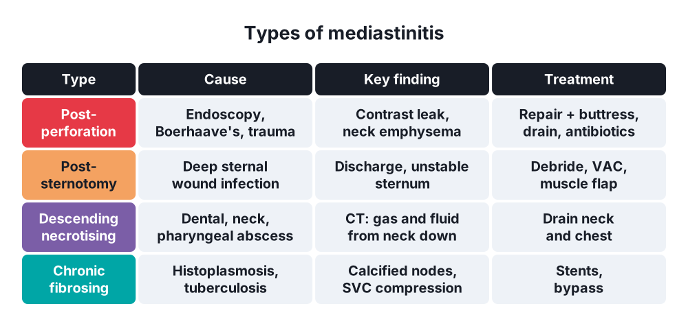
- Iatrogenic → endoscopy, dilatation (about 60%)
- Boerhaave's syndrome → rupture after forceful vomiting, left posterolateral distal oesophagus
- Foreign body, trauma, anastomotic leak after oesophagectomy
- Post-sternotomy → deep sternal wound infection:
- Diabetes, obesity, COPD, smoking
- Bilateral internal mammary artery grafts
- Long operation, re-exploration for bleeding
- Descending necrotising mediastinitis → spread from:
- Odontogenic infection, Ludwig's angina
- Retropharyngeal or peritonsillar abscess
- Tracheobronchial injury, spread from lung, pleura or vertebra
- Organisms → mixed aerobes and anaerobes from the mouth; S. aureus and S. epidermidis after sternotomy
Chronic mediastinitis
- Histoplasmosis (most common), tuberculosis
- Sarcoidosis, radiotherapy, IgG4-related disease, drugs (methysergide)
4. Pathogenesis
- Mediastinum has no barriers → infection spreads along fascial planes
- Neck to mediastinum → three routes:
- Pretracheal space → anterior mediastinum
- Perivascular (carotid sheath) → middle mediastinum
- Retropharyngeal and "danger space" → posterior mediastinum (most common, about 70%)
- Oesophageal perforation → saliva, gastric juice and bacteria enter the mediastinum → chemical then bacterial mediastinitis → sepsis
- Negative intrathoracic pressure draws contents in; pleura often breaks → empyema
- Fibrosing form → granulomatous inflammation → fibrosis that encases the SVC, airways, pulmonary vessels and oesophagus
5. Classification
- By course → acute, chronic
- By cause:
- Post-sternotomy (deep sternal wound infection)
- Post-perforation (oesophagus, airway)
- Descending necrotising (from the neck)
- Secondary (lung, pleura, spine, subphrenic)
- Post-sternotomy by time (El Oakley) → type I (within 2 weeks) to type V (recurrent)
- Descending necrotising (Endo) → type I above the carina; type IIA lower anterior; type IIB anterior and lower posterior
6. Clinical picture
Symptoms
- Retrosternal chest pain, often severe, spreading to the back or neck
- Fever > 38 °C, rigors
- Dysphagia, odynophagia
- Dyspnoea
- Boerhaave's → vomiting, then chest pain and collapse (Mackler's triad → vomiting, chest pain, subcutaneous emphysema)
- Post-sternotomy → wound discharge, sternal pain, sternal "click"
- Neck infection → neck pain, swelling, trismus
Signs
- Tachycardia, tachypnoea, hypotension → sepsis
- Subcutaneous emphysema of the neck
- Hamman's sign → crunching sound with the heartbeat (air in the mediastinum)
- Sternal instability, purulent drainage
- Pleural effusion, signs of pneumothorax
- Fibrosing form → SVC syndrome, cough, haemoptysis, dysphagia
7. Diagnosis
Laboratory
- Leucocytosis, high C-reactive protein, procalcitonin, lactate
- Blood cultures
- Wound swab, mediastinal fluid culture
- Pleural fluid with high amylase or food particles → oesophageal perforation
Imaging
- Chest X-ray → widened mediastinum, pneumomediastinum, air-fluid levels, left effusion
- Contrast CT of neck and chest → test of choice:
- Fluid collections, gas in the mediastinum
- Spread from the neck
- Sternal separation, retrosternal collection
- Water-soluble contrast swallow (Gastrografin), then barium if negative → site of perforation
- Flexible endoscopy → careful, in theatre; can show and treat the leak
- Fibrosing form → CT with calcified nodes; histoplasma serology; biopsy to exclude cancer
8. Differential diagnosis
- Myocardial infarction, aortic dissection
- Pulmonary embolism
- Pericarditis
- Perforated peptic ulcer, acute pancreatitis
- Spontaneous pneumomediastinum (no infection)
- Lymphoma, mediastinal tumour (fibrosing form)
9. Treatment
Conservative
- Resuscitation → IV fluids, oxygen, ICU
- Broad-spectrum IV antibiotics → cover aerobes, anaerobes and fungi (e.g. piperacillin-tazobactam or carbapenem ± antifungal)
- Nil by mouth, nasogastric tube, PPI
- Nutrition → feeding jejunostomy or parenteral
- Contained small perforation, no sepsis → non-operative care with antibiotics and drainage
- Fibrosing mediastinitis → no proven drug; stents for SVC, airway or pulmonary vessels
Surgical
- Aim → remove the source, debride and drain
- Oesophageal perforation:
- Early (< 24 h), healthy tissue → primary two-layer repair, buttressed with pleura, intercostal muscle, diaphragm or omentum; drain the chest
- Late or friable → T-tube drainage, or endoscopic stent or vacuum therapy
- Severe, with diseased oesophagus (cancer, achalasia) → oesophagectomy, or exclusion with cervical oesophagostomy and gastrostomy
- Approach → left thoracotomy for distal tears, right thoracotomy for mid-oesophagus, neck for cervical tears
- Descending necrotising:
- Neck drainage plus mediastinal drainage
- Below the carina → thoracotomy or VATS debridement
- Tracheostomy if the airway is threatened
- Post-sternotomy:
- Open the wound, remove wires, debride bone and cartilage
- Negative pressure wound therapy (VAC)
- Then closure with rewiring or pectoralis major or omental flap
- Fibrosing → surgery only for bypass of a blocked SVC or a resectable mass
10. Complications
- Septic shock, multiorgan failure
- Empyema, pericarditis, tamponade
- Erosion of great vessels → haemorrhage
- Tracheo-oesophageal fistula
- Sternal non-union, osteomyelitis
- Fibrosing → SVC syndrome, pulmonary hypertension, haemoptysis
Show as a list
Mediastinitis
- Acute
- Oesophageal perforation
- Boerhaave's
- Post-sternotomy
- Descending necrotising
- Chronic
- Histoplasmosis
- Tuberculosis
- Fibrosis
- Clinical
- Chest pain
- Fever, sepsis
- Subcutaneous emphysema
- Hamman's sign
- Diagnosis
- Contrast CT
- Contrast swallow
- Treatment
- Antibiotics
- Repair, drainage
- VAC, muscle flap
- Stents


