42. Mediastinal tumours and cysts, superior vena cava syndrome and mediastinal emphysema
High yield
Masses sorted by compartment; thymoma, germ cell, neurogenic tumours and cysts; SVC obstruction; air in the mediastinum.
Exam Prep · State examSurgery · General surgerySurgery · Surgical oncologyAnatomy · Thorax
1. Definition
- Mediastinal tumour or cyst → primary mass arising in the mediastinum, between the pleural sacs, from the sternum to the spine
- Superior vena cava (SVC) syndrome → symptoms from blocked venous return from the head, neck and arms through the SVC
- Mediastinal emphysema (pneumomediastinum) → free air in the mediastinum
2. Epidemiology
- Adults → thymoma, neurogenic tumours, cysts, germ cell tumours, lymphoma
- Children → neurogenic tumours, germ cell tumours, cysts, lymphoma; more often malignant
- Malignant → 25–50% of mediastinal masses in adults
- SVC syndrome → malignant in about 80% (lung cancer most often, then non-Hodgkin's lymphoma)
- Spontaneous pneumomediastinum → young men, 20–30 years
3. Aetiology and risk factors
Tumours and cysts
- Thymus → thymoma, thymic carcinoma, thymic hyperplasia (after chemotherapy, Graves'), thymic cyst
- Germ cells → teratoma, seminoma, non-seminomatous germ cell tumour (young men)
- Nerves → schwannoma, neurofibroma (neurofibromatosis), ganglioneuroma, neuroblastoma, paraganglioma
- Lymph nodes → Hodgkin's and non-Hodgkin's lymphoma, sarcoidosis, metastases
- Retrosternal goitre
- Foregut and mesothelial cysts → bronchogenic, oesophageal duplication, pericardial
SVC syndrome
- Malignant → lung cancer (small cell, right upper lobe), lymphoma, metastases, thymoma
- Benign → thrombosis around catheters and pacing leads (rising), fibrosing mediastinitis, goitre, aortic aneurysm
Pneumomediastinum
- Spontaneous → asthma, coughing, vomiting, Valsalva, labour, inhaled drugs
- Secondary → oesophageal or airway rupture, chest trauma, ventilation, dental or neck surgery
4. Pathogenesis
Compartments (key to diagnosis)
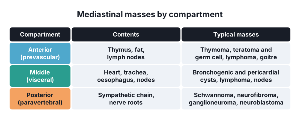
- Anterior (prevascular) → thymus, fat, nodes: the "4 Ts":
- Thymoma
- Teratoma and other germ cell tumours
- "Terrible" lymphoma
- Thyroid (retrosternal goitre)
- Middle (visceral) → heart, trachea, oesophagus, nodes: cysts, lymphoma, nodes
- Posterior (paravertebral) → sympathetic chain and nerves: neurogenic tumours
SVC syndrome
- Tumour compresses, invades or thromboses the SVC
- Venous pressure in the head and arms rises
- Collaterals open → azygos, internal mammary, lateral thoracic, vertebral veins
- Slow onset → collaterals compensate; fast onset → severe symptoms
Pneumomediastinum
- Macklin effect → alveoli rupture → air tracks along the bronchovascular sheaths to the hilum → mediastinum → neck, face and chest wall
5. Classification
Thymoma (WHO)
- Types A, AB, B1, B2, B3 → rising malignancy; thymic carcinoma (type C)
- Masaoka-Koga stage:
- I → encapsulated
- II → through the capsule into fat
- III → into nearby organs (pericardium, lung, great vessels)
- IVa → pleural or pericardial spread; IVb → lymph node or blood spread
- TNM 8th edition now also used
Germ cell tumours
- Mature teratoma → benign; hair, teeth, fat, calcium
- Seminoma → β-HCG may be mildly raised, AFP normal
- Non-seminomatous → AFP and β-HCG raised
Neurogenic tumours
- Nerve sheath → schwannoma (most common; Antoni A and B), neurofibroma, malignant nerve sheath tumour
- Ganglion cells → ganglioneuroma (benign, children) → ganglioneuroblastoma → neuroblastoma (malignant, infants)
- Paraganglia → paraganglioma, phaeochromocytoma (1% in the mediastinum)
- Dumbbell tumour → about 10% extend through the foramen into the spinal canal
Cysts (about 25% of masses)
- Pericardial cyst → "spring water" cyst at the right cardiophrenic angle
- Bronchogenic cyst → most common; near the carina
- Oesophageal duplication cyst, thymic cyst
6. Clinical picture
Symptoms
- Many → none; found on chest X-ray
- Mass effect → chest pain, cough, dyspnoea, stridor, dysphagia, hoarseness
- Myasthenia gravis → ptosis, diplopia, fatigable weakness: in about 40% of thymomas; 10–15% of myasthenia patients have a thymoma
- Other thymoma syndromes → pure red cell aplasia, hypogammaglobulinaemia
- Lymphoma → fever, night sweats, weight loss (B symptoms)
- Neurogenic → back pain, Horner's syndrome, cord compression (dumbbell)
- Phaeochromocytoma → hypertension, headache, sweating
- SVC syndrome:
- Swelling of face, neck and arms; worse on bending or lying flat
- Headache, dyspnoea, cough
- Severe → stridor, confusion (cerebral oedema)
- Pneumomediastinum → sudden retrosternal pain, neck pain, dysphagia, change in voice
Signs
- SVC syndrome → distended neck and chest wall veins, facial plethora, cyanosis, arm oedema; Pemberton's sign with goitre
- Pneumomediastinum:
- Subcutaneous emphysema → crepitus of the neck
- Hamman's sign → crunch with each heartbeat
7. Diagnosis
Imaging
- Chest X-ray → widened mediastinum, mass, lateral film shows the compartment
- Contrast CT chest → test of choice: site, fat, calcium, cyst, invasion
- MRI → neurogenic tumours (spinal canal), cysts, vascular invasion
- PET-CT → lymphoma, malignant tumours
- SVC syndrome → contrast CT shows level, thrombus and collaterals
- Pneumomediastinum → chest X-ray: lucent lines along the heart, continuous diaphragm sign, thymic "sail" sign in children; CT if unclear
- Contrast swallow or endoscopy → exclude oesophageal rupture when there is vomiting, fever or effusion
Laboratory
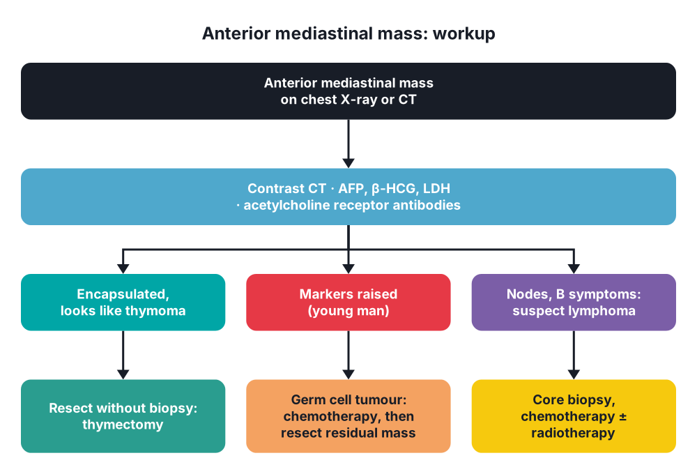
- Tumour markers in every young man with an anterior mass → AFP, β-HCG, LDH
- Acetylcholine receptor antibodies → myasthenia
- Urinary or plasma metanephrines → paraganglioma
- Thyroid function → goitre
Tissue
- Thymoma, clear on CT and resectable → no biopsy; operate (biopsy can seed the pleura)
- Suspected lymphoma or germ cell tumour → CT-guided core biopsy, mediastinoscopy, anterior mediastinotomy (Chamberlain), VATS biopsy
- SVC syndrome → tissue diagnosis before steroids or radiotherapy if the patient is stable
8. Differential diagnosis
- Aortic aneurysm, vascular anomalies
- Hiatus hernia, achalasia (dilated oesophagus)
- Lymph node metastases (lung cancer), sarcoidosis, tuberculosis
- Pericardial fat pad
- Morgagni or Bochdalek hernia
- Pneumomediastinum versus pneumothorax, pneumopericardium
9. Treatment
Surgical
- Thymoma → complete resection (thymectomy with the whole thymus and fat):
- Median sternotomy, or VATS or robotic approach for small, stage I–II tumours
- Preserve both phrenic nerves
- Myasthenia → control symptoms first; plasma exchange or IVIG before surgery
- Stage III → en bloc resection of pericardium, lung or vessels; adjuvant radiotherapy
- Mature teratoma → complete resection
- Non-seminomatous germ cell tumour → cisplatin chemotherapy, then resect any residual mass
- Neurogenic tumour → resection, usually VATS; dumbbell tumour with a neurosurgeon (laminectomy first)
- Cysts → resect if symptomatic, growing or unclear; pericardial cysts may be watched
- Retrosternal goitre → usually through a neck incision; sternotomy rarely
- SVC syndrome, benign cause → surgical bypass or reconstruction if stenting fails
Conservative
- Lymphoma → chemotherapy, radiotherapy; no resection
- Seminoma → chemotherapy, radiotherapy
- SVC syndrome:
- Sit the patient up, oxygen
- Steroids for lymphoma or oedema, diuretics
- Endovascular stent → fastest relief; first choice for severe symptoms
- Radiotherapy or chemotherapy for the cause (chemotherapy for small cell cancer and lymphoma)
- Thrombosis → anticoagulation, catheter-directed thrombolysis, remove the catheter
- Pneumomediastinum:
- Spontaneous → rest, analgesia, oxygen, avoid Valsalva; resolves in days
- Secondary → treat the cause (perforation → see mediastinitis)
- Tension pneumomediastinum (rare) → decompression
10. Complications
- Tumour → invasion of SVC, pericardium, phrenic and recurrent laryngeal nerves; airway collapse under anaesthesia
- Large anterior mass → airway and cardiac collapse at induction of general anaesthesia
- Thymectomy → myasthenic crisis, phrenic nerve palsy, bleeding
- SVC syndrome → cerebral oedema, laryngeal oedema, thrombosis
- Pneumomediastinum → pneumothorax, pneumopericardium (rare)
11. Prognosis
- Thymoma → 10-year survival about 90% (stage I) to under 50% (stage IV)
- Mature teratoma, schwannoma → cured by resection
- SVC syndrome → depends on the cause; median survival with cancer often < 1 year
Show as a list
Mediastinal masses
- Anterior
- Thymoma
- Teratoma, germ cell
- Lymphoma
- Retrosternal goitre
- Middle
- Bronchogenic cyst
- Pericardial cyst
- Nodes
- Posterior
- Schwannoma
- Neuroblastoma
- Dumbbell tumour
- SVC syndrome
- Lung cancer
- Catheter thrombosis
- Collaterals
- Stent, radiotherapy
- Pneumomediastinum
- Macklin effect
- Hamman's sign
- Rest, oxygen


