
83. Meckel's diverticulum
High yield
Remnant of the vitelline duct on the ileum; causes painless bleeding in children, obstruction and diverticulitis; resect when symptomatic.
Exam Prep · State examSurgery · General surgerySurgery · Emergency and traumaAnatomy · Abdomen
1. Definition
- Meckel's diverticulum → a true diverticulum (contains all layers of the bowel wall) on the antimesenteric border of the distal ileum, the persistent proximal end of the vitelline (omphalomesenteric) duct
- Most common congenital anomaly of the gastrointestinal tract
- Described by Johann Meckel (1809); Hildanus first noted it in 1598
- Other vitelline remnants → see question 55 (patent duct, cyst, fibrous band, umbilical polyp)
2. Epidemiology
- "Rule of 2s":
- Present in about 2% of the population
- About 2 feet (60 cm) from the ileocaecal valve (range 30–100 cm)
- About 2 inches (5 cm) long
- 2 types of ectopic mucosa → gastric (most common) and pancreatic
- Twice as common in males (symptomatic 2–3 : 1)
- Symptoms usually before age 2
- About 2% cause symptoms in their lifetime (4–6% in some series)
- Lifetime risk of complications → about 4%, falling with age (most complications in children)
- Children → bleeding most common; adults → obstruction most common
- Associated anomalies → exomphalos, oesophageal atresia, anorectal malformations, Down's syndrome, cardiac and nervous system defects
3. Aetiology and risk factors
Embryology (revision)
- Vitelline duct joins the midgut to the yolk sac in early foetal life
- Normally obliterates and disappears by weeks 5–9
- If the ileal (proximal) end persists → Meckel's diverticulum
- Other remnants → patent duct (umbilico-ileal fistula), vitelline cyst, fibrous band from the diverticulum to the umbilicus, umbilical sinus or polyp
Features
- Own blood supply → a remnant of the right vitelline artery (branch of the superior mesenteric artery); may form a mesodiverticular band
- Ectopic (heterotopic) tissue in about 50% of all and over 60% of symptomatic cases:
- Gastric mucosa (about 60–80% of ectopic tissue) → secretes acid → ulcers
- Pancreatic tissue (5–16%)
- Rarely duodenal, colonic, endometrial, hepatobiliary tissue
Risk factors for complications
- Male sex, age under 50 (especially under 2)
- Length over 2 cm, narrow neck (diverticulitis), wide base
- Ectopic tissue (palpable thickening)
- Fibrous band to the umbilicus or mesentery
4. Pathogenesis
- Bleeding:
- Ectopic gastric mucosa secretes acid
- Acid ulcerates the adjacent normal ileal mucosa (the ulcer is usually at the base or in the ileum next to it, not in the gastric tissue)
- Painless rectal bleeding, often large
- Obstruction (several mechanisms):
- Volvulus of small bowel around a fibrous band to the umbilicus (most dangerous)
- Internal hernia under the band or mesodiverticular band
- Intussusception → the inverted diverticulum is the lead point (ileo-ileal or ileocolic)
- Littré's hernia → diverticulum trapped in a hernia (inguinal, femoral, umbilical)
- Stricture after diverticulitis, enterolith, foreign body (fish bone)
- Diverticulitis → narrow neck blocked, like appendicitis → inflammation, gangrene, perforation
- Perforation → peptic ulcer, diverticulitis, foreign body
- Neoplasms → neuroendocrine (carcinoid) tumour most common (about 75%); adenocarcinoma (from gastric mucosa), GIST, lymphoma, leiomyoma
5. Classification
By presentation
- Asymptomatic (incidental) → most; found at surgery, imaging or autopsy
- Symptomatic:
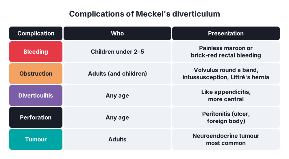
- Bleeding (30–50% of complications; children)
- Obstruction (about 35–40%; adults): volvulus, intussusception, internal hernia, Littré's hernia
- Diverticulitis (about 20–25%) ± perforation
- Umbilical abnormalities (fistula, sinus)
- Tumours (under 5%)
By content
- With ectopic mucosa (gastric, pancreatic) or without
By attachment
- Free (no band)
- Attached by a fibrous band to the umbilicus or mesentery
6. Clinical picture
Symptoms
- Most are silent
- Bleeding (children, under 2–5 years):
- Painless rectal bleeding
- Brick-red or maroon ("redcurrant"), sometimes bright red or melaena
- Can be massive, with anaemia and shock; episodes may recur
- Most common cause of major lower GI bleeding in children
- Obstruction (adults and children):
- Colicky central pain, bilious vomiting, distension, constipation
- Intussusception → colicky pain, vomiting, redcurrant jelly stool, sausage-shaped mass (older than usual intussusception age)
- Volvulus → sudden severe pain, shock (strangulation)
- Meckel's diverticulitis:
- Mimics appendicitis → periumbilical pain, then RIF pain; often more central or medial
- Fever, vomiting
- Perforation → peritonitis
- Umbilical discharge (persistent duct)
Signs
- Bleeding → pallor, tachycardia, shock; abdomen often soft and non-tender
- Obstruction → distension, high-pitched bowel sounds, tenderness if strangulated
- Diverticulitis → tenderness below and medial to the umbilicus; guarding
- Intussusception → palpable mass
- Littré's hernia → irreducible tender hernia without full obstruction at first
7. Diagnosis
- Silent diverticula are hard to find before surgery
For bleeding
- FBC (anaemia), coagulation, group and crossmatch
- Upper GI endoscopy and colonoscopy → exclude other sources
- Technetium-99m pertechnetate scan ("Meckel's scan") → test of choice in children:
- Pertechnetate is taken up by parietal cells of ectopic gastric mucosa → hot spot in the RIF
- Sensitivity about 85–90% in children, lower in adults (about 60%)
- Better with: H2 blockers (cimetidine or ranitidine-type agents; reduce washout), pentagastrin, glucagon
- False negatives → no gastric mucosa, too little, washout by bleeding
- False positives → duplication cysts with gastric mucosa, Barrett's, inflammation, urinary tract
- CT angiography or mesenteric angiography → active bleeding (over 0.5–1 mL/min); angiography can show the persistent vitelline artery
- Capsule endoscopy, double-balloon enteroscopy → adults with obscure bleeding
- Diagnostic laparoscopy → when tests are negative but suspicion is high
For obstruction and diverticulitis
- Abdominal X-ray → dilated small bowel loops
- CT → blind-ending tubular structure from the ileum with inflammation, target sign (intussusception), whirl sign (volvulus), transition point, band
- Ultrasound in children → intussusception, inflamed diverticulum
- Often diagnosed at operation
8. Differential diagnosis
- Bleeding in children → juvenile polyps, intussusception, anal fissure, infectious colitis, inflammatory bowel disease, vascular malformations, duplication cysts
- Diverticulitis → acute appendicitis, mesenteric adenitis, Crohn's disease, caecal diverticulitis
- Obstruction → adhesions (unlikely without previous surgery), malrotation with volvulus, intussusception from other causes, hernia
- Other remnants → urachal anomalies (urine from the umbilicus)
9. Treatment
Conservative
- Asymptomatic diverticulum found by chance → leave it in most adults
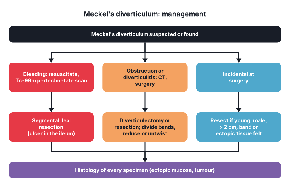
- Consider resection (incidental diverticulectomy) in → children, males under 50, diverticulum over 2 cm, palpable ectopic tissue or thickening, narrow neck, fibrous band (higher future risk)
- Debate → risk of complications from resection (about 2–5%) vs lifetime risk of complications (about 4–6%)
- Bleeding → resuscitation: IV access, fluids, blood transfusion, correct coagulopathy; bleeding usually stops, allowing planned surgery
- Obstruction → drip and suck (NG tube, IV fluids) while preparing for surgery
- Antibiotics → diverticulitis, perforation
Surgical
- Symptomatic Meckel's diverticulum → always resect
- Approach → laparoscopic (or laparoscopic-assisted: deliver through the umbilical port) or open (right lower transverse or midline incision)
- Options:
- Simple diverticulectomy (wedge excision):
- Stapled across the base, transverse to the ileum (avoids narrowing), or hand-sewn
- For diverticulitis or obstruction with a narrow-based diverticulum without bleeding
- Segmental (small bowel) resection with end-to-end anastomosis:
- Bleeding → the ulcer is in the adjacent ileum, so the ileum must be removed
- Wide base, palpable ectopic tissue at the base
- Perforation at the base, gangrene of nearby bowel (volvulus)
- Tumours → oncological resection with mesentery (neuroendocrine tumour: right hemicolectomy-type resection if large or nodal disease)
- Simple diverticulectomy (wedge excision):
- Divide fibrous bands and the mesodiverticular band; ligate the vitelline artery
- Intussusception → reduce if possible, then resect the diverticulum (lead point); resect if non-viable
- Volvulus → untwist, assess viability, resect dead bowel
- Littré's hernia → repair hernia + resect the diverticulum through the hernia or laparoscopically
- Always check for a Meckel's diverticulum when a normal appendix is found at surgery for suspected appendicitis (run 60–100 cm of ileum)
- Histology of every specimen (ectopic tissue, tumour)
10. Complications
Of the diverticulum
- Haemorrhage (most common in children) → anaemia, shock
- Intestinal obstruction (most common in adults) → volvulus with strangulation, intussusception, internal hernia, Littré's hernia
- Diverticulitis → gangrene, perforation, peritonitis, abscess
- Ulceration and perforation of the ileum
- Neoplasm → neuroendocrine tumour, adenocarcinoma, GIST
- Enteroliths, foreign body impaction
- Umbilical fistula, sinus
Of surgery
- Anastomotic or staple-line leak
- Stricture of the ileum (narrowing after longitudinal closure)
- Bleeding, wound infection, adhesive obstruction
- Recurrent bleeding if ectopic mucosa left behind (incomplete excision)
Prognosis
- Excellent after resection; mortality under 1% in elective cases, higher with strangulation and delay
Show as a list
Meckel's diverticulum
- Embryology
- Vitelline duct
- Closes by week 5-9
- True diverticulum
- Antimesenteric ileum
- Rule of 2s
- 2% people
- 2 feet from ICV
- 2 inches long
- 2 mucosa types
- 2:1 males
- Under 2 years
- Complications
- Bleeding: children
- Obstruction: adults
- Diverticulitis
- Littre's hernia
- Carcinoid
- Tests
- Tc-99m pertechnetate scan
- CT
- Angiography
- Laparoscopy
- Treatment
- Symptomatic: resect
- Diverticulectomy
- Segmental resection if bleeding
- Incidental: selective


