26. Maxillofacial injuries
High yield
Open and closed injuries of the face and jaws; airway first, then mandible, Le Fort, zygomatic and orbital fractures.
Exam Prep · State examSurgery · Emergency and traumaAnatomy · Head and neck
1. Definition
- Maxillofacial injuries → injuries of the soft tissues and bones of the face, jaws, teeth and oral cavity
- Causes → road traffic accidents, assault, falls, sports, gunshot
- Associated injuries → head injury, cervical spine injury (up to 10%), eye injury
2. Classification
By skin and mucosa integrity
- Closed → contusion, haematoma, closed fractures
- Open → wounds of the face, open fractures (all fractures through teeth-bearing bone are open into the mouth)
By tissue
- Soft tissue injuries
- Dental and alveolar injuries
- Bone injuries:
- Mandible
- Midface → maxilla (Le Fort), zygoma, nasal bones, naso-orbito-ethmoid complex
- Orbit
- Frontal sinus
- Combined
3. Pathogenesis
- Face → rich blood supply; heavy bleeding, fast healing, low infection rate
- Mandible → U-shaped bone; weak points fracture, often 2 fractures (ring principle)
- Midface → buttresses absorb force; protect brain and orbit
- Muscle pull → displaces fragments (masseter, pterygoids, suprahyoid muscles)
4. Clinical picture
Soft tissue injuries
- Heavy bleeding, swelling, early oedema
- Wounds of lips, eyelids, nose, ears
- Facial nerve injury → weakness of facial muscles; repair if lateral to the lateral canthus
- Parotid (Stensen's) duct injury → line from tragus to mid upper lip; saliva leak
Mandible fractures
- Sites → condyle, angle, body, symphysis, ramus, coronoid
- Signs:
- Malocclusion → teeth do not meet normally
- Step deformity, abnormal mobility, crepitus
- Sublingual haematoma → pathognomonic of mandible fracture
- Numb lower lip and chin → inferior alveolar nerve
- Trismus, pain on chewing
- Bilateral condyle fractures → anterior open bite
- Bilateral body fractures → tongue falls back, airway obstruction
Midface fractures (Le Fort)
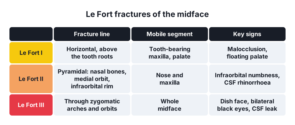
- Le Fort I → horizontal, above the teeth roots; mobile tooth-bearing maxilla (floating palate)
- Le Fort II → pyramidal, through nasal bones, medial orbit, infraorbital rim; mobile nose and maxilla
- Le Fort III → craniofacial dysjunction through zygomatic arches and orbits; whole midface mobile
- Signs → dish face (flattened midface), periorbital oedema, bilateral black eyes, malocclusion
- Cerebrospinal fluid (CSF) rhinorrhoea → Le Fort II and III, anterior cranial fossa fracture
Zygomatic complex fracture
- Flattened cheek, step in the infraorbital rim
- Infraorbital nerve numbness (cheek, upper lip)
- Trismus → depressed arch presses on coronoid process
- Subconjunctival haemorrhage without posterior limit
Orbital blow-out fracture
- Orbital floor fracture with entrapment of inferior rectus
- Diplopia on upward gaze, enophthalmos, infraorbital numbness
Nasal fractures
- Deformity, epistaxis
- Septal haematoma → drain urgently; otherwise septal necrosis and saddle nose
5. Diagnosis
- Inspection and palpation → symmetry, steps, occlusion, mobility, sensation, eye movements
- CT of the face with 3D reconstruction → standard for midface and orbit
- Orthopantomogram (OPG) and posteroanterior mandible view → mandible fractures
- CT of head and cervical spine
6. Treatment
Emergency (ATLS)
- Airway → clear blood, teeth, dentures; pull tongue forward; jaw thrust
- Intubation or surgical airway (cricothyroidotomy, tracheostomy) when needed
- Cervical spine protection
- Haemorrhage control → pressure, packing (anterior and posterior nasal packs), ligation, embolisation
- Exclude head injury, eye injury
Soft tissue
- Irrigation, minimal debridement → face tolerates delay up to 24 h
- Primary closure in layers; align landmarks first (vermilion border, eyebrow, eyelid margin)
- Repair facial nerve and parotid duct primarily
- Tetanus prophylaxis, antibiotics for bites and open fractures
Fractures
- Timing → within days, after swelling settles; airway and life-threatening injuries first
- Open reduction and internal fixation (ORIF) with titanium miniplates → standard
- Intermaxillary fixation (IMF) → restores occlusion; wires or arch bars
- Condyle fractures → often conservative (soft diet, elastics) or ORIF
- Orbital floor → repair with plate or mesh for entrapment, enophthalmos
- Nasal fracture → closed reduction at 5–10 days
7. Complications
- Airway obstruction, aspiration
- Haemorrhage
- Infection, osteomyelitis of the mandible
- Malunion, non-union, malocclusion
- Diplopia, enophthalmos, blindness
- CSF leak, meningitis
- Facial nerve palsy, sensory loss
- Ankylosis of the temporomandibular joint
- Facial asymmetry, scarring
Show as a list
Maxillofacial injuries
- Soft tissue
- Wounds
- Facial nerve
- Parotid duct
- Mandible
- Malocclusion
- Sublingual haematoma
- Lip numbness
- Midface
- Le Fort I
- Le Fort II
- Le Fort III
- Zygoma and orbit
- Flat cheek
- Diplopia
- Enophthalmos
- Treatment
- Airway first
- Primary closure
- ORIF, IMF