51. Lymphangitis, lymphadenitis and elephantiasis
High yield
Infection spreading along lymph vessels and nodes; chronic lymph obstruction causes lymphoedema and, at its end stage, elephantiasis.
Exam Prep · State examSurgery · General surgery
1. Definition
- Lymphangitis → acute inflammation of lymphatic vessels, usually bacterial, spreading from a skin wound or infection
- Lymphadenitis → inflammation of lymph nodes (acute or chronic), usually draining an infected area
- Lymphoedema → swelling from build-up of protein-rich fluid in the tissues, due to poor lymph drainage
- Elephantiasis → end stage of chronic lymphoedema: huge limb with thick, fibrotic, warty skin
2. Epidemiology
- Lymphangitis and acute lymphadenitis → common; follow minor wounds, cellulitis, abscesses
- Secondary lymphoedema → most common form worldwide:
- Developed countries → after cancer treatment (axillary or groin dissection, radiotherapy); about 20% after axillary clearance
- Worldwide → filariasis (over 100 million infected in tropical Asia, Africa, South America)
- Primary lymphoedema → rare (about 1 in 6,000–10,000); more in women
3. Aetiology and risk factors
Lymphangitis and acute lymphadenitis
- Streptococcus pyogenes (most), Staphylococcus aureus
- Entry → cuts, insect bites, tinea pedis (athlete's foot), ingrown nails, abscesses
- Risk factors → diabetes, immunosuppression, lymphoedema, venous disease
- Rare → cat-scratch disease (Bartonella), sporotrichosis, Pasteurella
Chronic lymphadenitis
- Tuberculosis (scrofula, neck) → most common cause of chronic lymphadenitis in endemic areas
- Atypical mycobacteria (children), actinomycosis, HIV, syphilis, toxoplasmosis
Lymphoedema
- Primary → failure of lymph vessel development:
- Congenital (< 1 year, 10%) → familial form = Milroy's disease
- Praecox (1–35 years, 80%) → familial form = Meige's disease, around puberty
- Tarda (> 35 years, 10%)
- Secondary:
- Surgery → axillary, groin or pelvic node dissection
- Radiotherapy
- Tumour invasion of nodes
- Filariasis → Wuchereria bancrofti, Brugia malayi; mosquito-borne
- Recurrent cellulitis and lymphangitis
- Trauma, burns
- Podoconiosis → barefoot contact with volcanic soil
- Obesity, chronic venous disease (phlebolymphoedema)
4. Pathogenesis
Lymphangitis and lymphadenitis
- Bacteria enter through a break in the skin
- Spread along superficial lymph vessels → red streaks towards the regional nodes
- Nodes swell, become tender → lymphadenitis
- Node may suppurate → abscess; bacteria pass the node → bacteraemia, sepsis
Lymphoedema
- Lymph flow blocked or absent
- Protein-rich fluid collects in the tissue
- Protein draws in more water; macrophages and fibroblasts arrive
- Chronic inflammation → fibrosis and fat deposition
- Skin thickens; immunity in the limb falls → recurrent infections
- Each infection destroys more lymphatics → vicious cycle → elephantiasis
Filariasis
- Adult worms live in lymph vessels and nodes → inflammation, blockage
- Acute attacks (filarial fever, lymphangitis) → chronic lymphoedema, hydrocele, chyluria
5. Classification
Lymphangitis
- Acute (reticular, tubular)
- Chronic → recurrent attacks in lymphoedema, fungal, mycobacterial
Lymphadenitis
- Acute (simple, suppurative → abscess)
- Chronic (non-specific, specific: tuberculosis, syphilis, actinomycosis)
Lymphoedema (International Society of Lymphology)
- Stage 0 → latent; lymph transport impaired, no swelling
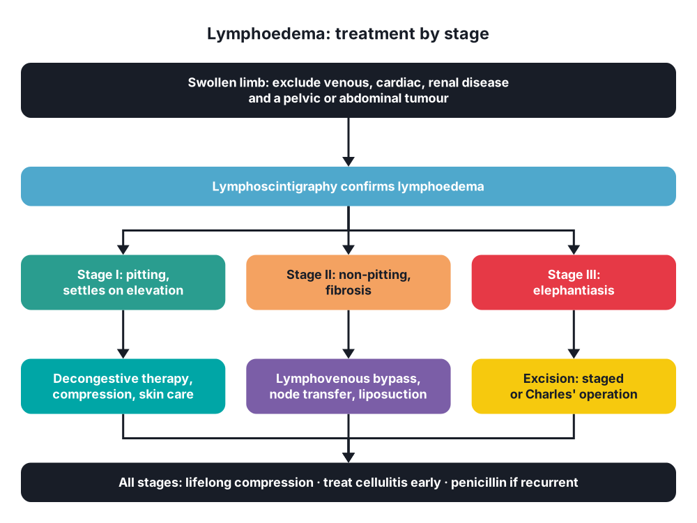
- Stage I → pitting oedema; settles with elevation
- Stage II → non-pitting; does not settle with elevation; fibrosis
- Stage III → elephantiasis: fibrosis, fat, warty skin (papillomatosis), skin folds
6. Clinical picture
Symptoms
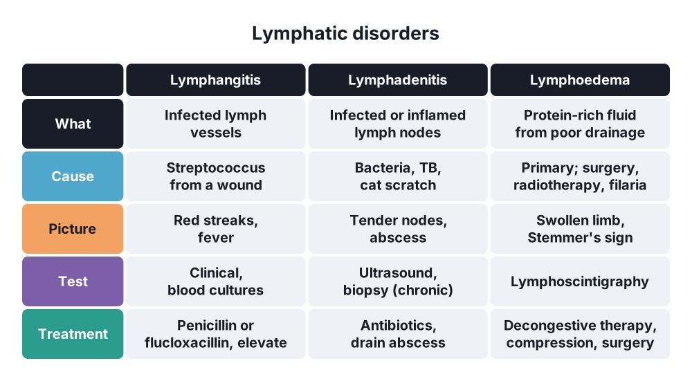
- Lymphangitis → painful red streaks spreading up the limb from a wound; fever, rigors, malaise
- Acute lymphadenitis → painful, tender swollen nodes (groin, axilla, neck)
- Abscess → throbbing pain, fluctuant swelling
- Tuberculous lymphadenitis → painless, slow-growing, matted neck nodes; may form a cold abscess and sinus ("collar-stud" abscess)
- Lymphoedema → heavy, swollen limb; starts distally; little pain
Signs
- Lymphangitis → tender red lines; surrounding cellulitis
- Lymphadenitis → tender, warm, enlarged nodes; fluctuation if suppurating
- Lymphoedema:
- Starts at the foot; swelling of the dorsum ("buffalo hump")
- Stemmer's sign → cannot pinch a fold of skin at the base of the second toe
- Square toes
- Pitting early; non-pitting later
- Thick, rough skin, hyperkeratosis, papillomas, deep folds
- Lymph leak (lymphorrhoea), fungal infection between toes
- No ulcers and no pigment (unlike venous disease)
7. Diagnosis
Acute infection
- Clinical diagnosis
- FBC, CRP; blood cultures if systemically unwell
- Swab or pus culture
- Ultrasound → node abscess
Chronic lymphadenitis
- Fine-needle aspiration or excision biopsy → tuberculosis, lymphoma, metastases
- Acid-fast stain, culture, PCR; chest X-ray
Lymphoedema
- Clinical in stages II–III
- Lymphoscintigraphy → test of choice: radioactive tracer injected into the web space; shows slow or absent transport, dermal backflow
- Indocyanine green (ICG) lymphography → maps superficial lymphatics before surgery
- MRI or CT → exclude pelvic or abdominal tumour (new lymphoedema in an adult)
- Duplex → exclude venous disease
- Filariasis → night blood smear (microfilariae), antigen test, ultrasound ("filarial dance" sign)
- Limb volume measurement → follow-up
8. Differential diagnosis
- Lymphangitis versus superficial thrombophlebitis, cellulitis, necrotising fasciitis
- Lymphadenitis versus lymphoma, metastatic nodes, hernia (groin), cyst
- Lymphoedema versus:
- Venous oedema (pigment, ulcers, varicose veins)
- Lipoedema → both legs, fat, spares the feet, women
- Heart, kidney, liver failure, hypoproteinaemia (both legs, pitting)
- Myxoedema
- Deep vein thrombosis
9. Treatment
Conservative
- Lymphangitis and acute lymphadenitis:
- Antibiotics → flucloxacillin or penicillin (co-amoxiclav, clindamycin in allergy)
- Rest, elevation, analgesia
- Treat the portal of entry (wound, tinea pedis)
- IV antibiotics if septic or not responding
- Tuberculous lymphadenitis → antituberculous drugs for 6 months
- Lymphoedema → no cure; aim to reduce swelling and prevent infection:
- Complete decongestive therapy:
- Manual lymphatic drainage (massage)
- Multilayer compression bandaging
- Exercise
- Skin care
- Then compression garments for life (class III–IV)
- Elevation, weight loss
- Intermittent pneumatic compression
- Skin hygiene, emollients, antifungals
- Prophylactic penicillin → recurrent cellulitis (≥ 2 attacks a year)
- Diuretics → no role (only short-term effect)
- Complete decongestive therapy:
- Filariasis → diethylcarbamazine, albendazole, ivermectin; mass drug treatment; hygiene and limb care
Surgical
- Lymph node abscess → incision and drainage; send pus for culture
- Tuberculous cold abscess → aspirate through a non-dependent site; excise nodes if drugs fail or sinus persists
- Lymphoedema surgery (stage II–III, when conservative care fails):
- Physiological (restore drainage; early disease):
- Lymphovenous anastomosis (bypass)
- Vascularised lymph node transfer
- Reductive / excisional (late disease):
- Liposuction → removes fat in non-pitting lymphoedema; needs lifelong compression
- Staged subcutaneous excision under flaps (Homans', Sistrunk)
- Charles' operation → excise all skin and subcutaneous tissue down to the deep fascia; cover with split-skin grafts; for elephantiasis
- Physiological (restore drainage; early disease):
- Filarial hydrocele → excision of the sac (hydrocelectomy)
10. Complications
- Lymphangitis → cellulitis, abscess, sepsis, necrotising fasciitis (rare)
- Lymphadenitis → abscess, sinus, scarring
- Lymphoedema:
- Recurrent cellulitis
- Disability, psychological distress
- Lymphorrhoea, skin breakdown
- Lymphangiosarcoma (Stewart-Treves syndrome) → rare, aggressive tumour after > 10 years of lymphoedema (classically arm after mastectomy)
- Charles' operation → poor cosmetic result, graft breakdown, hyperkeratosis, amputation in severe cases
Show as a list
Lymphatic disease
- Lymphangitis
- Streptococcus
- Red streaks
- Penicillin, elevation
- Lymphadenitis
- Acute: abscess
- Chronic: tuberculosis
- Drainage, biopsy
- Lymphoedema
- Primary: Milroy, praecox
- Secondary: surgery, filaria
- Stages 0-III
- Stemmer's sign
- Lymphoscintigraphy
- Treatment
- Decongestive therapy
- Compression
- Lymphovenous bypass
- Liposuction
- Charles' operation
- Elephantiasis
- Stage III
- Stewart-Treves


