38. Lung tumours
High yield
Hamartoma, carcinoid, non-small cell and small cell lung cancer; TNM staging; lobectomy for early, operable disease.
Exam Prep · State examSurgery · Surgical oncologyAnatomy · Thorax
1. Definition
- Lung tumour → benign or malignant neoplasm of the lung parenchyma or airways
- Lung cancer (bronchogenic carcinoma) → malignant epithelial tumour arising from the bronchial or alveolar epithelium
- Solitary pulmonary nodule (SPN) → single round opacity ≤ 3 cm surrounded by lung; > 3 cm → mass
2. Epidemiology
- Lung cancer → most common cause of cancer death in the UK and worldwide (1.8 million deaths in 2020)
- Rising incidence in women → about 25% of cancer deaths in women
- Age 50–70 years at diagnosis; most present at an advanced stage
- Benign tumours → under 5% of lung tumours
3. Aetiology and risk factors
- Cigarette smoking → about 85% of cases; risk rises with pack-years; passive smoking
- Radon → second most important cause
- Occupational → asbestos (synergistic with smoking), arsenic, chromium, nickel, polycyclic hydrocarbons, coal tar
- Previous chest radiotherapy (Hodgkin's lymphoma, breast cancer)
- Air pollution
- Pulmonary fibrosis, chronic obstructive pulmonary disease (COPD)
- Family history; never-smoker adenocarcinoma → EGFR, ALK mutations, women, Asian origin
4. Pathogenesis
- Carcinogens injure the bronchial epithelium
- Squamous metaplasia → dysplasia → carcinoma in situ → invasive carcinoma
- Spread:
- Local → bronchus, pleura, chest wall, mediastinum, nerves
- Lymphatic → hilar → mediastinal → supraclavicular nodes
- Blood → brain, bone, liver, adrenal glands, other lung
- Bone metastases → osteolytic
- Adenocarcinoma → spreads early, often to the brain
5. Classification
Benign tumours
- Hamartoma → most common benign lung tumour; cartilage, fat, epithelium; "popcorn" calcification
- Others → lipoma, leiomyoma, chondroma, papilloma, haemangioma
Low-grade malignant
- Carcinoid tumour → neuroendocrine, from Kulchitsky cells; 1–2% of lung cancers:
- Typical → indolent; 5-year survival about 90%
- Atypical → intermediate grade; more nodal spread
- Central, endobronchial ("cherry red" polyp); young adults; carcinoid syndrome rare
- Salivary gland-type → adenoid cystic carcinoma, mucoepidermoid carcinoma
Lung cancer (World Health Organization)
- Non-small cell lung cancer (NSCLC) → about 80–85%:
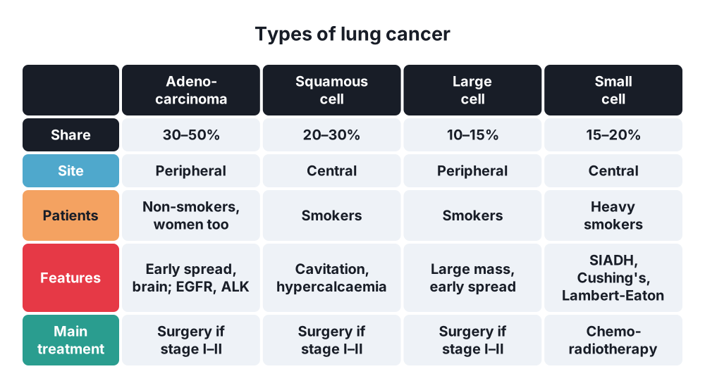
- Adenocarcinoma → 30–50%; most common type; peripheral (75%); non-smokers and women; early metastasis
- Squamous cell carcinoma → 20–30%; central (two-thirds); smokers; cavitation; hypercalcaemia; late metastasis
- Large cell carcinoma → about 10–15%; peripheral; poor prognosis
- Small cell lung cancer (SCLC) → about 15–20%:
- Neuroendocrine, central, aggressive
- Early spread to mediastinal nodes, brain, bone marrow
- Paraneoplastic syndromes
- Limited stage (one hemithorax, one radiotherapy field) or extensive stage
Secondary tumours
- Lung metastases → from colorectal cancer, kidney, breast, sarcoma, germ cell tumours, melanoma
TNM staging (8th edition, key points)
- T1 ≤ 3 cm (T1a ≤ 1, T1b 1–2, T1c 2–3 cm)
- T2 > 3–5 cm, main bronchus, visceral pleura, atelectasis
- T3 > 5–7 cm, chest wall, phrenic nerve, parietal pericardium, separate nodule same lobe
- T4 > 7 cm, mediastinum, heart, great vessels, trachea, carina, oesophagus, vertebra, recurrent laryngeal nerve, diaphragm, nodule in another ipsilateral lobe
- N1 → ipsilateral hilar; N2 → ipsilateral mediastinal or subcarinal; N3 → contralateral or supraclavicular
- M1a → malignant pleural or pericardial effusion, contralateral lung nodule; M1b → single extrathoracic metastasis; M1c → multiple
6. Clinical picture
Symptoms
- Cough, change in a chronic cough
- Haemoptysis
- Dyspnoea, wheeze, stridor
- Chest pain → pleural or chest wall invasion
- Recurrent or non-resolving pneumonia
- Weight loss > 5%, anorexia
- Local invasion:
- Hoarseness → left recurrent laryngeal nerve
- Raised hemidiaphragm → phrenic nerve
- Dysphagia → oesophagus
- Pleural effusion, pericardial effusion
- Superior vena cava (SVC) obstruction → swelling of face and arms, distended neck and chest veins
- Pancoast tumour (superior sulcus) → shoulder and arm pain (C8–T1), Horner's syndrome, small hand muscle wasting
- Metastatic → headache, seizures, bone pain, jaundice
Signs
- Finger clubbing, hypertrophic pulmonary osteoarthropathy
- Supraclavicular or cervical lymph nodes
- Signs of effusion or collapse
- Cachexia
Paraneoplastic syndromes
- SCLC → syndrome of inappropriate antidiuretic hormone (SIADH), ectopic adrenocorticotropic hormone (Cushing's), Lambert-Eaton myasthenic syndrome
- Squamous cell → hypercalcaemia (parathyroid hormone-related peptide)
- Adenocarcinoma → migratory thrombophlebitis (Trousseau's sign)
7. Diagnosis
- Aim → diagnosis and staging within 6–12 weeks of first suspicion
Laboratory
- Full blood count, calcium, sodium, liver function tests
- Sputum cytology → central squamous tumours
- Pleural fluid cytology
- Molecular tests on tissue → EGFR, ALK, ROS1, KRAS, PD-L1
Imaging
- Chest X-ray → mass, collapse, effusion, widened mediastinum
- CT chest and upper abdomen with contrast → T stage, nodes, liver, adrenals
- PET-CT → nodal and distant staging before radical treatment
- MRI brain → stage II–III before radical treatment, or symptoms
- MRI → Pancoast tumour, chest wall invasion
Tissue and nodal staging
- Bronchoscopy → central tumours; before any lung resection
- Endobronchial ultrasound (EBUS) with transbronchial needle aspiration → mediastinal nodes
- Mediastinoscopy → stations 2, 4, 7
- CT-guided percutaneous biopsy → peripheral lesions
- Biopsy of supraclavicular node → N3, stage at once
SPN
- Benign features → complete or "popcorn" calcification, stable for 2 years on CT
- Malignant features → spiculated edge, size > 8 mm, growth, upper lobe, smoker
- Management → CT surveillance, PET-CT, biopsy, or diagnostic wedge resection
Fitness for surgery
- Spirometry and transfer factor (DLCO)
- Predicted postoperative FEV1 and DLCO > 40% → acceptable risk
- Borderline → cardiopulmonary exercise testing; VO₂ max > 15 mL/kg/min favours resection
- Cardiac assessment, performance status
8. Differential diagnosis
- Pneumonia, lung abscess
- Tuberculosis, fungal infection (aspergilloma)
- Hamartoma, granuloma
- Hydatid cyst
- Metastasis
- Lymphoma, mediastinal mass
- Pulmonary infarction, organising pneumonia → mass that persists 6–8 weeks is resected to exclude cancer
9. Treatment
- Decided by lung cancer MDT; based on stage, histology, mutations, fitness
Surgical
- Indications → NSCLC stage I–II and selected stage IIIA; typical carcinoid; small peripheral SCLC (T1–2 N0)
- Principles:
- Lobectomy → standard operation, with mediastinal node dissection or sampling
- Pneumonectomy → central tumours not removable by lobectomy
- Sleeve lobectomy → central tumour, spares lung
- Segmentectomy or wedge → small peripheral tumours (≤ 2 cm) or poor lung function
- Clear margins (R0), no spillage, frozen section of the bronchial margin
- Approach → VATS or robotic lobectomy, or thoracotomy
- Operative mortality → lobectomy about 2%, pneumonectomy about 6%
- Contraindications → N3, M1 (except selected single metastasis), malignant effusion, SVC invasion, poor lung function
- Carcinoid → lobectomy or sleeve resection with nodal dissection
- Lung metastases → metastasectomy if primary controlled, all lesions removable, no other metastases, enough lung reserve
Conservative
- Stage I unfit for surgery → stereotactic ablative radiotherapy (SABR)
- Adjuvant chemotherapy (cisplatin-based) → resected stage II–IIIA
- Adjuvant osimertinib → resected EGFR-mutant NSCLC; immunotherapy → selected patients
- Stage III unresectable → concurrent chemoradiotherapy, then durvalumab
- Stage IV → targeted therapy (EGFR, ALK inhibitors), immunotherapy (pembrolizumab), chemotherapy, palliative radiotherapy
- SCLC → chemotherapy (platinum + etoposide) ± radiotherapy; prophylactic cranial irradiation if response
- Palliation:
- SVC obstruction → stent, steroids, radiotherapy
- Airway obstruction → endobronchial stent, laser, debulking
- Malignant effusion → drainage, talc pleurodesis, indwelling pleural catheter
- Bone pain → radiotherapy
10. Complications
Of disease
- Haemoptysis, airway obstruction, post-obstructive pneumonia, lung abscess
- SVC obstruction
- Malignant pleural and pericardial effusion
- Spinal cord compression, hypercalcaemia, SIADH
Of surgery
- Prolonged air leak (> 5 days)
- Atelectasis, pneumonia, respiratory failure
- Atrial fibrillation
- Bronchopleural fistula → after pneumonectomy, especially right
- Empyema
- Haemorrhage
- Recurrent laryngeal or phrenic nerve injury
- Chylothorax
- Post-thoracotomy pain
11. Prognosis
- 5-year survival by stage → I about 75% (IA1 92%), II about 40–55%, III under 30%, IV near 0–10%
- SCLC → 5-year survival about 5%
- Typical carcinoid → about 90% at 5 years
Show as a list
Lung tumours
- Benign
- Hamartoma
- Lipoma, chondroma
- Low grade
- Typical carcinoid
- Atypical carcinoid
- NSCLC
- Adenocarcinoma
- Squamous cell
- Large cell
- SCLC
- Central, aggressive
- Paraneoplastic
- Clinical
- Cough, haemoptysis
- Pancoast, Horner's
- SVC obstruction
- Clubbing
- Staging
- CT, PET-CT
- EBUS
- TNM
- Treatment
- Lobectomy
- SABR
- Chemoradiotherapy
- Targeted, immunotherapy


