37. Lung cysts and pulmonary echinococcosis
High yield
Congenital cysts, sequestration and bullae; hydatid lung cyst from Echinococcus granulosus, removed by cystotomy and capitonnage without spillage.
Exam Prep · State examSurgery · General surgeryAnatomy · Thorax
1. Definition
- Lung cyst → air- or fluid-filled cavity in the lung with a thin epithelial or fibrous wall (< 4 mm)
- Bulla → air space > 1 cm in emphysematous lung
- Bleb → subpleural air collection < 2 cm
- Pulmonary echinococcosis (hydatid disease) → cyst of the larval stage of the tapeworm Echinococcus granulosus in the lung
2. Epidemiology
- Congenital cystic lesions → most found in children, on antenatal ultrasound or with infection
- Hydatid disease → endemic in sheep-raising regions: Mediterranean, Balkans (including Bulgaria), Middle East, South America
- Lung → second most common site after the liver (adults 20–30%; children more often lung than liver)
3. Aetiology and risk factors
Congenital
- Abnormal budding of the foregut and bronchial tree
Acquired
- Emphysema, smoking → bullae
- Infection → pneumatocoele after Staphylococcus pneumonia, abscess cavity
- Tuberculosis → cavities
- Trauma → traumatic pseudocyst
Hydatid
- Definitive host → dog
- Intermediate host → sheep, cattle; humans → accidental intermediate host
- Contact with dogs, eating vegetables contaminated with dog faeces
- Rural life, sheep farming
4. Pathogenesis
Hydatid life cycle
- Adult tapeworm in the dog's small intestine
- Eggs passed in the faeces
- Human swallows eggs → embryo (oncosphere) penetrates the bowel wall
- Portal blood → liver (first filter) → lung (second filter)
- Larva grows into a cyst → about 1–5 cm a year
Cyst wall (3 layers)
- Pericyst → host fibrous layer
- Laminated membrane (ectocyst) → white, acellular
- Germinal layer (endocyst) → produces brood capsules, protoscolices ("hydatid sand") and daughter cysts
- Lung cysts → thin pericyst, elastic tissue → grow faster than liver cysts, rupture more often, rarely calcify
5. Classification
Congenital
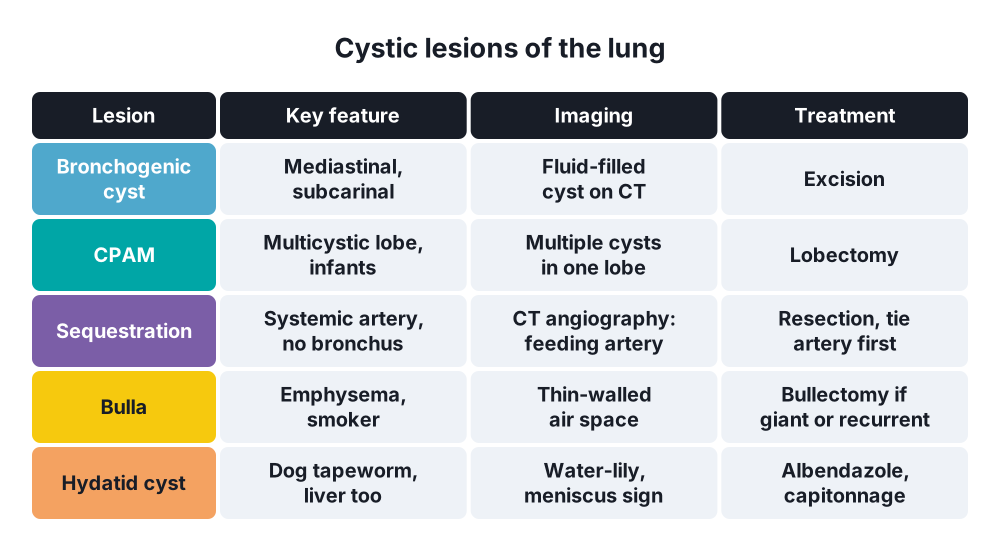
- Bronchogenic cyst → most common mediastinal cyst; usually subcarinal or paratracheal; may be intrapulmonary
- Congenital pulmonary airway malformation (CPAM) → formerly congenital cystic adenomatoid malformation; multicystic lobe with airway connection
- Pulmonary sequestration → non-functioning lung with systemic arterial supply (usually from the aorta), no normal bronchial connection:
- Intralobar → within the pleura of a lobe (75%), usually left lower lobe; drains to pulmonary veins; recurrent infection
- Extralobar → own pleural covering; drains to systemic veins; neonates; other anomalies
- Congenital lobar emphysema → overinflated lobe (left upper lobe most often) from bronchial cartilage defect; neonatal respiratory distress
Acquired
- Bullae, blebs
- Pneumatocoele
- Post-infective, post-traumatic cysts
- Hydatid cyst → intact, complicated (ruptured or infected)
6. Clinical picture
Symptoms
- Congenital:
- One-third → no symptoms
- One-third → cough
- One-third → recurrent infection
- Neonates → respiratory distress (lobar emphysema, large CPAM)
- Intact hydatid cyst → often none; found on chest X-ray
- Large hydatid cyst → cough, chest pain, dyspnoea, haemoptysis
- Ruptured into a bronchus:
- Sudden cough with salty fluid and pieces of membrane ("grape skins")
- Haemoptysis
- Anaphylaxis, urticaria
- Ruptured into pleura → pleuritic pain, hydropneumothorax, empyema
- Infected cyst → fever, purulent sputum; looks like a lung abscess
Signs
- Often no signs
- Large cyst → dullness, reduced breath sounds
- Infected sequestration → signs of lower-lobe pneumonia, recurrent at the same site
7. Diagnosis
Laboratory
- Eosinophilia → under 25% of cases
- Serology → enzyme-linked immunosorbent assay (ELISA), indirect haemagglutination; less sensitive for lung than liver cysts
- Never needle-aspirate a suspected hydatid cyst → anaphylaxis, spread
Imaging
- Chest X-ray:
- Intact → round, smooth, homogeneous opacity ("cannon ball")
- Meniscus (crescent) sign → air between pericyst and laminated membrane
- Cumbo's sign (onion peel) → air-fluid levels inside and outside the endocyst
- Water-lily sign → collapsed membrane floating on fluid after rupture
- CT → fluid density, daughter cysts, wall; screens the liver
- Ultrasound of the abdomen → always look for liver cysts; both organs involved in some patients
- Sequestration → CT angiography shows the systemic feeding artery
- Bronchogenic cyst → CT or MRI; fluid-filled, mediastinal
- Bronchoscopy → floating membranes in a ruptured hydatid cyst
8. Differential diagnosis
- Lung abscess
- Tuberculous cavity
- Lung carcinoma → cavitating squamous cell carcinoma
- Benign tumours → hamartoma
- Bullous emphysema
- Pneumatocoele
- Aspergilloma
9. Treatment
Conservative
- Small asymptomatic congenital cyst → observation; resect if it enlarges or gets infected
- Asymptomatic bullae → stop smoking, observe
- Hydatid → albendazole 10–15 mg/kg a day:
- Before surgery → reduces viability and recurrence
- After surgery → 1–3 months; for spillage, multiple cysts or inoperable disease
- Alone → small cysts in patients unfit for surgery
- Percutaneous aspiration (PAIR) → not used in the lung
Surgical
- Congenital cysts, CPAM, sequestration → anatomical resection (segmentectomy or lobectomy), often by video-assisted thoracoscopic surgery (VATS)
- Sequestration → identify and ligate the systemic artery first (may arise below the diaphragm)
- Bronchogenic cyst → complete excision
- Giant bulla (> 1/3 of hemithorax) or recurrent pneumothorax → bullectomy
- Hydatid cyst → lung-sparing surgery:
- Single-lung ventilation; positive pressure kept until the cyst is out
- Pack the field with swabs soaked in scolicidal agent (10–20% hypertonic saline)
- Aspirate the cyst through a needle, or enucleate intact cysts (Ugon's method)
- Open the cyst (cystotomy), remove membrane and daughter cysts
- Close bronchial openings in the cyst wall
- Capitonnage → obliterate the cavity with purse-string sutures
- Lobectomy → only for destroyed lobe, large infected cysts, bronchiectasis
- Bilateral cysts → staged thoracotomies or median sternotomy
- Lung and liver cysts → treat the lung first (unless liver is complicated), or one-stage through the right diaphragm
10. Complications
- Hydatid:
- Rupture into a bronchus or pleura
- Anaphylaxis
- Secondary infection → abscess
- Spillage → recurrence, secondary echinococcosis
- Postoperative air leak, bronchopleural fistula, empyema
- Congenital cysts → infection, pneumothorax, haemorrhage, rare malignant change in CPAM
- Sequestration → recurrent pneumonia, haemorrhage from the systemic artery
Show as a list
Lung cysts and echinococcosis
- Congenital
- Bronchogenic cyst
- CPAM
- Sequestration
- Lobar emphysema
- Acquired
- Bullae, blebs
- Pneumatocoele
- Hydatid cyst
- Echinococcus granulosus
- Dog, sheep
- Liver then lung
- Three-layer wall
- Signs on imaging
- Meniscus sign
- Water-lily sign
- Cumbo's sign
- Treatment
- Albendazole
- Cystotomy
- Capitonnage
- Resection for congenital


