39. Lung abscess, lung gangrene and bronchiectasis
High yield
Suppurative lung disease; abscess after aspiration, spreading gangrene, permanent bronchial dilatation; antibiotics first, resection for failure or haemoptysis.
Exam Prep · State examSurgery · General surgeryAnatomy · Thorax
1. Definition
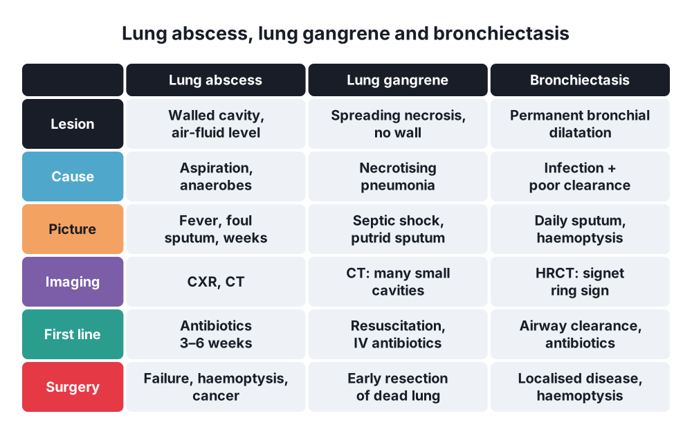
- Lung abscess → circumscribed cavity of pus or necrosis in the lung parenchyma caused by infection, > 2 cm, often with an air-fluid level
- Lung gangrene → diffuse putrid necrosis of lung tissue with no clear demarcation from living lung; extreme form of necrotising pneumonia
- Bronchiectasis → chronic disease with permanent, abnormal dilatation of the bronchi, chronic cough, sputum and infection
2. Epidemiology
- Lung abscess → men, age 40–60 years, alcohol misuse, poor dental hygiene
- Lung gangrene → rare; debilitated or immunocompromised patients; high mortality
- Bronchiectasis → more common in women and older adults; cystic fibrosis in the young
3. Aetiology and risk factors
Lung abscess
- Aspiration → most common cause:
- Reduced consciousness → alcohol, drugs, seizures, anaesthesia, stroke
- Dysphagia, oesophageal disease
- Poor dental hygiene, gingivitis
- Organisms → anaerobes from the mouth (Peptostreptococcus, Prevotella, Bacteroides, Fusobacterium), often polymicrobial
- Others → Staphylococcus aureus, Klebsiella pneumoniae, Streptococcus anginosus (milleri), Pseudomonas, Nocardia, fungi in the immunocompromised
- Bronchial obstruction → tumour, foreign body
- Septic emboli → right-sided endocarditis, intravenous drug use
- Infected cyst, bulla or infarct
Lung gangrene
- Severe pneumonia with Klebsiella, S. aureus, anaerobes, Pseudomonas
- Diabetes, alcohol misuse, immunosuppression
- Thrombosis of pulmonary vessels in the infected area
Bronchiectasis
- Two factors → infection + impaired drainage, obstruction or poor host defence
- Post-infective → childhood pneumonia, measles, whooping cough, tuberculosis
- Cystic fibrosis
- Primary ciliary dyskinesia → Kartagener's syndrome (situs inversus, sinusitis, bronchiectasis)
- Immunodeficiency → hypogammaglobulinaemia
- Allergic bronchopulmonary aspergillosis (ABPA)
- Obstruction → foreign body, tumour, nodes (middle lobe syndrome)
- α1-antitrypsin deficiency, rheumatoid arthritis, inflammatory bowel disease
- Organisms → Haemophilus influenzae, Pseudomonas aeruginosa, Moraxella, S. aureus
4. Pathogenesis
Lung abscess
- Aspiration of infected oropharyngeal material
- Pneumonitis in a dependent segment
- Necrosis and liquefaction over 1–2 weeks
- Drainage into a bronchus → cavity with air-fluid level
- Fibrous wall forms → chronic abscess after 6 weeks
- Supine → posterior segment of the upper lobe, apical (superior) segment of the lower lobe; right more often (straighter right main bronchus)
- Upright → basal segments of the lower lobes
Lung gangrene
- Severe necrotising pneumonia
- Vascular thrombosis
- Spreading putrid necrosis with no capsule
- Sloughs of dead lung in the cavity; sepsis
Bronchiectasis
- Infection and poor clearance
- Neutrophilic inflammation → elastase destroys the bronchial wall
- Bronchial dilatation, mucus plugging
- More infection → "vicious cycle"
- Hypertrophied bronchial arteries → haemoptysis
5. Classification
Lung abscess
- By duration → acute (< 6 weeks), chronic (> 6 weeks)
- By cause → primary (aspiration, pneumonia in a healthy host) or secondary (obstruction, immunosuppression, emboli)
- By number → single or multiple
Bronchiectasis
- By shape:
- Cylindrical (tubular) → mildest
- Varicose → beaded
- Cystic (saccular) → most severe
- By extent → localised (one lobe) or diffuse (bilateral)
6. Clinical picture
Symptoms
- Lung abscess:
- Fever, rigors, night sweats, weight loss over weeks
- Cough with large volumes of foul-smelling (putrid) sputum after drainage into a bronchus
- Pleuritic chest pain, haemoptysis
- Lung gangrene:
- Severe sepsis, septic shock
- Very foul, dark, copious sputum with pieces of lung tissue
- Bronchiectasis:
- Chronic productive cough, daily purulent sputum, worse in the morning and on lying down
- Recurrent chest infections
- Haemoptysis → mild to massive
- Dyspnoea, fatigue, sinusitis
Signs
- Poor dentition, foul breath
- Dullness, bronchial breathing over consolidation
- Amphoric breath sounds over a large cavity
- Bronchiectasis → coarse crackles over the affected area, finger clubbing, wheeze
7. Diagnosis
Laboratory
- Full blood count → leucocytosis; C-reactive protein
- Sputum Gram stain, culture, acid-fast bacilli (tuberculosis)
- Blood cultures
- Bronchiectasis work-up → sweat test (cystic fibrosis), immunoglobulins, Aspergillus serology, α1-antitrypsin
Imaging
- Chest X-ray → thick-walled cavity with air-fluid level (abscess); "tram lines" and ring shadows (bronchiectasis)
- CT chest → separates abscess from empyema, shows cavitating cancer
- Abscess on CT → round, thick wall, acute angle with the chest wall; empyema → lenticular, obtuse angle, "split pleura" sign
- Gangrene → spreading consolidation with multiple small cavities and sloughs
- High-resolution CT (HRCT) → gold standard for bronchiectasis:
- Signet ring sign → bronchus wider than the adjacent artery
- Bronchi seen within 1 cm of the pleura
- No tapering of bronchi
Bronchoscopy
- Exclude obstructing tumour or foreign body, especially over 50 years or in smokers
- Bronchoalveolar lavage for culture
- Locate the bleeding source in haemoptysis
8. Differential diagnosis
- Cavitating lung cancer (squamous cell)
- Tuberculosis
- Empyema with bronchopleural fistula
- Infected hydatid cyst or bulla
- Granulomatosis with polyangiitis (Wegener's)
- Pulmonary infarction
- Aspergilloma
- Bronchiectasis versus COPD, chronic bronchitis
9. Treatment
Conservative
- Lung abscess:
- Prolonged antibiotics → covering anaerobes: β-lactam with β-lactamase inhibitor (co-amoxiclav, ampicillin-sulbactam), or carbapenem, or clindamycin; IV then oral
- Duration → 3–6 weeks or until the cavity resolves
- Postural drainage, physiotherapy
- Treat the cause → dental care, swallowing assessment
- 85–95% respond to medical treatment; complete healing in 3–4 months
- Poor response if → symptoms > 3 months, cavity > 4–6 cm, obstructed bronchus
- Not responding → CT-guided percutaneous drainage or bronchoscopic drainage
- Lung gangrene → resuscitation, broad-spectrum IV antibiotics, organ support; early surgery if sepsis is not controlled
- Bronchiectasis:
- Airway clearance → physiotherapy, active cycle of breathing, postural drainage
- Antibiotics for exacerbations → 14 days, guided by sputum culture
- Frequent exacerbations (≥ 3 a year) → long-term macrolide (azithromycin), inhaled antibiotics for Pseudomonas
- Bronchodilators, vaccination, treat cause (immunoglobulin, ABPA)
- Massive haemoptysis → bronchial artery embolisation first
Surgical
- Lung abscess → resection (usually lobectomy):
- Persistent thick-walled cavity ≥ 2 cm after treatment
- Sepsis not cleared after 8 weeks of medical treatment
- Massive haemoptysis
- Suspected carcinoma
- Complications → rupture into pleura (empyema), bronchopleural fistula
- Lung gangrene → anatomical resection (lobectomy or pneumonectomy) of dead lung once stabilised
- Bronchiectasis:
- Localised disease with frequent exacerbations despite best medical treatment
- Massive haemoptysis not controlled by embolisation
- Destroyed lobe
- Usually segmentectomy or lobectomy, VATS preferred
- Bilateral, diffuse disease → relative contraindication; lung transplant in end-stage disease (cystic fibrosis)
- Massive haemoptysis (> 150 mL in 24 h or > 100 mL/h):
- Protect the airway, oxygen
- Lie the patient bleeding side down
- Large-bore endotracheal tube or single-lung intubation
- Bronchoscopy to locate and control (cold saline, adrenaline, balloon)
- Bronchial artery embolisation
- Resection if bleeding continues
- Lung resection for sepsis → double-lumen tube to protect the healthy lung from spillage
10. Complications
- Empyema, pyopneumothorax
- Bronchopleural fistula
- Massive haemoptysis → death from asphyxia, not blood loss (mortality 7–30%)
- Spread to the other lung
- Brain abscess
- Septic shock, multiorgan failure
- Secondary amyloidosis (chronic suppuration)
- Respiratory failure, cor pulmonale (bronchiectasis)
Show as a list
Suppurative lung disease
- Lung abscess
- Aspiration
- Anaerobes
- Dependent segments
- Air-fluid level
- Lung gangrene
- Necrotising pneumonia
- No demarcation
- Septic shock
- Bronchiectasis
- Permanent dilatation
- Cystic fibrosis
- Kartagener's
- HRCT signet ring
- Treatment
- Prolonged antibiotics
- Postural drainage
- Percutaneous drainage
- Lobectomy
- Embolisation
- Complications
- Empyema
- Haemoptysis
- Bronchopleural fistula


