
72. Tumours of the liver, gallbladder and bile ducts
High yield
Benign liver lesions, metastases, hepatocellular carcinoma, gallbladder cancer and cholangiocarcinoma: diagnosis, staging and resection.
Exam Prep · State examSurgery · HPBSurgery · Surgical oncologySurgical Anatomy · Hepatobiliary
1. Definition
- Benign liver tumours → haemangioma, focal nodular hyperplasia (FNH), hepatocellular adenoma, simple cysts, biliary cystadenoma
- Malignant liver tumours:
- Secondary (metastases) → most common malignant liver tumour (colorectal, neuroendocrine, breast, lung, stomach, pancreas)
- Primary → hepatocellular carcinoma (HCC), intrahepatic cholangiocarcinoma, fibrolamellar HCC, hepatoblastoma (children), angiosarcoma
- Gallbladder carcinoma → adenocarcinoma of the gallbladder
- Cholangiocarcinoma → adenocarcinoma of the bile duct epithelium: intrahepatic, perihilar (Klatskin) or distal
2. Epidemiology
- Metastases → about 20 times more common than primary liver cancer in the West; 50% of colorectal cancer patients develop liver metastases
- Haemangioma → most common benign liver tumour (up to 5–20% at autopsy); women
- FNH → second most common; young women
- Adenoma → rare; women on the oral contraceptive pill, anabolic steroids, glycogen storage disease, obesity
- HCC → 6th most common cancer, 3rd cause of cancer death worldwide; Asia and Africa (hepatitis B); men 3 : 1; 80–90% in cirrhosis
- Gallbladder cancer → most common biliary cancer; women 2–3 : 1; over 65; Chile, India (north), Japan, Eastern Europe
- Cholangiocarcinoma → rare in the West (1–2 per 100,000); perihilar 50–60%, distal 20–30%, intrahepatic 10–20%; common in Thailand (liver flukes)
3. Aetiology and risk factors
Benign
- Haemangioma → congenital vascular malformation; may grow in pregnancy
- FNH → hyperplastic response to an abnormal artery; not hormone-driven
- Adenoma → oestrogens, androgens, glycogen storage disease, metabolic syndrome; subtypes (HNF1A, inflammatory, beta-catenin mutated → highest cancer risk, sonic hedgehog → bleeding)
HCC
- Cirrhosis from any cause
- Hepatitis B (can cause HCC without cirrhosis) and hepatitis C
- Alcohol
- Metabolic dysfunction-associated steatotic liver disease (MASLD), diabetes, obesity (rising)
- Aflatoxin (Aspergillus on stored grain), haemochromatosis, alpha-1 antitrypsin deficiency, Wilson's disease, tyrosinaemia
Gallbladder cancer
- Gallstones (in 70–90%); large stones (over 3 cm)
- Porcelain gallbladder (selective mucosal calcification carries more risk)
- Gallbladder polyps over 1 cm
- Anomalous pancreaticobiliary duct junction, choledochal cyst
- Chronic typhoid carriage (Salmonella), obesity, female sex
Cholangiocarcinoma
- Primary sclerosing cholangitis (lifetime risk 10–20%; often with ulcerative colitis)
- Choledochal cysts, Caroli's disease
- Liver flukes → Opisthorchis viverrini, Clonorchis sinensis
- Hepatolithiasis, recurrent pyogenic cholangitis
- Hepatitis B and C, cirrhosis, alcohol, MASLD (intrahepatic)
- Thorotrast (historical), biliary-enteric anastomosis
4. Pathogenesis
- HCC → chronic injury, regeneration and fibrosis → dysplastic nodules → HCC; arterial blood supply (portal supply lost) → arterial enhancement with washout on imaging; invades the portal vein (tumour thrombus); spreads to lung, bone, adrenal
- Liver metastases → arrive by the portal vein (gut) or hepatic artery (other sites); mostly supplied by the hepatic artery
- Gallbladder cancer → dysplasia-carcinoma sequence in chronic inflammation; spreads early:
- Direct into liver segments IVb and V, duodenum, colon
- Lymph nodes (cystic, pericholedochal, then coeliac)
- Peritoneum; port sites after laparoscopic cholecystectomy
- Cholangiocarcinoma → chronic inflammation and stasis → dysplasia → cancer; grows along and around the ducts (periductal infiltrating), into the portal vein and hepatic artery, along nerves
- Hilar tumours block both lobes → painless obstructive jaundice
5. Classification
HCC staging: Barcelona Clinic Liver Cancer (BCLC, 2022)
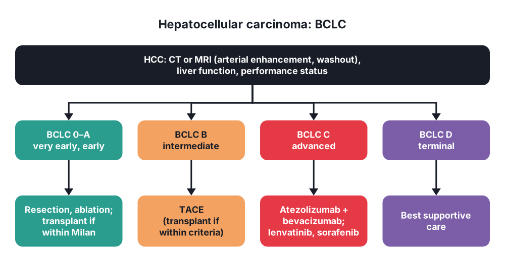
- 0 (very early) → single tumour < 2 cm, Child-Pugh A, performance status 0 → ablation or resection
- A (early) → single tumour, or up to 3 nodules all ≤ 3 cm, liver function preserved → resection, transplantation or ablation
- B (intermediate) → multinodular, preserved liver function, PS 0 → TACE (transplantation if within extended criteria)
- C (advanced) → portal vein invasion or spread outside the liver, PS 1–2 → systemic therapy (atezolizumab-bevacizumab, durvalumab-tremelimumab; sorafenib, lenvatinib)
- D (terminal) → end-stage liver function or PS > 2 → best supportive care
Transplantation criteria
- Milan criteria → one tumour ≤ 5 cm, or 2–3 tumours each ≤ 3 cm; no vascular invasion or spread → 5-year survival over 70%
Cholangiocarcinoma
- By site → intrahepatic, perihilar (Klatskin), distal
- Bismuth-Corlette (perihilar):
- I → below the confluence
- II → at the confluence
- IIIa → into the right hepatic duct; IIIb → into the left
- IV → both secondary ducts, or multifocal
Gallbladder cancer (TNM 8th)
- T1a lamina propria; T1b muscle; T2 perimuscular tissue (T2a peritoneal side, T2b liver side); T3 serosa or liver invasion; T4 main portal vein, hepatic artery or two or more organs
6. Clinical picture
Symptoms
- Benign lesions → usually none; found by chance ("incidentaloma"); large ones cause RUQ fullness or pain
- Adenoma → rupture and bleeding (sudden pain, shock), especially in pregnancy
- Giant haemangioma → rarely Kasabach-Merritt syndrome (platelet consumption)
- HCC:
- Often found on surveillance ultrasound in cirrhosis
- RUQ pain, weight loss, anorexia
- Decompensation of known cirrhosis → ascites, jaundice, encephalopathy, variceal bleeding
- Rupture → haemoperitoneum
- Paraneoplastic → hypoglycaemia, polycythaemia, hypercalcaemia
- Gallbladder cancer → found in about 1 in 200 cholecystectomy specimens (incidental); otherwise late: RUQ pain, weight loss, jaundice (spread to the hilum), mass
- Cholangiocarcinoma → painless progressive jaundice, itch, dark urine, pale stools, weight loss; cholangitis after stenting; intrahepatic → pain, mass, no jaundice
Signs
- Hepatomegaly, hard irregular liver
- Bruit over the liver (HCC, vascular tumour)
- Signs of chronic liver disease
- Jaundice; palpable gallbladder only in distal duct tumours (Courvoisier); in hilar tumours the gallbladder is collapsed
- Ascites, Virchow's node, Sister Mary Joseph nodule (advanced)
7. Diagnosis
Blood tests
- LFT, INR, albumin, platelets (liver function and portal hypertension)
- Alpha-fetoprotein (AFP) → HCC (raised in about 60%); also hepatoblastoma, germ cell tumours
- CA 19-9 → cholangiocarcinoma, gallbladder cancer (also high in cholangitis and jaundice); CEA
- Hepatitis B and C serology
- Child-Pugh and MELD scores
Imaging
- Ultrasound → first test; 6-monthly surveillance ± AFP in cirrhosis and high-risk hepatitis B
- Multiphase contrast CT or MRI:
- HCC → arterial hyperenhancement with portal venous or delayed washout and a capsule (LI-RADS 5) → diagnosis without biopsy in cirrhosis
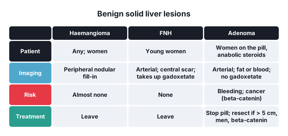
- Haemangioma → peripheral nodular enhancement filling in centripetally
- FNH → arterial enhancement with a central scar; retains hepatobiliary (gadoxetate) contrast
- Adenoma → arterial enhancement; fat or bleeding; no gadoxetate uptake (most)
- MRCP → level and extent of biliary tumours (Bismuth type)
- CT chest, abdomen and pelvis → staging; PET-CT in selected
- Liver volumetry → future liver remnant before major hepatectomy
Tissue
- Biopsy → when imaging is not typical; metastases of unknown primary; before systemic therapy; not needed for resectable typical HCC
- ERCP with brushings or cholangioscopy biopsy → biliary strictures (low sensitivity); EUS-FNA for distal tumours
- Staging laparoscopy → gallbladder cancer, perihilar cholangiocarcinoma (peritoneal spread)
Liver function before surgery
- Child-Pugh class, MELD, hepatic venous pressure gradient (≥ 10 mmHg → high risk), indocyanine green clearance
8. Differential diagnosis
- Liver lesions → abscess, hydatid cyst, simple cyst, regenerative nodules in cirrhosis, focal fat, metastases
- Biliary stricture → benign (PSC, IgG4 cholangiopathy, after surgery, Mirizzi), pancreatic cancer, gallbladder cancer at the hilum
- Gallbladder → xanthogranulomatous cholecystitis, adenomyomatosis, cholesterol polyps
9. Treatment
Conservative
- Haemangioma and FNH → no treatment, no follow-up when the diagnosis is certain; FNH → may continue the oral contraceptive pill
- Adenoma:
- Stop the oral contraceptive pill, lose weight; repeat MRI in 6–12 months
- Under 5 cm in women → observe
- Gallbladder polyps → ultrasound follow-up if 6–9 mm (more often with risk factors)
- HCC non-surgical:
- Ablation (radiofrequency or microwave) → tumours ≤ 3 cm; as good as resection for small tumours
- TACE (transarterial chemoembolisation) → intermediate stage; bridge to transplant
- TARE / SIRT (yttrium-90 radioembolisation), stereotactic radiotherapy
- Systemic → atezolizumab with bevacizumab or durvalumab with tremelimumab first-line; lenvatinib or sorafenib
- Treat the liver disease → antivirals for hepatitis B and C, stop alcohol
- Cholangiocarcinoma and gallbladder cancer:
- Adjuvant capecitabine for 6 months after resection (BILCAP)
- Advanced → gemcitabine, cisplatin and durvalumab (TOPAZ-1) or pembrolizumab
- Targeted therapy for FGFR2 fusions (pemigatinib) and IDH1 mutations (ivosidenib) in intrahepatic tumours
- Biliary drainage for jaundice and cholangitis → metal stent (ERCP or PTBD); photodynamic therapy or radiofrequency ablation of the stent
Surgical
- Adenoma → resect if over 5 cm, in men (any size), beta-catenin mutated, growing, or bleeding (embolise first if ruptured)
- Liver metastases (colorectal):
- Resect if all disease can be removed with clear margins and an adequate future liver remnant (over 20–25% in a normal liver, 30% after chemotherapy, 40% in cirrhosis)
- Perioperative chemotherapy (FOLFOX); 5-year survival after resection about 40–50%
- Small liver remnant → portal vein embolisation, two-stage hepatectomy, ALPPS
- Ablation for small deep lesions
- Neuroendocrine metastases → resection or debulking
- HCC:
- Resection → single tumour, Child-Pugh A, no clinically significant portal hypertension, normal bilirubin; anatomical resection preferred
- Liver transplantation → within Milan criteria with cirrhosis; treats the tumour and the cirrhosis; 5-year survival over 70%
- Gallbladder cancer:
- T1a found after cholecystectomy → cholecystectomy is enough (if margins and cystic duct are clear)
- T1b or more → re-resection: segment IVb and V resection (or wedge of 2 cm of liver bed) with regional lymphadenectomy (hepatoduodenal ligament), ± bile duct resection if the cystic duct margin is positive
- Suspected cancer before surgery → no laparoscopic cholecystectomy; open radical cholecystectomy
- Port-site excision → no longer routine
- Cholangiocarcinoma:
- Perihilar → hepatectomy (extended right or left) with caudate lobe and bile duct resection, lymphadenectomy and Roux-en-Y hepaticojejunostomy; preoperative drainage of the future liver remnant and portal vein embolisation
- Liver transplantation for selected unresectable perihilar tumours (Mayo protocol: chemoradiotherapy then transplant)
- Distal → pancreaticoduodenectomy (Whipple)
- Intrahepatic → hepatectomy with lymphadenectomy
- Unresectable → biliary bypass rarely (when found at laparotomy)
10. Complications
- Disease → rupture and bleeding (adenoma, HCC), obstructive jaundice, cholangitis, liver failure, portal vein thrombosis, cachexia
- Liver resection:
- Post-hepatectomy liver failure (small remnant, cirrhosis) → main cause of death
- Bile leak, biloma
- Bleeding, subphrenic abscess, pleural effusion
- Transplantation → rejection, infection from immunosuppression, recurrence of HCC (about 10–15%)
- TACE → post-embolisation syndrome (fever, pain, nausea), liver failure
Prognosis
- HCC → 5-year survival under 20% in all patients; 60–80% after resection or transplant for early disease
- Gallbladder cancer → 5-year survival under 10–20% in all; good for T1 tumours
- Cholangiocarcinoma → 5-year survival 20–40% after R0 resection; median survival under 1 year if unresectable
Show as a list
Liver and biliary tumours
- Benign
- Haemangioma: leave
- FNH: central scar, leave
- Adenoma: pill, resect > 5 cm
- Metastases
- Commonest malignancy
- Colorectal: resect
- Future liver remnant
- PVE, ALPPS
- HCC
- Cirrhosis, hepatitis B, C
- AFP
- Arterial washout
- BCLC staging
- Resect, ablate, transplant
- TACE
- Atezolizumab + bevacizumab
- Gallbladder cancer
- Gallstones, porcelain
- T1b+: re-resection IVb, V
- Cholangiocarcinoma
- PSC, flukes, cysts
- Klatskin: Bismuth I-IV
- Hepatectomy + caudate
- Distal: Whipple
- Gem-cis + durvalumab


