69. Liver and biliary trauma
High yield
Most liver injuries in stable patients are managed without surgery; unstable patients need damage control laparotomy and packing.
Exam Prep · State examSurgery · HPBSurgery · Emergency and traumaSurgical Anatomy · Hepatobiliary
1. Definition
- Liver trauma → injury to the liver parenchyma, capsule, hepatic veins, retrohepatic vena cava or portal structures from blunt or penetrating force
- Biliary trauma → injury to the gallbladder or bile ducts, from external trauma or during surgery (iatrogenic bile duct injury)
2. Epidemiology
- Liver → most commonly injured solid organ in penetrating trauma; second after the spleen in blunt trauma (in many series now the most common in blunt too)
- Blunt (road traffic collisions, falls) → about 80% in Europe; penetrating (stab, gunshot) more in some countries
- Right lobe injured far more than the left (larger, fixed, near the ribs)
- Non-operative management → succeeds in over 80–90% of haemodynamically stable blunt injuries
- Extrahepatic bile duct injury from external trauma → rare (under 1% of abdominal trauma), often with duodenal, pancreatic or vascular injuries
- Iatrogenic bile duct injury → 0.3–0.5% of laparoscopic cholecystectomies (most common cause of bile duct injury)
- Mortality → about 10% in all; over 50% with retrohepatic caval or hepatic vein injury
3. Aetiology and risk factors
Anatomy (revision)
- Couinaud segments I–VIII; Cantlie's line (gallbladder fossa to IVC) divides right and left lobes
- Dual blood supply → portal vein (75% of flow) and hepatic artery (25%)
- Hepatic veins drain into the retrohepatic IVC; short and hard to reach
- Portal triad in the hepatoduodenal ligament (free edge of the lesser omentum) → CBD (lateral), hepatic artery (medial), portal vein (posterior); Pringle manoeuvre clamps it
- Ligaments → falciform, coronary, triangular; bare area
- Gallbladder fixed in its fossa; CBD in the hepatoduodenal ligament
Mechanisms
- Blunt:
- Direct crush against the ribs and spine
- Deceleration → shear at the ligaments and hepatic veins (tears between segments, caval injuries)
- Penetrating → stab (lower energy), gunshot (cavitation, more damage)
- Iatrogenic → liver biopsy, PTC, laparoscopic cholecystectomy (misidentification of the CBD as the cystic duct, thermal injury)
- Risk factors → lower right rib fractures, seat-belt injuries, coagulopathy, cirrhosis, large liver
4. Pathogenesis
- Bleeding → main early killer; portal and hepatic venous bleeding is low-pressure, so many stop on their own; arterial bleeding continues
- Subcapsular haematoma → may rupture later (delayed bleeding)
- Bile leak → biloma, bile peritonitis (chemical then infected)
- Haemobilia → fistula between an artery (or pseudoaneurysm) and a bile duct → bleeding into the bile ducts
- Bilhaemia → bile into the hepatic veins (rare)
- Devitalised liver → necrosis, abscess
- Lethal triad in massive bleeding → hypothermia, acidosis, coagulopathy (with hypocalcaemia, the "lethal diamond")
5. Classification
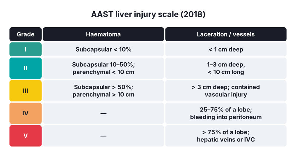
AAST liver injury scale (2018)
- Grade I → subcapsular haematoma < 10% surface; laceration < 1 cm deep
- Grade II → subcapsular haematoma 10–50%; intraparenchymal haematoma < 10 cm; laceration 1–3 cm deep, < 10 cm long
- Grade III → subcapsular haematoma > 50% or expanding; intraparenchymal haematoma > 10 cm; laceration > 3 cm deep; any vascular injury or active bleeding contained in the liver
- Grade IV → parenchymal disruption of 25–75% of a lobe; active bleeding beyond the liver into the peritoneum
- Grade V → disruption > 75% of a lobe; juxtahepatic venous injury (retrohepatic IVC, central hepatic veins)
- (Grade VI, hepatic avulsion, removed in the 2018 revision)
WSES (World Society of Emergency Surgery)
- Minor (AAST I–II), moderate (III), severe (IV–V or any haemodynamically unstable patient)
Bile duct injury (iatrogenic)
- Strasberg → A (cystic duct or duct of Luschka leak), B (occluded aberrant right duct), C (transected aberrant right duct), D (lateral injury to the CBD), E1–E5 (transection or stricture of the main duct by level: Bismuth types 1–5)
6. Clinical picture
Symptoms
- RUQ pain, right shoulder tip pain (Kehr's sign on the right; diaphragm irritation)
- Abdominal pain, distension
- Symptoms of blood loss → thirst, dizziness, confusion
- Biliary injury → pain, distension, fever, jaundice days later (biloma, bile peritonitis)
- Haemobilia → Quincke's triad: RUQ pain, jaundice, upper GI bleeding (haematemesis or melaena), often weeks later
Signs
- Haemorrhagic shock → tachycardia, hypotension, cool skin
- RUQ tenderness, guarding; peritonism; distension
- Lower right rib fractures, seat-belt bruising
- Penetrating wounds of the lower right chest or RUQ (thoracoabdominal)
- Bile leak → bile in a drain, ascites, jaundice, fever
7. Diagnosis
- ATLS primary survey (ABCDE); FBC, crossmatch, coagulation, lactate, base deficit, blood gas
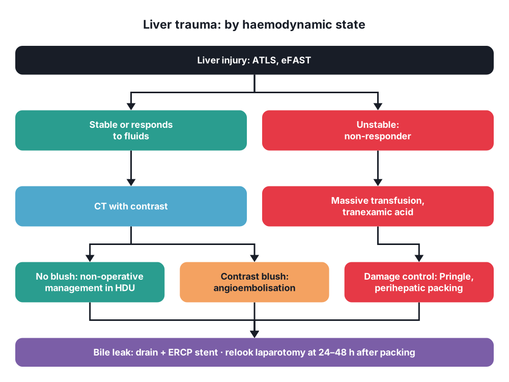
- eFAST → free fluid in the abdomen (Morison's pouch) in an unstable patient → straight to theatre
- CT with IV contrast (arterial and portal phases) → stable patients; gold standard:
- Grade of injury
- Contrast blush (active arterial bleeding) or pseudoaneurysm → angioembolisation
- Other injuries (spleen, kidneys, bowel)
- Delayed (excretory) phase → bile leak
- Bile leak → HIDA scan; MRCP; ERCP (diagnosis and treatment)
- Haemobilia → CT angiography, angiography (diagnostic and therapeutic); endoscopy shows blood from the ampulla
- Diagnostic peritoneal lavage → rarely now (when FAST not available)
- Laparoscopy → penetrating thoracoabdominal wounds (diaphragm)
8. Differential diagnosis
- Splenic injury, renal injury, pancreatic or duodenal injury
- Hollow viscus injury (bowel, mesentery)
- Rib fractures alone, chest injury (haemothorax)
- Retroperitoneal haematoma, pelvic fracture bleeding
9. Treatment
Conservative
- Non-operative management (NOM) → standard for haemodynamically stable patients, any grade:
- Conditions → stable (or responds to fluids), no peritonitis, no other injury needing laparotomy, CT available, ICU or HDU monitoring, surgeon and theatre available 24 hours
- Serial examination, serial haemoglobin, bed rest (short), VTE prophylaxis after 24–48 hours if no bleeding
- Failure of NOM → about 5–10%; most in the first 24–72 hours
- Angioembolisation → active contrast blush on CT in a stable patient, pseudoaneurysm, haemobilia, or after packing (hybrid approach)
- Selected stab wounds in stable patients without peritonitis → NOM in expert centres
- Bile leak or biloma → percutaneous drainage plus ERCP with sphincterotomy and stent (lowers duct pressure; leak heals)
- Follow-up CT → only for symptoms or high grades; avoid contact sport for 3–6 months (grades III–V)
Surgical
- Indications for laparotomy → haemodynamic instability (non-responder), peritonitis, other injuries needing surgery, failed NOM, gunshot wounds in most centres
- Damage control resuscitation → massive transfusion protocol (red cells, plasma, platelets 1 : 1 : 1), tranexamic acid within 3 hours, permissive hypotension, warming, calcium
- Damage control laparotomy:
- Midline laparotomy; pack all four quadrants
- Manual compression of the liver
- Perihepatic packing → packs above, below and behind the liver to compress it (not packs inside a laceration)
- Pringle manoeuvre → clamp the hepatoduodenal ligament; bleeding stops → arterial or portal source; continues → hepatic veins or retrohepatic IVC
- Control bleeding → ligation of vessels in the wound (hepatotomy and direct ligation), suture, haemostatic agents, balloon tamponade for tracts (gunshot)
- Resectional debridement of dead liver (not formal anatomical resection in the acute phase)
- Temporary abdominal closure (vacuum dressing); ICU to correct the lethal triad
- Relook in 24–48 hours → remove packs, definitive repair
- Severe juxtahepatic venous injury → packing; total vascular exclusion, atriocaval shunt (Schrock) → high mortality
- REBOA and hybrid theatre → selected centres
- Liver transplantation → rare, for total devastation
- Gallbladder injury → cholecystectomy
- Extrahepatic bile duct injury:
- Small lateral tear → primary repair over a T-tube or stent
- Transection or loss of length → Roux-en-Y hepaticojejunostomy (best when patient is stable; drain first in damage control)
- Iatrogenic injury found at surgery → call an HPB surgeon; if not available, drain and transfer
- Late strictures → hepaticojejunostomy in an HPB centre
10. Complications
- Bleeding → early death; delayed bleeding after NOM
- Bile leak, biloma, bile peritonitis
- Haemobilia, pseudoaneurysm
- Liver abscess, infected haematoma
- Abdominal compartment syndrome (after packing and massive transfusion)
- Hepatic necrosis (after embolisation), gallbladder ischaemia
- Biliary stricture, secondary biliary cirrhosis (bile duct injury)
- Lethal triad, multiorgan failure, ARDS
Show as a list
Liver and biliary trauma
- Anatomy
- Couinaud segments
- Portal triad
- Hepatic veins, IVC
- Mechanism
- Blunt: crush, deceleration
- Penetrating
- Iatrogenic: lap chole
- Grading
- AAST I-V
- WSES: unstable = severe
- Tests
- eFAST: unstable
- CT: stable
- Contrast blush
- Stable
- Non-operative
- Angioembolisation
- ERCP stent for leak
- Unstable
- Massive transfusion
- Damage control
- Pringle
- Perihepatic packing
- Relook 24-48 h
- Biliary
- Gallbladder: cholecystectomy
- Duct: hepaticojejunostomy
- Haemobilia: Quincke


