46. Limb ischaemia and acute arterial insufficiency
High yield
Chronic claudication to critical ischaemia; acute embolus or thrombosis with the six Ps; graded by Rutherford, treated by revascularisation.
Exam Prep · State examSurgery · General surgerySurgery · Emergency and trauma
1. Definition
- Peripheral arterial disease (PAD) → narrowing or occlusion of the arteries of the limbs, mostly by atherosclerosis
- Chronic limb ischaemia → slow loss of perfusion, from claudication to rest pain and tissue loss
- Chronic limb-threatening ischaemia (CLTI) → rest pain, ulcer or gangrene for > 2 weeks with proven arterial insufficiency (formerly "critical limb ischaemia")
- Acute limb ischaemia (ALI) → sudden fall in limb perfusion that threatens viability; symptoms < 2 weeks
2. Epidemiology
- PAD → about 20% of people over 60 years; many have no symptoms
- Intermittent claudication → 4% of people over 55; mostly men
- Claudication course → one-third improve, one-third stay the same, one-third get worse; only about 2% need amputation
- ALI → about 1 in 6,000 a year; death about 20%, limb loss up to 40%
- 40% of PAD patients have coronary or cerebral disease → most die of myocardial infarction or stroke
3. Aetiology and risk factors
Chronic
- Atherosclerosis → most cases
- Risk factors:
- Smoking → the strongest
- Diabetes mellitus
- Hypertension, hyperlipidaemia
- Age, male sex, family history
- Chronic kidney disease
- Rare → Buerger's disease, vasculitis (Takayasu's), popliteal entrapment, cystic adventitial disease
Acute
- Thrombosis in situ (about 60%) → on an atherosclerotic plaque; dehydration, low blood pressure, cancer, polycythaemia, thrombophilia; bypass graft or stent thrombosis
- Embolism (about 30%):
- Heart in 80% → atrial fibrillation (left atrial appendage), mural thrombus after MI, valve disease, endocarditis
- Aneurysms (aorta, popliteal) → 10%
- Paradoxical embolus → patent foramen ovale
- Others → trauma, iatrogenic (catheters), aortic dissection, thrombosed popliteal aneurysm, intra-arterial drug injection
4. Pathogenesis
Chronic
- Plaque narrows the artery
- Flow at rest is enough; exercise needs more → muscle ischaemia → claudication
- Collaterals develop over time
- More narrowing → flow is too low at rest → rest pain (worse at night, relieved by hanging the leg down)
- Tissue loss → ulcers, gangrene
Acute
- No time for collaterals → severe ischaemia
- Tolerance → nerves and muscle are most sensitive; irreversible muscle damage after about 6 h
- Emboli lodge at bifurcations → common femoral (most common), popliteal, brachial, aortic bifurcation ("saddle embolus")
- Reperfusion injury → potassium, myoglobin, acid and free radicals enter the circulation → hyperkalaemia, acidosis, renal failure, arrhythmia; muscle swelling → compartment syndrome
5. Classification
Chronic: Fontaine
- I → no symptoms
- IIa → mild claudication (> 200 m); IIb → moderate to severe claudication (< 200 m)
- III → ischaemic rest pain
- IV → ulcer or gangrene
Chronic: Rutherford categories
- 0 none; 1–3 mild, moderate, severe claudication; 4 rest pain; 5 minor tissue loss; 6 major tissue loss
- WIfI (wound, ischaemia, foot infection) → staging of limb threat in CLTI
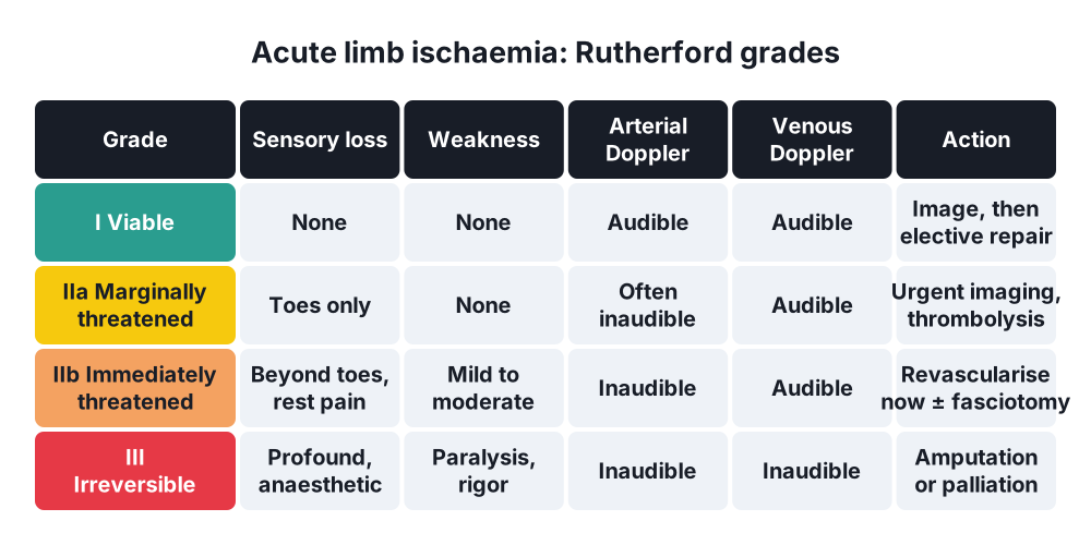
Acute: Rutherford
- I Viable → no sensory loss, no weakness; arterial Doppler audible
- IIa Marginally threatened → minimal sensory loss (toes); salvageable if treated promptly
- IIb Immediately threatened → sensory loss beyond toes, rest pain, mild to moderate weakness; needs revascularisation now
- III Irreversible → anaesthetic, paralysed, rigid muscles, fixed mottling; no arterial or venous Doppler signal
6. Clinical picture
Symptoms
- Intermittent claudication → cramping muscle pain on walking, relieved by rest within minutes; same distance each time
- Calf → femoropopliteal disease
- Buttock and thigh → aortoiliac disease; with impotence → Leriche syndrome
- Rest pain → burning pain in the forefoot at night; patient sleeps in a chair or with the leg over the bed
- Ulcers, gangrene of toes
- ALI → the six Ps:
- Pain
- Pallor
- Pulselessness
- Paraesthesia
- Paralysis
- Perishingly cold
Signs
- Absent or weak pulses; bruits over iliac and femoral arteries
- Cold, pale, hairless, shiny skin; thick nails
- Buerger's test → leg pales when raised (angle < 20° severe); rubor on hanging down
- Slow capillary refill
- Ischaemic ulcers → toes, heel, pressure points; punched out, painful
- ALI → compare with the other leg:
- Embolus → sudden onset, a source (AF), normal pulses in the other leg
- Thrombosis → past claudication, slower onset, weak pulses in both legs
- Late ALI → fixed mottling, muscle tenderness, woody rigid muscles
7. Diagnosis
Bedside
- Ankle-brachial pressure index (ABPI) → ankle systolic ÷ brachial systolic:
- 0.9–1.3 → normal
- 0.5–0.9 → claudication
- < 0.5 → CLTI; ankle pressure < 50 mmHg
- > 1.3 → calcified, incompressible vessels (diabetes, kidney disease) → use toe pressures
- Fall in ABPI after exercise → confirms claudication
- Handheld Doppler signals (ALI grading)
Imaging
- Duplex ultrasound → first imaging; site and length of stenosis
- CT angiography → plan for revascularisation; acute cases when the limb allows the delay
- MR angiography → kidney impairment
- Digital subtraction angiography → at the time of endovascular treatment
- ALI source → ECG (AF), echocardiography, CT for aneurysm
Laboratory
- FBC, U&E, glucose and HbA1c, lipids, clotting
- ALI → creatine kinase, potassium, lactate, group and save, troponin
8. Differential diagnosis
- Claudication versus spinal stenosis (neurogenic claudication: relieved by bending forward, not standing still)
- Hip or knee osteoarthritis
- Sciatica, nerve root pain
- Venous claudication (after iliofemoral DVT)
- Popliteal artery entrapment (young athletes)
- ALI versus DVT with phlegmasia, spinal cord compression, low-output shock
9. Treatment
Conservative: chronic
- Best medical therapy for all:
- Stop smoking
- Antiplatelet → clopidogrel or aspirin; low-dose rivaroxaban + aspirin in selected patients
- High-intensity statin
- Control blood pressure and diabetes
- Supervised exercise → 2 h a week for 3 months; first-line for claudication
- Naftidrofuryl if exercise does not help
- Foot care
Surgical: chronic
- Claudication → intervene only for severe lifestyle limits after exercise therapy
- CLTI → revascularise whenever possible
- Endovascular → angioplasty ± stent:
- Aortoiliac → > 90% success
- Short femoropopliteal lesions
- First choice in high-risk patients
- Open surgery:
- Aortobifemoral bypass → aortoiliac disease; 5-year patency > 90%
- Femorofemoral crossover or axillobifemoral → unilateral disease or unfit
- Common femoral endarterectomy with patch
- Femoropopliteal and femorodistal bypass → long occlusions; autologous great saphenous vein is the best conduit (BEST-CLI)
- Amputation → unreconstructable CLTI, non-viable limb, uncontrolled sepsis
Surgical: acute limb ischaemia
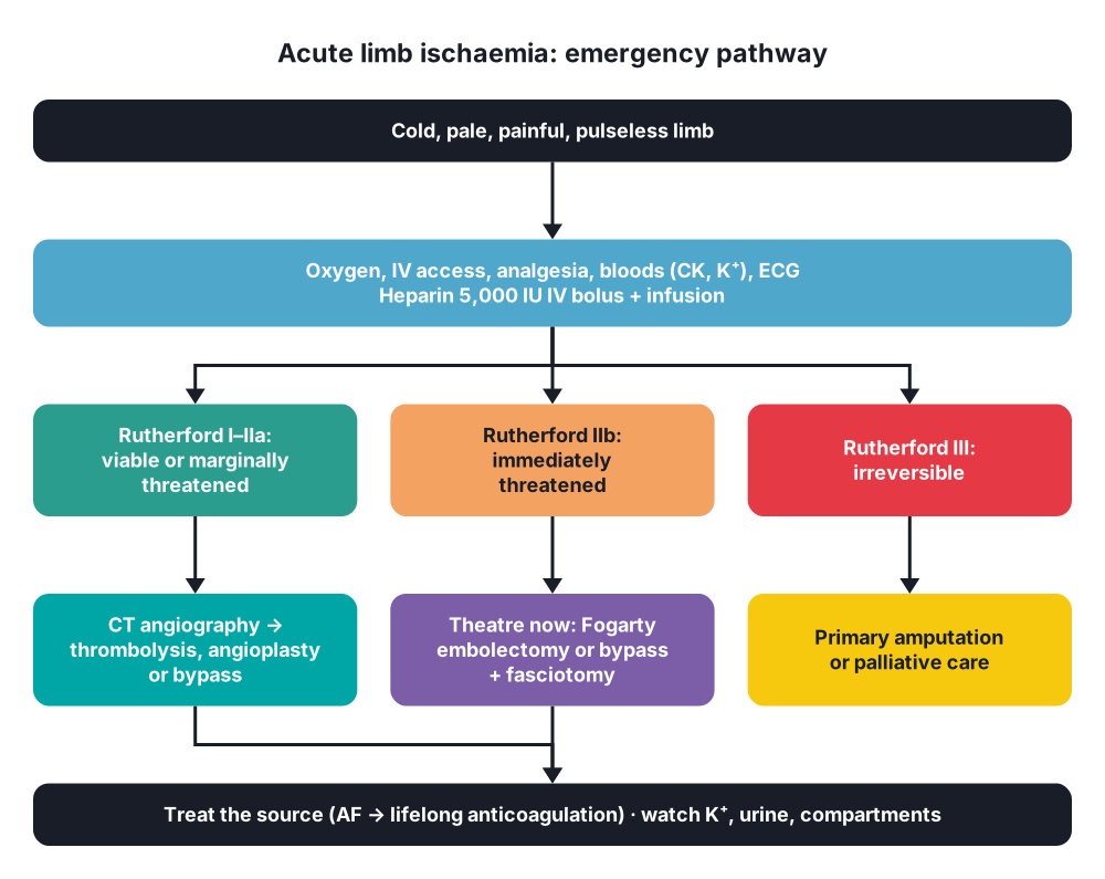
- Oxygen, IV access, fluids, analgesia (opiate)
- Bloods, ECG; call senior help
- Heparin 5,000 IU IV bolus, then infusion (unless contraindicated)
- Grade by Rutherford → decides the timing
- I and IIa → imaging (CT angiography), then:
- Catheter-directed thrombolysis (tPA) → thrombosis, graft occlusion; takes hours; bleeding and stroke risk
- Angioplasty or bypass for the underlying lesion
- IIb → surgery at once:
- Embolectomy with a Fogarty balloon catheter through a groin (common femoral) or brachial arteriotomy
- Success → fresh clot out, good back-bleeding and inflow; on-table angiogram
- Thrombosis on plaque → bypass, endarterectomy or stent
- Fasciotomy of all four calf compartments if ischaemia > 4–6 h or swelling
- III → primary amputation or palliation; revascularisation can kill through reperfusion
- After an embolus → lifelong anticoagulation and treat the source
10. Complications
- Reperfusion syndrome → hyperkalaemia, acidosis, myoglobinuria, acute kidney injury, arrhythmia
- Compartment syndrome → anterior compartment most often
- Re-occlusion, graft thrombosis
- Bleeding → thrombolysis, anticoagulation; stroke
- Limb loss, death (MI)
- Chronic → ulcers, gangrene, infection, amputation
- Graft → infection, false aneurysm, intimal hyperplasia
Show as a list
Limb ischaemia
- Chronic
- Atherosclerosis
- Claudication
- Rest pain
- Fontaine I-IV
- ABPI
- Acute
- Embolus: AF
- Thrombosis: plaque
- Six Ps
- Rutherford I-III
- Treatment chronic
- Stop smoking
- Statin, antiplatelet
- Exercise
- Angioplasty
- Bypass
- Treatment acute
- Heparin
- Thrombolysis
- Fogarty embolectomy
- Fasciotomy
- Amputation


