109. Principles of laparoscopic surgery
High yield
Surgery through small ports with a camera and CO2 pneumoperitoneum; less pain and faster recovery, with its own risks.
Exam Prep · State examSurgery · General surgerySurgery · Operative techniques
1. Definition
- Laparoscopic surgery → operations inside the abdomen through small incisions (ports, usually 5–12 mm), using a camera (laparoscope) and long instruments, with the abdomen distended by gas (pneumoperitoneum)
- Part of minimally invasive surgery (MIS), together with thoracoscopy, endoscopic surgery, robotic surgery and natural orifice surgery (NOTES)
- Diagnostic laparoscopy → to look and take biopsies
- Therapeutic laparoscopy → to operate
- Hand-assisted laparoscopy → one hand inside through a sealed port
- Single-incision laparoscopic surgery (SILS) → all instruments through one umbilical incision
- Robotic-assisted surgery → the surgeon controls articulated instruments from a console (3D view, wrist movement, tremor filtering)
2. Epidemiology
- History:
- Kelling (1901) looked into a dog's abdomen after insufflating air
- Jacobaeus (1910) did the first human laparoscopy
- Veress (1938) designed the spring-loaded needle
- Semm (1980) did the first laparoscopic appendicectomy
- Mühe (1985) and Mouret (1987) did the first laparoscopic cholecystectomies
- Hasson introduced the open entry technique
- Today the standard approach for cholecystectomy, appendicectomy, fundoplication, bariatric surgery, many hernia repairs, colorectal and adrenal surgery, and splenectomy
- Over 90% of cholecystectomies are done laparoscopically
3. Aetiology and risk factors
Indications
- Diagnostic → acute abdomen with unclear cause (especially young women with right iliac fossa pain), staging of cancer (stomach, pancreas, liver, peritoneal metastases), chronic pain, infertility, trauma (diaphragm injury in stable patients)
- Therapeutic → cholecystectomy, appendicectomy, hernia repair (TAPP, TEP), fundoplication and hiatus hernia repair, perforated ulcer repair, adhesiolysis, colectomy and rectal resection, bariatric surgery, splenectomy, adrenalectomy, nephrectomy, gynaecological surgery
Absolute contraindications
- Haemodynamic instability that cannot tolerate pneumoperitoneum (shock, uncontrolled bleeding)
- Patient unfit for general anaesthesia or for a possible conversion to open surgery
- Massive abdominal distension that leaves no working space
Relative contraindications
- Severe cardiopulmonary disease (heart failure, severe COPD), raised intracranial pressure
- Many previous abdominal operations (adhesions)
- Uncorrected coagulopathy
- Late pregnancy (safe in the second trimester), morbid obesity (technically harder, but laparoscopy is still preferred), portal hypertension, large hernias, generalised peritonitis with gross distension
4. Pathogenesis
Physiology of CO2 pneumoperitoneum
- CO2 is used because it is non-flammable, cheap, very soluble in blood (low risk of fatal gas embolism) and quickly breathed out
- Working pressure → 10–15 mmHg in adults (lower in children); over 20–25 mmHg is harmful
- Cardiovascular:
- Raised IAP compresses the IVC → less venous return and lower preload
- Raised systemic vascular resistance (mechanical, plus catecholamines and vasopressin)
- Cardiac output may fall by 10–30%; usually well tolerated in healthy patients
- Bradycardia and asystole from vagal stretch of the peritoneum during insufflation
- Head-down (Trendelenburg) raises venous return; head-up (reverse Trendelenburg) lowers it further
- Respiratory:
- Diaphragm pushed up → lower lung compliance, higher airway pressure, atelectasis
- CO2 absorption → hypercapnia and respiratory acidosis → the anaesthetist raises minute ventilation
- Renal → less renal blood flow and urine output during the operation (pressure on renal veins, ADH release)
- Splanchnic → reduced liver and gut blood flow at high pressure
- Neurological → raised intracranial pressure (avoid in head injury)
- Metabolic and immune → smaller stress response, less inflammation and better preserved immunity than open surgery
- Thermal → cold dry gas causes heat loss; warmed humidified CO2 helps
- Pain after surgery → shoulder tip pain from diaphragmatic irritation by CO2 (reduced by evacuating the gas fully and low-pressure surgery)
5. Classification
Methods of abdominal access
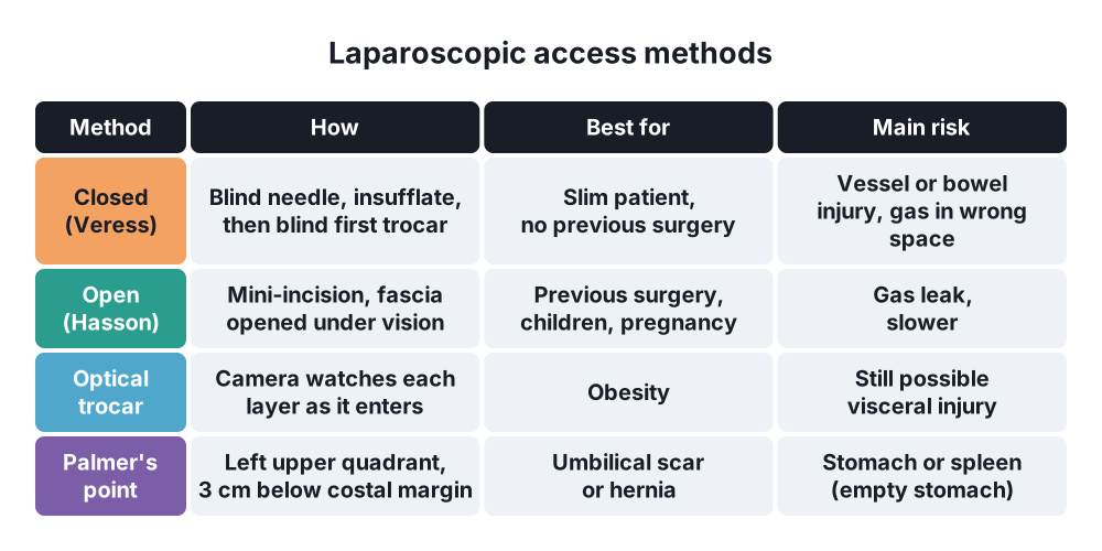
- Closed (Veress needle) → blind insertion at the umbilicus or Palmer's point (left upper quadrant, 3 cm below the costal margin in the midclavicular line), then a blind first trocar
- Confirm position → hanging drop or saline test, low opening pressure (under 8–10 mmHg), free flow
- Open (Hasson) technique → mini-laparotomy at the umbilicus, fascia opened under vision, blunt trocar fixed with stay sutures; recommended by UK and many other guidelines, especially after previous surgery and in children
- Optical (visual) entry trocar → the camera watches each layer as the trocar goes in; useful in obesity
- Direct trocar entry without insufflation → used by some surgeons
Gasless laparoscopy
- Abdominal wall lift devices → for patients who cannot tolerate CO2; poorer view
Generations of minimally invasive surgery
- Conventional multiport laparoscopy, single-incision, robotic-assisted, NOTES, endoluminal
6. Clinical picture
Advantages over open surgery
- Less pain and lower analgesic needs
- Shorter hospital stay and faster return to work and normal activity
- Fewer wound infections and fewer incisional hernias
- Less ileus, earlier eating
- Fewer adhesions, smaller scars, better cosmesis
- Better view of the whole abdomen and the pelvis (magnification)
- Fewer chest complications (smaller incision)
Disadvantages
- Loss of tactile feedback (no hand in the abdomen)
- Two-dimensional view (3D and robotic systems help), limited field of view and movement (fulcrum effect, counter-intuitive movements)
- Longer learning curve and often longer operating time
- Higher equipment costs
- Harder to control major bleeding
- Unique complications → access injuries, gas embolism, thermal injury outside the view, port-site hernias
7. Diagnosis
Before surgery (patient selection)
- History of previous operations and scars, hernias, ascites, coagulopathy
- Cardiopulmonary assessment → ability to tolerate pneumoperitoneum and head-up or head-down positions
- Consent includes the possibility of conversion to open surgery and access-related injuries
Equipment and set-up
- Imaging chain → laparoscope (0° or 30°, 5 or 10 mm), camera, cold light source (LED or xenon), fibre-optic cable, HD or 4K monitor, recording
- White balance, focus and warming (anti-fog) before starting
- Insufflator → set pressure 12–15 mmHg, flow rate; high-flow insufflator for smoke evacuation
- Trocars (ports) → 5, 10 and 12 mm; reusable or disposable; bladed or blunt
- Instruments → graspers, dissectors (Maryland), scissors, hook, needle holders, clip applicators, staplers, suction-irrigation, retrieval bags
- Energy devices:
- Monopolar diathermy → cheap; risk of stray current
- Bipolar diathermy
- Advanced bipolar (LigaSure) and ultrasonic (Harmonic) devices → seal vessels up to 7 mm
- Theatre layout → monitor in line with the surgeon's view, opposite the target organ
Principles of port placement
- Triangulation → camera between two working ports, with the target in front
- Baseball diamond layout; ports about 8–10 cm (a hand's breadth) apart and about 15 cm from the target
- Ideal angle between instruments 60–90°
- Camera port usually at the umbilicus
- All secondary ports inserted under direct vision, avoiding the inferior epigastric vessels (transillumination, view from inside)
- Positioning and gravity → Trendelenburg for pelvic organs, reverse Trendelenburg for upper GI and biliary surgery, side tilt
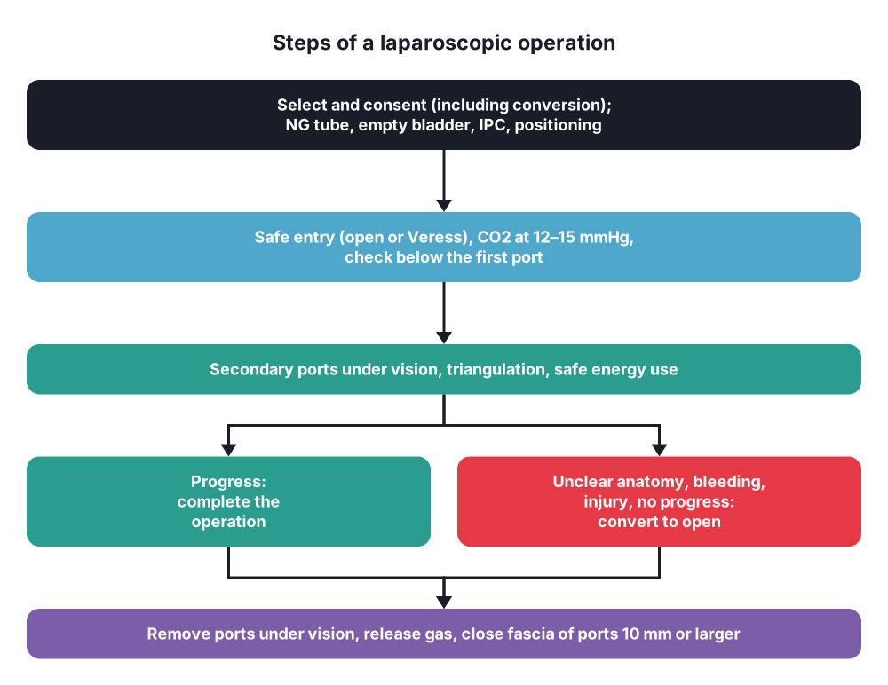
- NG tube to empty the stomach, urinary catheter or voiding to empty the bladder
8. Differential diagnosis
- Choosing the approach is the real decision:
- Laparoscopic → most elective and many emergency operations in stable patients
- Open → unstable patients, massive distension, hostile abdomen, some trauma, failed progress
- Robotic → narrow spaces (pelvis, rectal cancer, prostate), complex reconstruction
- Endoscopic or radiological → drainage, stenting, haemostasis
- Conversion to open surgery is a judgement, not a failure:
- Unclear anatomy (for example, the critical view of safety cannot be achieved in cholecystectomy)
- Bleeding that cannot be controlled
- Injury to bowel, bile duct or vessels that cannot be repaired laparoscopically
- Dense adhesions, no progress, patient intolerance of pneumoperitoneum
- Conversion rates → about 5% in elective cholecystectomy, higher in acute cholecystitis
9. Treatment
Conservative
- Anaesthesia → general anaesthesia with intubation and muscle relaxation; capnography for CO2; monitoring of airway pressure
- ERAS principles → short fasting, carbohydrate drinks, multimodal analgesia, early feeding and mobilisation
- Thromboprophylaxis → IPC and LMWH as for open surgery (pneumoperitoneum and head-up position slow venous return)
- Low-pressure pneumoperitoneum (8–10 mmHg) with deep neuromuscular block → less shoulder tip pain
- Local anaesthetic at port sites and in the peritoneum
Surgical
- Basic steps:
- Positioning, safety checks (WHO checklist), equipment check
- Safe entry (open or closed), insufflation, inspection of the area below the first port for injury
- Secondary ports under vision
- Operation: exposure, dissection, haemostasis, specimen removal in a bag (prevents port-site infection and tumour seeding)
- Check haemostasis at low pressure, irrigate if needed
- Remove ports under vision, release all gas
- Close fascia of ports 10 mm or larger (port-site hernia), close skin
- Safe use of energy:
- Keep the active tip in view; use the lowest power needed
- Avoid direct coupling (touching another metal instrument) and capacitive coupling (avoid hybrid metal-plastic trocars)
- Check insulation of instruments
- Common laparoscopic operations:
- Cholecystectomy → critical view of safety before clipping
- Appendicectomy
- Hernia repair (TAPP, TEP)
- Fundoplication (Nissen, Toupet), Heller myotomy
- Colectomy, rectal resection (TME)
- Sleeve gastrectomy and gastric bypass
- Perforated peptic ulcer repair, splenectomy, adrenalectomy
- Training → box trainers, virtual reality simulators, animal and cadaver models; FLS certification in the USA; supervised learning curve
10. Complications
Access-related (about half of major complications)
- Vascular injury → aorta, iliac vessels, IVC (rare, about 0.05%, but high mortality); inferior epigastric vessels from secondary ports
- Bowel injury (0.1–0.4%) → often missed at the time → peritonitis days later
- Bladder injury, stomach injury
- Preperitoneal insufflation, omental emphysema
Related to pneumoperitoneum
- Hypercapnia, acidosis, arrhythmias, bradycardia
- Gas (CO2) embolism → sudden fall in end-tidal CO2, hypotension, "mill-wheel" murmur → stop insufflation, left lateral head-down position, hyperventilate with 100% oxygen, aspirate from a central line
- Subcutaneous emphysema, pneumothorax, pneumomediastinum
- Raised intracranial pressure, reduced urine output
Related to energy
- Thermal injury to bowel or bile duct outside the field of view (direct and capacitive coupling, insulation failure)
After surgery
- Shoulder tip pain (30–50%)
- Port-site hernia (about 1%, mostly 10–12 mm umbilical ports) → Richter's hernia can strangulate
- Port-site infection, bleeding
- Port-site metastases (rare, under 1%)
- Bile duct injury in cholecystectomy (0.3–0.5%, higher than open in the early era)
- VTE, shoulder or nerve injuries from positioning
Prognosis
- In experienced hands, laparoscopic operations are at least as safe as open ones, with faster recovery
Show as a list
Laparoscopic surgery
- Pneumoperitoneum
- CO2 at 12-15 mmHg
- Less venous return
- Hypercapnia
- Shoulder tip pain
- Access
- Veress (closed)
- Hasson (open)
- Optical trocar
- Palmer's point
- Set-up
- Triangulation
- Ports under vision
- Gravity and tilt
- Advantages
- Less pain
- Short stay
- Fewer wound problems
- Contraindications
- Shock
- Unfit for GA
- Severe cardiopulmonary disease
- Complications
- Vascular, bowel injury
- Gas embolism
- Thermal injury
- Port-site hernia


