
92. Intussusception
High yield
One bowel segment telescopes into the next; infants need air enema reduction, adults need resection for a lead point.
Exam Prep · State examSurgery · General surgerySurgery · Emergency and trauma
1. Definition
- Intussusception → telescoping (invagination) of a proximal segment of bowel (the intussusceptum) into the lumen of the next distal segment (the intussuscipiens)
- The intussusceptum drags its mesentery with it → venous then arterial compromise → a mixed obstruction: obstructive and strangulating at the same time
- Lead point → a lesion at the apex that the bowel pushes forward like a bolus (polyp, tumour, Meckel's diverticulum, enlarged Peyer's patch)
- Parts → apex (leading tip), neck (where the outer tube folds), three layers: entering (inner), returning (middle) and outer tube (sheath)
- Idiopathic (primary) → no structural lead point (most children)
- Secondary → a pathological lead point (most adults)
2. Epidemiology
Children
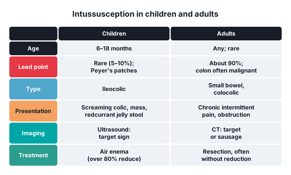
- Most common cause of intestinal obstruction between 3 months and 3 years (after the neonatal period)
- Peak 6–18 months (about 60% in the first year)
- Boys more than girls (about 2–3 : 1)
- Incidence about 1–4 per 1000 live births
- More in spring and winter (viral infections); a small rise after rotavirus vaccine
- About 90–95% idiopathic; lead point in 5–10% (more likely under 3 months or over 5 years)
Adults
- Rare → about 5% of all intussusceptions, 1–5% of bowel obstruction in adults
- Lead point in about 90%
- Small bowel → mostly benign lead points (lipoma, polyps, Meckel's, adhesions) but metastases (melanoma) too
- Colon → about 50–65% malignant (adenocarcinoma, lymphoma)
- Transient small bowel intussusception → often seen on CT, short, no lead point, resolves alone (coeliac disease, Crohn's)
3. Aetiology and risk factors
Children (idiopathic)
- Hypertrophy of Peyer's patches in the terminal ileum after a viral infection (adenovirus, rotavirus, URTI, gastroenteritis) → acts as a lead point
- Weaning, changes in diet (debated)
Pathological lead points in children
- Meckel's diverticulum (most common), polyps (juvenile, Peutz-Jeghers), duplication cyst
- Henoch-Schönlein purpura (submucosal haematoma; often small bowel)
- Lymphoma (Burkitt's, older children), cystic fibrosis (inspissated stool), appendix
- Postoperative intussusception (small bowel, after retroperitoneal surgery)
Adults
- Benign → lipoma (most common benign colonic and small bowel cause), adenoma, hamartoma (Peutz-Jeghers), GIST, Meckel's, inflammatory fibroid polyp, adhesions, feeding tubes, after gastric bypass (jejunojejunal)
- Malignant → colorectal adenocarcinoma, lymphoma, metastases (melanoma, lung, breast)
- Idiopathic → about 10%
4. Pathogenesis
- A lead point (or uneven peristalsis around a swollen Peyer's patch) is gripped by peristalsis and pushed distally
- The proximal bowel telescopes into the distal bowel, pulling its mesentery
- Obstruction → colicky pain, vomiting, distension
- Compression of the mesentery between the layers → venous obstruction first → oedema, mucosal congestion → mucus and blood mix → "redcurrant jelly" stool
- Arterial obstruction → ischaemia of the intussusceptum → gangrene → perforation, peritonitis, sepsis
- In infants the process can progress to gangrene within 24–48 hours
- In adults the onset is slower and often chronic or intermittent (partial obstruction)
- Ileocolic is the most common type: the ileum passes through the ileocaecal valve into the colon; it may travel as far as the rectum or prolapse through the anus
5. Classification
By site
- Ileocolic → most common (about 80–90% in children); the ileum enters the colon
- Ileo-ileocolic → ileo-ileal that then enters the colon
- Ileo-ileal and jejuno-jejunal (small bowel) → HSP, Peutz-Jeghers, postoperative, adults
- Caecocolic, colocolic → adults (often malignant)
- Retrograde (rare) → distal bowel into proximal; after gastrojejunostomy (jejunogastric)
By cause
- Idiopathic (primary) → children
- Secondary → lead point (benign or malignant)
By course
- Acute → infants; sudden onset
- Chronic or recurrent → older children and adults; weeks of intermittent pain
- Transient → short small bowel segments on CT, no lead point
6. Clinical picture
Infant (classic)
- Previously well, often after a viral illness
- Sudden screaming episodes of colicky pain every 15–20 minutes, drawing the legs up, pallor during attacks; quiet and lethargic between attacks
- Vomiting → first food or milk, later bilious
- "Redcurrant jelly" stool (blood and mucus) → a late sign (about 50–60%)
- Lethargy may be the main sign (can mimic encephalitis or sepsis)
- Classic triad (pain, palpable mass, redcurrant jelly stool) in under a quarter of children
Signs
- Sausage-shaped mass, often in the right upper quadrant or epigastrium, curved, firm; changes shape during spasms
- Dance's sign → empty right iliac fossa (the caecum has moved up)
- Rectal examination → blood and mucus on the glove; rarely the apex is felt or prolapses through the anus (must not be confused with rectal prolapse)
- Dehydration, tachycardia; shock and peritonitis late (gangrene, perforation)
Adults
- Chronic, intermittent colicky pain over weeks, nausea, vomiting, change in bowel habit
- Bleeding per rectum, weight loss (tumour)
- Palpable mass in some; often found on CT for vague pain
- Acute complete obstruction in a minority
7. Diagnosis
Children
- Ultrasound → investigation of choice (sensitivity and specificity about 98–100%):
- Target (doughnut) sign on transverse view → rings of bowel wall
- Pseudokidney sign on longitudinal view
- Shows a lead point, free fluid, trapped fluid between layers, and loss of colour Doppler flow (ischaemia, lower chance of reduction)
- Plain abdominal X-ray → not needed for diagnosis; may show a soft tissue mass, empty right iliac fossa, crescent sign (gas around the apex), SBO, or free gas (perforation)
- Bloods → FBC, U&E, CRP, blood gas, group and save; IV access before reduction
- Contrast or air enema → diagnostic and therapeutic (coiled spring sign with contrast; claw sign)
Adults
- CT abdomen and pelvis with IV contrast → investigation of choice:
- Target sign, "sausage" mass with fat and vessels of the mesentery inside, lead point, length, ischaemia
- Short (under 3.5 cm) small bowel intussusception without a lead point or obstruction → usually transient
- Colonoscopy → colonic lesions (biopsy), but not in acute obstruction
- Capsule endoscopy or enteroscopy → small bowel polyps (Peutz-Jeghers)
8. Differential diagnosis
- Children:
- Gastroenteritis (diarrhoea, less pain, no mass)
- Infantile colic, constipation
- Malrotation with volvulus (bilious vomiting in a neonate)
- Incarcerated inguinal hernia (examine the groins)
- Meckel's diverticulum (painless bleeding)
- HSP (rash, joints; may cause intussusception)
- Rectal prolapse (vs prolapsed intussusception: the finger can pass between the mass and the anal wall in intussusception)
- Sepsis, meningitis (lethargy)
- Appendicitis, testicular torsion
- Adults → colorectal cancer, adhesive SBO, Crohn's disease, IBS, mesenteric ischaemia
9. Treatment
Conservative
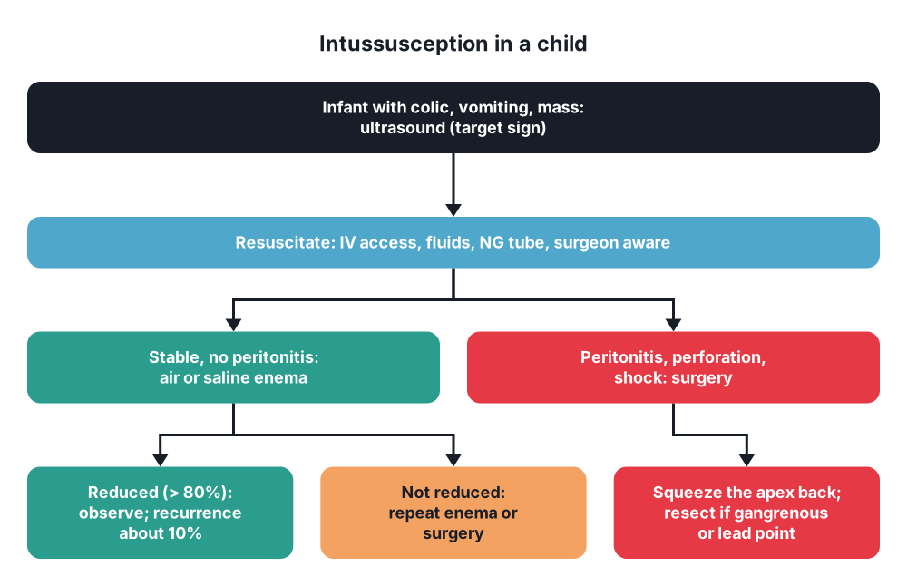
- Resuscitation → IV access, fluids (often 20 mL/kg boluses), NG tube if vomiting, analgesia; antibiotics in some protocols; surgeon informed before reduction
- Non-operative reduction in children → treatment of choice if no contraindication:
- Pneumatic (air) enema under fluoroscopy (preferred: cleaner, faster, less harm if perforation) or hydrostatic (saline or contrast) enema under ultrasound or fluoroscopy
- Air pressure up to about 120 mmHg; rule of threes (classic hydrostatic method): bag at 3 feet (about 1 m) above the table, up to 3 attempts, 3 minutes each
- Success → over 80% (higher with early presentation)
- Proof of reduction → free reflux of air or contrast into the terminal ileum, mass disappears, symptoms settle
- Delayed repeat attempt after 30 minutes to a few hours if partial reduction and the child is stable
- Perforation risk about 0.5–1%; a surgeon and needle decompression must be ready (tension pneumoperitoneum)
- Contraindications to enema reduction:
- Peritonitis
- Perforation (free gas)
- Shock not responding to resuscitation
- Known pathological lead point (relative)
- Recurrence after enema → about 10% (usually within 24–72 hours); a repeat enema is usually tried; surgery after several recurrences or if a lead point is suspected
- Observe for about 12–24 hours after reduction (some centres discharge early); fever after reduction is common
- Transient small bowel intussusception in adults (no lead point, no obstruction) → observation and repeat imaging
Surgical
- Indications (children) → failed enema reduction, peritonitis, perforation, shock, pathological lead point, multiple recurrences, small bowel intussusception not reducible by enema
- Approach → laparoscopy (selected) or right transverse supraumbilical incision
- Manual reduction (Hutchinson's manoeuvre) → squeeze the apex back from the distal end (like squeezing toothpaste), never pull on the proximal bowel
- After reduction → assess viability (warm packs); appendicectomy is often done
- Resection with primary anastomosis if irreducible, gangrenous, perforated, or a lead point is found
- Adults:
- Resection is the rule (lead point in about 90%, cancer risk in the colon)
- Colonic or ileocolic intussusception → oncological resection without reduction (right hemicolectomy, segmental colectomy with mesentery): reduction risks tumour spread, perforation and seeding through ischaemic tissue
- Small bowel intussusception with a probable benign cause → careful reduction may be tried to save bowel, then limited resection of the lead point
- Laparoscopic resection where feasible
- Peutz-Jeghers → remove polyps by enteroscopy or intraoperative enteroscopy to prevent recurrence (bowel-sparing)
10. Complications
Of the disease
- Bowel ischaemia, gangrene, perforation, peritonitis
- Sepsis, hypovolaemic shock, death (rare in children with early treatment)
- Recurrence → about 10% after enema, 2–5% after surgery
- Adults → complications of the underlying tumour
Of treatment
- Enema perforation (0.5–1%) → tension pneumoperitoneum (needle decompression), peritonitis
- Incomplete reduction
- Surgery → wound infection, anastomotic leak, adhesive obstruction, short bowel after large resection
Prognosis
- Children → excellent with early diagnosis; mortality under 1% in developed countries, higher with delay
- Adults → depends on the cause (malignancy)
Show as a list
Intussusception
- Children
- 6-18 months
- Peyer's patches
- Idiopathic 90%
- Ileocolic
- Clinical
- Screaming colic
- Vomiting
- Sausage mass
- Redcurrant jelly
- Tests
- US: target sign
- Pseudokidney
- AXR if perforation
- Treatment
- Resuscitate
- Air enema >80%
- Surgery if peritonitis
- Squeeze, don't pull
- Adults
- Lead point 90%
- Colon: cancer
- CT diagnosis
- Resect, no reduction


