
89. Intestinal obstruction
High yield
Mechanical or functional block to bowel flow; adhesions, hernias and cancer cause most; resuscitate, CT, operate if strangulated.
Exam Prep · State examSurgery · General surgerySurgery · Emergency and trauma
1. Definition
- Intestinal obstruction (ileus in the Central and Eastern European sense) → failure of the normal passage of bowel contents towards the anus
- Mechanical obstruction → a physical block (in the lumen, in the wall, or outside it)
- Functional (dynamic) obstruction → no physical block; the bowel does not move contents
- Paralytic ileus → bowel paralysed (in UK and US use, "ileus" means only this)
- Spastic ileus → rare; prolonged spasm (lead poisoning, porphyria)
- Pseudo-obstruction → acute colonic (Ogilvie's) or chronic
- Simple obstruction → the blood supply is intact
- Strangulated obstruction → the blood supply is cut off (see question 90)
- Closed-loop obstruction → a segment blocked at both ends (volvulus, hernia, colon cancer with a competent ileocaecal valve)
- Specific types → strangulation and volvulus (question 90), obstructive ileus (question 91), intussusception (question 92)
2. Epidemiology
- About 15% of emergency surgical admissions with abdominal pain
- Small bowel obstruction (SBO) → about 80%; large bowel obstruction (LBO) → about 20%
- Causes of SBO in the West:
- Adhesions → about 60–75% (after appendicectomy, colorectal and gynaecological surgery)
- Hernias → about 10–15% (most common cause worldwide and in patients without previous surgery)
- Tumours → about 10% (mostly metastases or colon cancer; primary small bowel tumours are rare)
- Crohn's disease → about 5%
- Causes of LBO:
- Colorectal cancer → about 60–80% in the West; 15–20% of colon cancers present with obstruction
- Volvulus → about 5–10% in the West, but up to a third or more in Eastern Europe, Africa, the Middle East and India
- Diverticular stricture → about 10%
- Children → intussusception, hernias, malrotation with volvulus, Meckel's band
- Mortality → about 3–5% for simple SBO, up to 25–30% with strangulation or delay
3. Aetiology and risk factors
Mechanical causes by site
- In the lumen (obturation) → gallstone, bezoar, food bolus, foreign body, faecal impaction, meconium, worms (Ascaris)
- In the wall → tumours (colorectal, small bowel), strictures (Crohn's, diverticular, ischaemic, radiation, TB), congenital (atresia, Hirschsprung's), intussusception
- Outside the wall → adhesions and bands, hernias (external and internal), volvulus, extrinsic tumour or abscess, carcinomatosis, endometriosis
Functional causes
- Paralytic ileus → after abdominal surgery (normal for 24–72 hours), peritonitis, retroperitoneal haematoma (spine fracture), hypokalaemia, hyponatraemia, hypomagnesaemia, uraemia, opioids, anticholinergics, sepsis, pancreatitis
- Pseudo-obstruction (Ogilvie's) → elderly, after hip or spinal surgery, heart failure, Parkinson's disease, electrolyte imbalance
Risk factors
- Previous abdominal or pelvic surgery (open more than laparoscopic), peritonitis, radiotherapy
- Groin and umbilical hernias, especially femoral
- Age, constipation, neurological disease, institutional care (sigmoid volvulus)
4. Pathogenesis
- Above the block → peristalsis increases (colicky pain, loud bowel sounds), later the bowel tires and dilates
- Distension → swallowed air (about two thirds) and gas from bacterial fermentation + fluid
- Fluid loss → vomiting, fluid pooled in the bowel lumen and wall (third space), less absorption, more secretion → hypovolaemia, oliguria, raised urea and haematocrit
- Electrolytes → high obstruction: loss of acid → hypochloraemic, hypokalaemic metabolic alkalosis; low obstruction: smaller changes; late: metabolic acidosis (shock, ischaemia)
- Bacterial overgrowth → counts rise from 10³–10⁵ to 10⁹–10¹⁰ per mL (E. coli, Klebsiella, Enterococcus) → faeculent vomiting, bacterial translocation, sepsis
- Rising wall pressure → venous return falls → oedema → arterial flow falls → ischaemia, necrosis, perforation (fastest in a closed loop)
- Systemic effects → raised intra-abdominal pressure, splinted diaphragm (basal atelectasis, aspiration), less venous return, multiple organ failure
- Large bowel → with a competent ileocaecal valve (about a third) the colon forms a closed loop; the caecum (widest diameter) bursts first (Laplace's law: wall tension = pressure × radius); caecum over 9–12 cm is a danger sign
- Incompetent valve → small bowel decompresses the colon; slower course, faeculent vomiting
5. Classification
By cause
- Mechanical → obturation (lumen), wall, outside the wall
- Functional (dynamic) → paralytic, spastic, pseudo-obstruction
By the Central European school (ileus)
- Dynamic ileus → paralytic, spastic
- Mechanical ileus:
- Obturation (obstructive) ileus → lumen blocked, blood supply intact (tumour, gallstone, bezoar)
- Strangulation ileus → blood supply cut off (volvulus, knotting, incarcerated hernia)
- Mixed → intussusception, adhesive obstruction
By level
- High small bowel (duodenum, jejunum), low small bowel (ileum), large bowel
By degree
- Complete (no flatus, no stool) vs partial (incomplete, subacute)
By onset
- Acute, chronic, acute on chronic (narrowing tumour suddenly blocked)
By blood supply
- Simple, strangulated, closed loop
By time from onset (stages)
- Early (first 12 hours) → colic, reflex vomiting, loud bowel sounds
- Intermediate (12–24 hours) → distension, dehydration, fluid levels
- Late (over 24–36 hours) → peritonitis, sepsis, silent abdomen, shock
6. Clinical picture
The four cardinal symptoms
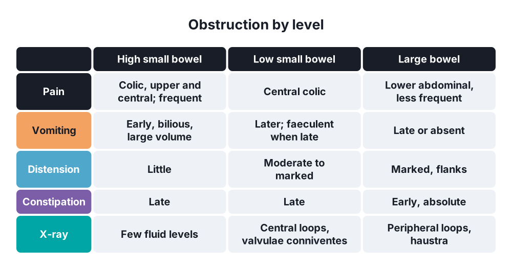
- Colicky abdominal pain → waves every few minutes (every 4–5 minutes in SBO, less often in LBO); central in SBO, lower abdominal in LBO
- Vomiting → early and bilious in high obstruction; late and faeculent in low obstruction
- Distension → slight in high obstruction, marked in low SBO and LBO
- Absolute constipation (no flatus or stool) → early in LBO, late in SBO; stool and flatus below the block can still pass early on, and diarrhoea does not exclude obstruction
Signs
- Dehydration → dry tongue, tachycardia, low BP, low urine output
- Distension, visible peristalsis (thin patients), scars of previous surgery
- Hernial orifices → always examine the groins (small femoral hernias are missed)
- Bowel sounds → loud, high-pitched, tinkling ("obstructive") early; silent late or in paralytic ileus
- Tympanic percussion; succussion splash
- Rectal examination → empty rectum, tumour, faecal impaction, blood (intussusception, ischaemia, cancer)
- Palpable mass → tumour, intussusception, volvulus, abscess
Signs suggesting strangulation (see question 90)
- Constant pain instead of colic, fever, tachycardia, localised tenderness, guarding, rebound
- Raised WCC and lactate, metabolic acidosis
- No combination of these signs reliably excludes strangulation
Paralytic ileus
- No colic, mild discomfort, distension, vomiting, silent abdomen, no flatus; usually after surgery or with peritonitis
7. Diagnosis
Bloods
- FBC (WCC, haematocrit), U&E (potassium, urea, creatinine), glucose, CRP
- Arterial or venous gas → lactate, pH, base excess (ischaemia; a normal lactate does not exclude it)
- Magnesium, calcium, amylase (pancreatitis), group and save, clotting
Imaging
- Plain abdominal X-ray (supine):
- SBO → central dilated loops over 3 cm, valvulae conniventes (cross the whole width: "stack of coins"), no gas in the colon
- LBO → peripheral dilated loops over 6 cm (caecum over 9 cm), haustra (do not cross the whole width)
- Gasless abdomen (fluid-filled loops), stretch sign, string of beads on the erect film
- Clues to the cause → coffee bean (sigmoid volvulus), Rigler's triad (gallstone ileus: pneumobilia, small bowel obstruction, ectopic stone), hernia gas below the inguinal ligament
- Erect X-ray → multiple air–fluid levels (more than 5, longer than 2.5 cm, stepped); erect chest X-ray → free gas (perforation)
- CT abdomen and pelvis with IV contrast → investigation of choice:
- Confirms obstruction, finds the transition point, level and cause
- Small bowel faeces sign (just above the transition point)
- Closed loop → U-shaped or C-shaped loop, two transition points close together, whirl sign (twisted mesentery), beak sign
- Ischaemia → reduced wall enhancement, wall thickening over 3 mm, mesenteric oedema, free fluid, pneumatosis, portal venous gas
- LBO → tumour, volvulus, competent ileocaecal valve, caecal diameter, distant spread
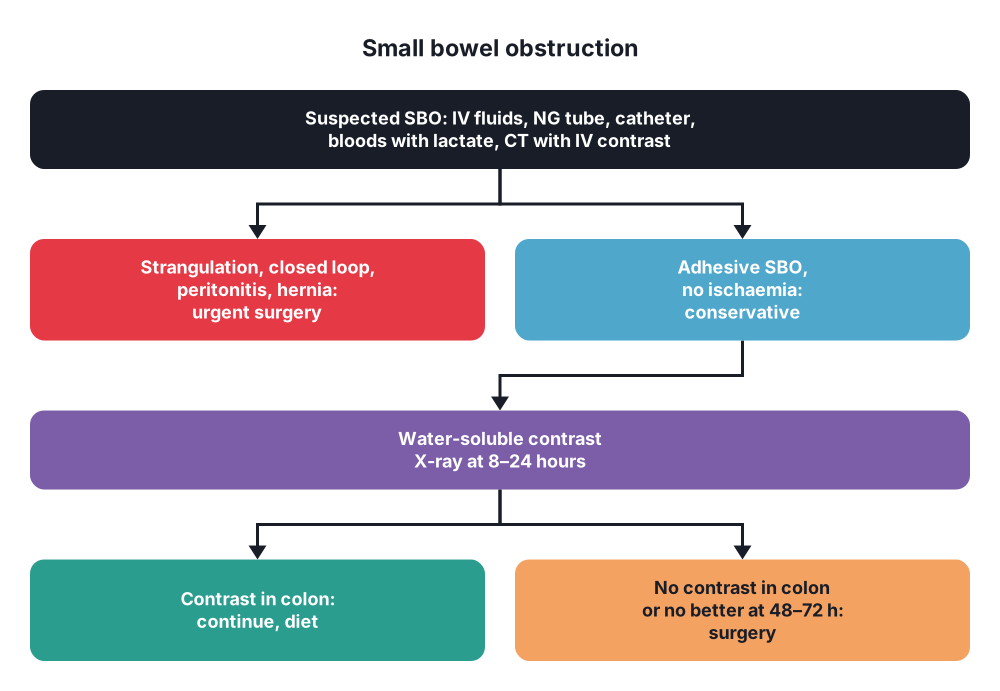
- Water-soluble contrast (Gastrografin) challenge in adhesive SBO:
- 100 mL orally or through the NG tube after decompression
- X-ray at 8–24 hours: contrast in the colon → conservative treatment will probably work (positive predictive value about 99%)
- No contrast in the colon at 24 hours → conservative treatment will probably fail → surgery
- Also therapeutic (draws fluid into the lumen, less wall oedema) → shortens hospital stay and lowers the need for surgery
- Water-soluble contrast enema or CT with rectal contrast → LBO vs pseudo-obstruction
- Ultrasound → children (intussusception), pregnancy, fluid-filled loops, to-and-fro peristalsis
- Flexible sigmoidoscopy or colonoscopy → LBO: biopsy of a tumour, decompression of volvulus or pseudo-obstruction, stent placement
- MRI → pregnancy
8. Differential diagnosis
- Paralytic ileus (no colic, silent abdomen, after surgery or with sepsis)
- Pseudo-obstruction (Ogilvie's) → colonic dilatation without a block on CT or contrast enema
- Mesenteric ischaemia (pain out of proportion to signs, atrial fibrillation)
- Acute pancreatitis (amylase, lipase), biliary colic
- Gastroenteritis (vomiting with diarrhoea; no distension)
- Perforated viscus, peritonitis
- Gastric outlet obstruction (vomiting without bile, succussion splash)
- Renal colic, ruptured abdominal aortic aneurysm
- Inferior myocardial infarction, diabetic ketoacidosis, hypercalcaemia, porphyria
9. Treatment
Conservative
- Initial treatment for all → "drip and suck":
- IV fluids → balanced crystalloid (Hartmann's or Ringer's lactate, or 0.9% saline) to restore volume, then maintenance; add potassium once urine flows
- Nasogastric tube on free drainage with aspiration → empties the stomach, lowers the risk of aspiration, measures losses
- Urinary catheter → hourly urine output (target over 0.5 mL/kg/h)
- Nil by mouth, analgesia (opioids are fine), antiemetics, VTE prophylaxis
- Correct electrolytes; fluid balance chart; serial examination and lactate
- Who can be managed without surgery:
- Adhesive SBO without signs of strangulation → trial of 48–72 hours with a water-soluble contrast challenge; about 70–85% settle (Bologna guidelines)
- Partial obstruction, early postoperative obstruction (most resolve), Crohn's flare (steroids), radiation enteritis
- Paralytic ileus → treat the cause (potassium, magnesium, sepsis, stop opioids), mobilise
- Sigmoid volvulus without ischaemia → endoscopic decompression (see question 90)
- Pseudo-obstruction → correct the cause; neostigmine IV (with ECG monitoring and atropine ready) if the caecum is over 10–12 cm or no response in 24–48 hours; colonoscopic decompression
- Faecal impaction → enemas, manual removal
- Malignant LBO → self-expanding metal stent as a bridge to elective surgery (selected left-sided cancers) or for palliation
- Malignant SBO in end-stage cancer → octreotide, steroids, antiemetics, venting gastrostomy
- Antibiotics → only if surgery is planned or there are signs of ischaemia or sepsis
- Parenteral nutrition if nil by mouth for more than 5–7 days
Surgical
- Indications for urgent surgery:
- Signs of strangulation or peritonitis (constant pain, fever, tachycardia, guarding, raised lactate, CT signs of ischaemia)
- Closed-loop obstruction on CT
- Obstructed or strangulated hernia
- Complete LBO (closed loop risk), caecum over 10–12 cm with tenderness
- No adhesions possible ("virgin abdomen") → often surgery, as a hernia or tumour is likely (CT may allow a trial first)
- Failure of conservative treatment → no contrast in the colon at 24 hours, or no improvement after 48–72 hours
- Preparation → resuscitate, catheter, NG tube, antibiotics, VTE prophylaxis, consent for a stoma, mark a stoma site
- Laparotomy or laparoscopy:
- Laparoscopy in selected patients (mild distension, single band likely, first episode) → shorter stay, fewer complications; convert if needed
- Find the transition point (collapsed bowel below, dilated above)
- Treat the cause:
- Adhesions or band → adhesiolysis (divide only those that cause obstruction; avoid enterotomies)
- Hernia → reduce, assess the bowel, repair the defect
- Tumour → resection with anastomosis, or bypass if not resectable
- Gallstone ileus → enterotomy above the stone and removal (question 91)
- Volvulus → untwist, assess, resect
- Assess viability of every compromised segment (see question 90)
- Resect non-viable bowel → anastomosis, or stoma if the patient is unstable or contaminated
- Decompress dilated bowel (milk contents back to the NG tube, or a suction trocar through an enterotomy) only if needed to close the abdomen
- Inspect the whole bowel from the duodenojejunal flexure to the rectum
- Large bowel obstruction:
- Right-sided cancer → right or extended right hemicolectomy with primary anastomosis
- Left-sided cancer → resection with primary anastomosis ± defunctioning ileostomy (stable patients), Hartmann's procedure (unstable), stent as a bridge, or a defunctioning stoma first
- Caecal perforation or ischaemia → subtotal colectomy with ileostomy or ileorectal anastomosis
- Second-look laparotomy after 24–48 hours if bowel viability was uncertain
- Prevention of adhesions → laparoscopic surgery, gentle handling, avoid glove powder and foreign material, cover the bowel with omentum; anti-adhesion barriers (hyaluronate–carboxymethylcellulose) reduce adhesions but their effect on obstruction is modest
10. Complications
Of the obstruction
- Dehydration, acute kidney injury, electrolyte imbalance
- Strangulation, gangrene, perforation, faecal or purulent peritonitis
- Aspiration pneumonia (vomiting)
- Sepsis, multiple organ failure, death
- Caecal perforation in LBO with a competent valve
Of surgery
- Anastomotic leak, enterocutaneous fistula (inadvertent enterotomy)
- Short bowel syndrome after extensive resection
- Wound infection, dehiscence, incisional hernia
- Recurrent adhesive obstruction (about 20–30% after a first episode; higher after conservative treatment than after surgery)
- Prolonged postoperative ileus, VTE, chest infection
Prognosis
- Simple obstruction → good; strangulation, age, delay over 24 hours and comorbidity raise mortality
Show as a list
Intestinal obstruction
- Types
- Mechanical
- Functional: paralytic
- Simple vs strangulated
- Closed loop
- Causes
- Adhesions: SBO
- Hernias
- Cancer: LBO
- Volvulus
- Clinical
- Colicky pain
- Vomiting
- Distension
- Absolute constipation
- Tests
- Bloods, lactate
- AXR: >3 cm SB, >6 cm colon
- CT: transition point
- Gastrografin challenge
- Treatment
- Drip and suck
- Surgery if strangulated
- Adhesiolysis
- Resection
- Complications
- Perforation
- Sepsis
- Recurrence


