56. Incisional, Spigelian, Petit's and Grynfeltt's hernias
High yield
Hernias through scars, the Spigelian aponeurosis and the two lumbar triangles; most are repaired with mesh.
Exam Prep · State examSurgery · General surgerySurgical Anatomy · Abdominal wall and hernia
1. Definition
- Incisional hernia → hernia through a defect in the scar of a previous abdominal incision (or port site)
- Spigelian hernia → hernia through the Spigelian aponeurosis, along the linea semilunaris, usually at or below the arcuate line
- Lumbar hernia → hernia through the posterolateral abdominal wall:
- Petit's → through the inferior lumbar triangle
- Grynfeltt's (Grynfeltt-Lesshaft) → through the superior lumbar triangle
2. Epidemiology
- Incisional hernia → 10–20% of midline laparotomies (up to 30% at 5 years with risk factors); most appear within the first 2–3 years
- Port-site hernia → about 1%, mostly at umbilical ports 10 mm or more
- Second commonest abdominal wall hernia after inguinal
- Spigelian → rare (1–2% of hernias); age 40–70; slightly more in women
- Lumbar → very rare (about 300 cases each in the old literature); superior more common than inferior; about 20% congenital, most acquired (flank incision, trauma)
3. Aetiology and risk factors
Incisional hernia
- Patient factors:
- Obesity
- Wound infection (strongest single factor; doubles or triples the risk)
- Smoking, diabetes, malnutrition, anaemia, jaundice, uraemia
- Steroids, chemotherapy, immunosuppression
- Chronic cough, constipation, prostatism, ascites
- Collagen disorders, abdominal aortic aneurysm
- Technical factors:
- Midline incision (more than transverse or paramedian)
- Poor closure: wrong suture, large bites, too much tension
- Emergency surgery, re-opening, drains through the wound
- Prevention → small bites (5 mm bites, 5 mm apart), slowly absorbable continuous suture, suture to wound length ratio at least 4 : 1 (STITCH trial); prophylactic mesh in high-risk patients (for example aortic aneurysm, BMI over 27)
Spigelian hernia
- Weak Spigelian aponeurosis (internal oblique and transversus) below the arcuate line, where no posterior rectus sheath
- Obesity, pregnancy, rapid weight loss, COPD, laparoscopic ports
Lumbar hernias
- Anatomy (revision):
- Inferior (Petit's) triangle → iliac crest (below), latissimus dorsi (posterior), external oblique (anterior); floor internal oblique
- Superior (Grynfeltt's) triangle → 12th rib (above), quadratus lumborum and erector spinae (medial), internal oblique (lateral); floor transversalis fascia; roof latissimus dorsi
- Acquired → flank incisions (nephrectomy), iliac crest bone graft, trauma (seat belt), infection
- Congenital → in children, with other defects
4. Pathogenesis
- Incisional:
- Early fascial separation (often in the first weeks, silent)
- Scar stretches, sac of peritoneum forms
- Defect enlarges with time; contents adhere to the sac
- Large hernias → loss of domain (contents live outside the abdomen; cannot return without raising pressure)
- Many defects in one scar → "Swiss cheese" pattern
- Spigelian → interparietal: sac lies under the intact external oblique aponeurosis; narrow neck → high risk of incarceration (about 20%)
- Lumbar → wide neck mostly; incarceration rare (about 10%)
5. Classification
Incisional (European Hernia Society)
- Midline (M) → M1 subxiphoid, M2 epigastric, M3 umbilical, M4 infraumbilical, M5 suprapubic
- Lateral (L) → L1 subcostal, L2 flank, L3 iliac, L4 lumbar
- Width → W1 < 4 cm, W2 4–10 cm, W3 > 10 cm
- Recurrent or not; length
Lumbar
- Superior (Grynfeltt) or inferior (Petit)
- Diffuse (after a flank incision)
- Congenital or acquired (primary or secondary)
6. Clinical picture
Symptoms
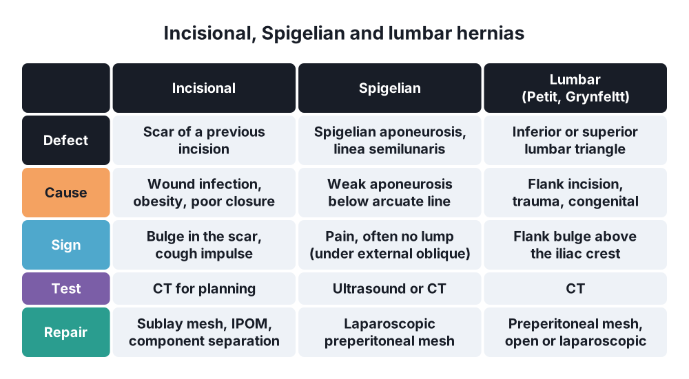
- Incisional → bulge in a scar, larger on standing and coughing; ache, dragging; skin changes in large hernias
- Spigelian → pain at the lateral edge of rectus, below the umbilicus; lump often hard to feel (lies under external oblique)
- Lumbar → flank or back bulge, back ache; may be mistaken for a lipoma
- Complications → painful, irreducible lump, vomiting, distension
Signs
- Incisional → reducible lump in the scar, cough impulse; feel the edges of the defect lying down; look for several defects
- Large hernia → thin skin, ulceration, intertrigo; loss of domain
- Spigelian → tender spot on the linea semilunaris, often no lump; ask the patient to lift the head (tense the wall)
- Lumbar → reducible flank swelling above the iliac crest, cough impulse; examine sitting up
7. Diagnosis
- Clinical in most incisional hernias
- CT → test of choice for planning: size and number of defects, loss of domain (ratio of hernia volume to abdominal volume), muscle quality, contents
- Ultrasound → small or occult hernias, Spigelian (dynamic, with straining)
- Spigelian → ultrasound or CT; laparoscopy confirms
- Lumbar → CT (defect, contents, rule out a mass)
- Preoperative → weight, smoking, diabetes control, nutrition
8. Differential diagnosis
- Divarication of the recti (no defect, no sac)
- Lipoma, desmoid tumour, abscess, haematoma in the scar
- Rectus sheath haematoma (Spigelian area)
- Endometriosis in a caesarean scar
- Lumbar → lipoma, abscess, kidney tumour, flank bulge from nerve injury (denervation) without a defect
9. Treatment
Conservative
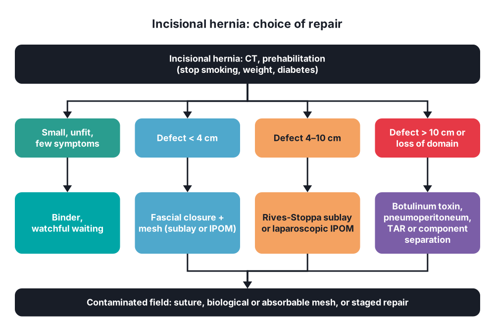
- Small, asymptomatic, wide-necked hernias in unfit patients → watchful waiting; abdominal binder or corset
- Prehabilitation before elective repair → stop smoking (4–6 weeks), weight loss (BMI under 35 ideally), control diabetes (HbA1c), nutrition
- Loss of domain → progressive preoperative pneumoperitoneum or botulinum toxin A into the lateral muscles (2–6 weeks before)
Surgical: incisional hernia
- Principles:
- Reduce the contents, excise or keep the sac
- Close the fascia (restore the linea alba) where possible
- Reinforce with mesh, overlap at least 5 cm beyond the defect
- Defects under 1–2 cm → sutured repair can be enough; above that, mesh (lower recurrence: suture alone recurs in up to 50%)
- Mesh positions:
- Sublay (retromuscular, Rives-Stoppa) → mesh behind rectus, in front of the posterior sheath; preferred open position
- Onlay → on the anterior sheath; more seroma and infection
- Preperitoneal → below the arcuate line
- Intraperitoneal onlay mesh (IPOM) → laparoscopic; coated mesh against bowel; fixed with tacks or sutures
- Inlay (bridging) → only when the fascia cannot be closed; high recurrence
- Laparoscopic IPOM → less wound infection, shorter stay; best for defects up to about 10 cm
- Component separation for large defects:
- Anterior → divide the external oblique lateral to the linea semilunaris (Ramirez); perforator-sparing or endoscopic forms reduce skin necrosis
- Posterior: transversus abdominis release (TAR) → extends the retromuscular space laterally; large sublay mesh
- Robotic eTEP and TAR → newer options
- Contaminated field → sutured repair, biological or absorbable synthetic mesh, or staged repair
- Strangulation → emergency repair; resect dead bowel
Surgical: Spigelian hernia
- All are repaired (narrow neck, incarceration risk)
- Laparoscopic TAPP or TEP with preperitoneal mesh (preferred), or IPOM
- Open → incision over the defect, open external oblique, reduce the sac, close the defect, mesh preperitoneally
Surgical: lumbar hernia
- Small, primary → open repair with sublay or preperitoneal mesh
- Large or diffuse → laparoscopic or robotic transabdominal preperitoneal mesh; fix to the iliac crest and 12th rib with wide overlap
- Watch the subcostal, iliohypogastric and ilioinguinal nerves
10. Complications
- Hernia → incarceration, strangulation, skin ulceration, loss of domain
- Repair:
- Seroma (common, mostly settles), haematoma
- Wound infection, skin necrosis (anterior component separation)
- Mesh infection, mesh erosion, enterocutaneous fistula (IPOM)
- Recurrence → up to 50% after suture alone; about 10–20% with mesh
- Abdominal compartment syndrome after reducing a large hernia
- Bowel injury (adhesiolysis)
- Chronic pain
Show as a list
Incisional and rare hernias
- Incisional
- 10-20% of laparotomies
- Wound infection
- Obesity, smoking
- Loss of domain
- Prevention
- Small bites
- 4:1 ratio
- Prophylactic mesh
- Repair
- Sublay: Rives-Stoppa
- Laparoscopic IPOM
- Component separation
- TAR
- Spigelian
- Linea semilunaris
- Under external oblique
- Incarceration
- Laparoscopic mesh
- Lumbar
- Petit: inferior
- Grynfeltt: superior
- CT
- Mesh repair


