
70. Hydatid disease of the liver
High yield
Echinococcus granulosus cysts in the liver; staged by WHO ultrasound types and treated with albendazole, PAIR or surgery.
Exam Prep · State examSurgery · HPBSurgery · General surgery
1. Definition
- Hydatid disease (echinococcosis) → zoonotic infection by the larval stage of the tapeworm Echinococcus
- Cystic echinococcosis → E. granulosus; slow-growing fluid-filled cysts, most in the liver
- Alveolar echinococcosis → E. multilocularis; infiltrating, tumour-like mass; behaves like a cancer
- Polycystic (neotropical) → E. vogeli, E. oligarthrus (South America; rare)
2. Epidemiology
- Endemic in sheep-farming regions → Mediterranean (including Bulgaria, Greece, Turkey), Middle East, Central Asia, China, North Africa, South America (Argentina, Chile, Peru), Australia
- Bulgaria → one of the highest rates in the European Union
- Over 1 million people infected worldwide
- Liver about 70%, lung about 20%; rest → spleen, kidney, brain, bone, muscle, heart
- Single cyst in about 75%; right lobe more than left
- Alveolar → Central Europe (Alps), China, Russia, Alaska; rarer, but more lethal (untreated mortality over 90% at 10 years)
3. Aetiology and risk factors
Life cycle (E. granulosus)
- Definitive host → dog (and other canids); adult tapeworm (3–6 mm) in the small bowel
- Eggs pass in dog faeces onto grass and soil
- Intermediate host → sheep (cattle, goats, pigs) eats eggs
- Embryo (oncosphere) hatches in the gut, crosses the wall, reaches the liver through the portal vein (first filter) or the lung (second filter)
- Larva forms a hydatid cyst
- Dog eats infected sheep offal → cycle completes
- Humans → accidental intermediate host (dead end); infected by eating eggs from contact with dogs, contaminated food, water, vegetables
- Alveolar (E. multilocularis) → foxes (definitive), rodents (intermediate)
Risk factors
- Rural life, sheep farming, close contact with dogs
- Home slaughter of animals; feeding offal to dogs
- Poor hygiene, untreated water
4. Pathogenesis
- Cyst structure (three layers):
- Pericyst (adventitia) → fibrous layer made by the host; not part of the parasite
- Laminated membrane (ectocyst) → acellular, white, elastic
- Germinal layer (endocyst) → inner living layer; makes brood capsules, protoscolices and daughter cysts
- Hydatid fluid → clear, rock-water; highly antigenic; contains hydatid sand (protoscolices)
- Grows slowly → about 1–5 cm a year; symptoms after years
- Rupture:
- Into the bile ducts (most common; 5–25%) → cholangitis, obstructive jaundice, biliary colic
- Into the peritoneum → anaphylaxis, secondary peritoneal hydatidosis (protoscolices seed)
- Into the chest → bronchobiliary fistula (coughing up bile and membranes), empyema
- Into the bowel, pericardium, vessels (rare)
- Secondary infection → liver abscess
- Each spilled protoscolex can grow into a new cyst → reason for scolicidal agents and careful surgery
- Alveolar → no limiting pericyst; buds infiltrate the liver like a cancer; spreads to lung and brain
5. Classification
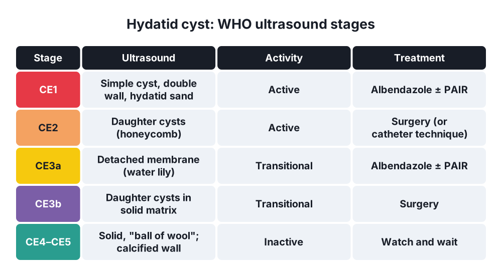
WHO-IWGE ultrasound classification (cystic echinococcosis)
- CL → cystic lesion, no visible wall (uncertain; may not be hydatid)
- CE1 → active; simple cyst with visible double wall; "hydatid sand" (snowflake sign)
- CE2 → active; multivesicular with daughter cysts ("honeycomb", "rosette")
- CE3a → transitional; detached membrane ("water lily sign")
- CE3b → transitional; daughter cysts inside a solid matrix
- CE4 → inactive; solid, heterogeneous ("ball of wool")
- CE5 → inactive; solid with calcified wall
- (Gharbi classification I–V is the older equivalent)
Alveolar echinococcosis
- WHO PNM staging (parasite extension, neighbouring organs, metastases)
6. Clinical picture
Symptoms
- Most are asymptomatic for years; found by chance on ultrasound
- RUQ pain or discomfort, fullness, mass
- Dyspepsia, nausea (pressure)
- Complications:
- Jaundice, fever, colic → rupture into the bile ducts (cholangitis)
- Anaphylaxis → urticaria, wheeze, shock after rupture or trauma
- Fever, rigors → infected cyst
- Cough with bile (biloptysis) → bronchobiliary fistula
- Portal hypertension, Budd-Chiari (large cysts near the veins; rare)
- Alveolar → RUQ pain, jaundice, weight loss, hepatomegaly (like liver cancer)
Signs
- Hepatomegaly or smooth RUQ mass
- "Hydatid thrill" (historical)
- Jaundice, fever
- Urticaria, eosinophilia signs
7. Diagnosis
- Ultrasound → first test; WHO stage; daughter cysts, membranes, calcification; screening in endemic areas
- CT → size, number, site, relation to vessels and ducts, extrahepatic cysts; surgical planning
- MRI and MRCP → cyst-biliary communication, membrane detail
- Serology → ELISA, indirect haemagglutination, immunoblot (Western blot) for confirmation
- Negative in up to 10–40% of liver cysts (higher in lung, calcified or intact cysts); a negative test does not exclude it
- FBC → eosinophilia (in 25–40%; more after leakage)
- LFT → cholestasis with biliary rupture
- Chest X-ray or CT chest → lung cysts (in 20%)
- ERCP → biliary rupture: membranes in the CBD; clears the duct
- Diagnostic aspiration is not done blindly (anaphylaxis, spillage); only as part of PAIR with albendazole cover
- Casoni skin test → obsolete
8. Differential diagnosis
- Simple liver cyst (thin wall, no septa; serology negative)
- Liver abscess (pyogenic or amoebic)
- Biliary cystadenoma or cystadenocarcinoma (mucinous cystic neoplasm)
- Polycystic liver disease
- Haematoma, biloma
- Necrotic tumour, metastases
- Alveolar → hepatocellular carcinoma, cholangiocarcinoma
9. Treatment
- Choice depends on WHO stage, size, site, complications, patient fitness and local expertise (WHO-IWGE expert consensus)
Conservative
- Albendazole (benzimidazole; first-line drug):
- 10–15 mg/kg/day (about 400 mg twice daily) with a fatty meal
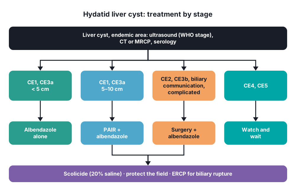
- Alone → small CE1 and CE3a cysts under 5 cm; inoperable or multiple cysts; cure in about 30%, response in 50–70%
- Perioperative cover → start 1 week to 1 month before surgery or PAIR, continue 1–6 months after (lowers recurrence from spillage)
- Side effects → raised liver enzymes, neutropenia, alopecia; monitor LFT and FBC; avoid in pregnancy
- Mebendazole, praziquantel (added before surgery) → second-line
- Watch and wait → inactive CE4 and CE5 (uncomplicated): ultrasound follow-up, no treatment
- PAIR (percutaneous) → CE1 and CE3a, 5–10 cm; unfit for surgery or patient's choice:
- Puncture under ultrasound (through a rim of liver tissue)
- Aspiration of fluid; check for bile (bile-stained fluid → stop; cyst-biliary communication)
- Injection of a scolicide → hypertonic saline 20% or 95% ethanol, left 15–20 minutes
- Re-aspiration
- Under albendazole cover; resuscitation ready for anaphylaxis
- Contraindications → CE2 and CE3b (multivesicular), cysts communicating with bile ducts, superficial cysts, inactive cysts, lung cysts
- Modified catheterisation techniques for larger or multivesicular cysts in expert centres
- Anaphylaxis → adrenaline, steroids, antihistamines, fluids
Surgical
- Indications → large CE2–CE3b cysts, multiple daughter cysts, cyst-biliary communication, superficial cysts at risk of rupture, infected or ruptured cysts, pressure on major structures, failed PAIR
- Protection of the field:
- Pack around the cyst with swabs soaked in a scolicide (hypertonic saline 20%; avoid formalin)
- Aspirate first through a closed system, inject scolicide, wait, then open
- Conservative (tissue-sparing) surgery:
- Evacuate the contents, laminated membrane and daughter cysts (endocystectomy)
- Inspect for bile leaks; suture any biliary openings
- Partial pericystectomy (deroofing; unroofing)
- Deal with the cavity → omentoplasty (fill with omentum), capitonnage (sutured closure), external drainage
- Simpler, but higher recurrence and bile leak
- Radical surgery:
- Total pericystectomy (closed or open) → removes the cyst with its pericyst
- Liver resection (segmentectomy, hepatectomy) → peripheral or large cysts that destroy a segment
- Lower recurrence; more bleeding risk near major vessels
- Laparoscopic → selected peripheral, anterior cysts in expert hands
- Biliary rupture → ERCP with sphincterotomy to clear membranes from the CBD (before or after surgery); persistent biliary fistula → ERCP stent
- Lung and liver cysts together → right thoracotomy for both, or staged (lung first)
- Alveolar echinococcosis → radical liver resection with clear margins (as for cancer) plus albendazole for at least 2 years; liver transplantation in selected unresectable cases; lifelong albendazole if not resectable
Conservative: prevention
- Deworm dogs (praziquantel); do not feed offal to dogs; control slaughter; hand washing; vaccination of sheep (EG95)
10. Complications
- Rupture into the bile ducts → cholangitis, obstructive jaundice, pancreatitis
- Intraperitoneal rupture → anaphylaxis, secondary hydatidosis
- Infection → liver abscess
- Bronchobiliary fistula, pleural empyema
- Portal hypertension, Budd-Chiari syndrome (rare)
- Treatment:
- Recurrence (2–25%; higher after conservative surgery)
- Postoperative bile leak and external biliary fistula (most common surgical complication)
- Residual cavity infection, abscess
- Sclerosing cholangitis → scolicide (formalin, hypertonic saline) passing into the ducts
- Anaphylaxis during PAIR or surgery
- Albendazole → liver toxicity, bone marrow suppression
Show as a list
Hydatid liver
- Parasite
- E. granulosus
- Dog: definitive host
- Sheep: intermediate
- Human: dead end
- Cyst layers
- Pericyst: host
- Laminated membrane
- Germinal layer
- Hydatid sand
- WHO stages
- CE1-2: active
- CE3: transitional
- CE4-5: inactive
- Tests
- Ultrasound
- CT, MRCP
- Serology
- Eosinophilia
- Treatment
- Albendazole
- PAIR: CE1, CE3a
- Surgery: CE2, CE3b
- Watch: CE4-5
- Scolicide: 20% saline
- Complications
- Biliary rupture
- Anaphylaxis
- Recurrence
- Bile fistula


