
63. Gastric cancer
High yield
Adenocarcinoma linked to H. pylori; found late; staged by CT, EUS and laparoscopy; treated with FLOT and D2 gastrectomy.
Exam Prep · State examSurgery · Upper GISurgery · Surgical oncology
1. Definition
- Gastric cancer → malignant tumour of the stomach; over 90% adenocarcinoma
- Other malignant tumours → lymphoma, GIST, neuroendocrine tumours (see question 64)
- Early gastric cancer → limited to the mucosa or submucosa (T1), with or without node spread
- Advanced gastric cancer → invades the muscularis propria or deeper (T2 or more)
2. Epidemiology
- About 5th most common cancer and 4th–5th cause of cancer death worldwide
- Highest in East Asia (Japan, Korea, China), Eastern Europe, South America; falling in the West
- Men : women about 2 : 1; peak 60–80 years
- Distal (antral) cancers falling; proximal (cardia) cancers rising in the West (reflux, obesity)
- 5-year survival → about 20–30% in the West (late diagnosis); over 60% in Japan and Korea (screening, early cancers)
3. Aetiology and risk factors
- Helicobacter pylori → class I carcinogen; most important cause of non-cardia cancer
- Diet → salt, smoked and pickled foods, nitrates (nitrosamines); low fruit and vegetables
- Smoking, alcohol
- Chronic atrophic gastritis, intestinal metaplasia, dysplasia
- Pernicious anaemia (autoimmune gastritis)
- Previous partial gastrectomy (gastric remnant cancer, 15–20 years later)
- Adenomatous polyps, Ménétrier's disease
- Epstein-Barr virus
- Blood group A
- Obesity and reflux → cardia cancer
- Genetic:
- Hereditary diffuse gastric cancer → CDH1 (E-cadherin) mutation; lifetime risk up to 70%; prophylactic total gastrectomy
- Lynch syndrome, familial adenomatous polyposis, Peutz-Jeghers, Li-Fraumeni
4. Pathogenesis
- Correa cascade (intestinal type):
- Normal mucosa
- Chronic active gastritis (H. pylori)
- Atrophic gastritis
- Intestinal metaplasia
- Dysplasia (low then high grade)
- Adenocarcinoma
- Diffuse type → loss of E-cadherin; single cells spread through the wall; no clear precancer stage
- Spread:
- Direct → pancreas, transverse colon, liver, spleen, oesophagus, duodenum
- Lymphatic → perigastric nodes, then along the coeliac axis branches; left supraclavicular (Virchow's node, Troisier's sign)
- Haematogenous → liver (portal vein), lung, bone
- Transcoelomic (peritoneal) → peritoneal seeding, ascites, Krukenberg tumours (ovaries), Sister Mary Joseph nodule, Blumer's shelf (pouch of Douglas)
5. Classification
Histology
- Lauren:
- Intestinal → glands; older men; distal; H. pylori; better prognosis; liver spread
- Diffuse → signet ring cells; younger patients, women; whole stomach (linitis plastica, "leather bottle stomach"); peritoneal spread; worse prognosis
- Mixed
- WHO → tubular, papillary, mucinous, poorly cohesive (including signet ring)
Macroscopic
- Borrmann (advanced):
- I polypoid
- II ulcerated with raised edges
- III ulcerated, infiltrating
- IV diffusely infiltrating (linitis plastica)
- Japanese early gastric cancer → type I protruded, II superficial (a raised, b flat, c depressed), III excavated
Site
- Cardia (Siewert III junctional), body, antrum and pylorus (most common in the past)
- Siewert (junctional tumours) → I distal oesophagus, II true cardia, III subcardial
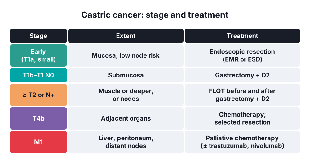
Staging (TNM 8th edition)
- T1 → lamina propria or muscularis mucosae (T1a), submucosa (T1b)
- T2 → muscularis propria
- T3 → subserosa
- T4a → serosa; T4b → adjacent structures
- N → N1 1–2 nodes, N2 3–6, N3 7 or more (at least 16 nodes needed for staging)
- M1 → distant spread, including positive peritoneal cytology
Lymph node stations (Japanese)
- Stations 1–6 → perigastric (N1 tier)
- Stations 7–12 → along the left gastric, common hepatic, coeliac, splenic and proper hepatic arteries
- D1 → perigastric nodes; D2 → D1 plus stations 7–12a
6. Clinical picture
Symptoms
- Early → often none, or vague dyspepsia
- Alarm features (urgent endoscopy):
- Dysphagia (cardia)
- Weight loss
- Persistent vomiting (pyloric obstruction)
- Upper GI bleeding, iron deficiency anaemia
- New dyspepsia over 55 (NICE), over 45 in Oxford
- Epigastric mass
- Early satiety (linitis plastica), anorexia, fatigue
Signs
- Often none
- Epigastric mass, hepatomegaly (metastases), ascites, jaundice
- Virchow's node (left supraclavicular, Troisier's sign)
- Sister Mary Joseph nodule (umbilicus)
- Pallor, cachexia
- Paraneoplastic → acanthosis nigricans, Leser-Trélat sign (sudden seborrhoeic keratoses), Trousseau's sign (migratory thrombophlebitis)
7. Diagnosis
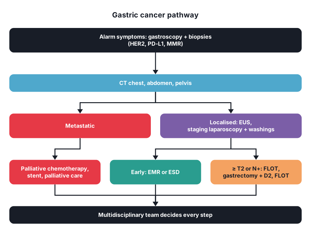
- Gastroscopy with multiple biopsies (at least 6–8) → diagnosis; HER2 status, PD-L1 (CPS), mismatch repair (MSI) status
- Staging:
- CT chest, abdomen and pelvis → metastases, nodes, local invasion
- Endoscopic ultrasound (EUS) → T and N stage; early cancers
- Staging laparoscopy with peritoneal washings → for T3 or more; finds peritoneal disease missed on CT (about 20–30%)
- PET-CT → selected (not reliable in diffuse or mucinous types)
- Bloods → FBC (anaemia), LFT, albumin
- Tumour markers → CEA, CA 19-9, CA 72-4 (follow-up only; not for diagnosis)
- Fitness → cardiopulmonary exercise testing, nutrition
- Multidisciplinary team decision for every patient
8. Differential diagnosis
- Benign gastric ulcer (biopsy every gastric ulcer and repeat endoscopy at 6–8 weeks)
- Gastric lymphoma, GIST, neuroendocrine tumour
- Ménétrier's disease
- Gastritis, functional dyspepsia
- Pancreatic cancer, oesophageal cancer
- Metastases to the stomach (breast, melanoma)
9. Treatment
Conservative
- Prevention → eradicate H. pylori (lowers risk, especially in relatives of patients); endoscopic surveillance of extensive intestinal metaplasia; screening in high-risk countries (Japan, Korea)
- Endoscopic resection of early cancer (low risk of node spread):
- Endoscopic mucosal resection (EMR) or endoscopic submucosal dissection (ESD)
- Criteria → intestinal type, T1a, no ulceration, ≤ 2 cm (extended criteria allow larger in ESD), no lymphovascular invasion
- Perioperative chemotherapy → standard in Europe for ≥ T2 or N+:
- FLOT (docetaxel, oxaliplatin, fluorouracil, leucovorin) → 4 cycles before and 4 after surgery; better survival than ECF (FLOT4 trial)
- Older regimen → ECF / ECX (MAGIC trial)
- Adjuvant chemotherapy (Asia) after D2 resection; chemoradiotherapy in selected cases
- Immunotherapy (durvalumab with FLOT) now added in some units (MATTERHORN)
- Palliative (metastatic):
- Chemotherapy → platinum plus fluoropyrimidine
- Add trastuzumab if HER2-positive (about 15–20%)
- Add nivolumab or pembrolizumab (PD-L1 positive)
- Zolbetuximab (claudin 18.2 positive)
- Bleeding → radiotherapy, endoscopic therapy, embolisation
- Obstruction → endoscopic stent, gastrojejunostomy
- Nutrition, analgesia, palliative care
Surgical
- Aim → R0 resection (clear margins: ≥ 3 cm for intestinal, ≥ 5 cm for diffuse type) with D2 lymphadenectomy
- Distal (subtotal) gastrectomy → antral and distal body tumours:
- Remove distal two-thirds to four-fifths of the stomach with greater and lesser omentum
- Divide the right gastric, right gastroepiploic and left gastric arteries at their origins
- D2 lymph node dissection
- Reconstruction → Roux-en-Y gastrojejunostomy (less bile reflux), or Billroth I or II
- Total gastrectomy → proximal and body tumours, linitis plastica, diffuse type near the margin, hereditary diffuse cancer:
- Remove the whole stomach and omentum
- D2 lymphadenectomy; spleen kept (splenectomy only for direct invasion)
- Roux-en-Y oesophagojejunostomy
- Junctional tumours → Siewert I and II as oesophageal cancer (oesophagectomy); Siewert III → extended total gastrectomy
- D2 vs D1 → D2 lowers local recurrence and cancer death at 15 years (Dutch D1D2 trial) if done without splenectomy and pancreatectomy
- Laparoscopic and robotic gastrectomy → equal cancer results to open in early and many advanced cancers (KLASS, CLASS trials)
- Not resectable → peritoneal metastases (except selected trials: HIPEC), liver metastases (selected only), fixed T4b
- Palliative gastrectomy → only for bleeding or obstruction
10. Complications
Of the disease
- Bleeding, gastric outlet obstruction, perforation, malignant ascites, jaundice
Of surgery
- Early:
- Anastomotic leak (oesophagojejunal), duodenal stump blowout
- Bleeding, pancreatic fistula, abscess
- Pneumonia
- Late (postgastrectomy syndromes):
- Dumping syndrome (early and late)
- Afferent loop syndrome (Billroth II), bile reflux gastritis
- Vitamin B12 deficiency (lifelong injections after total gastrectomy), iron and folate deficiency
- Weight loss, osteoporosis, small stomach syndrome
- Postoperative mortality → about 2–5%
Prognosis
- Depends on stage: stage I about 70–90% 5-year survival; stage IV under 5%
- Early gastric cancer after resection → over 90%
Show as a list
Gastric cancer
- Causes
- H. pylori
- Salt, nitrates
- Atrophic gastritis
- CDH1
- Types
- Intestinal: older, distal
- Diffuse: signet ring
- Linitis plastica
- Spread
- Lymph: Virchow
- Liver
- Peritoneum: Krukenberg
- Staging
- Gastroscopy, biopsy
- CT
- EUS
- Laparoscopy, washings
- Treatment
- EMR, ESD: early
- FLOT perioperative
- Distal gastrectomy
- Total gastrectomy
- D2 nodes
- Trastuzumab if HER2
- Complications
- Leak
- Dumping
- B12 deficiency


