65. Gallstone disease
High yield
Cholesterol, pigment and mixed stones; most are silent; biliary colic and its complications are treated by laparoscopic cholecystectomy.
Exam Prep · State examSurgery · HPBSurgical Anatomy · Hepatobiliary
1. Definition
- Cholelithiasis → stones in the gallbladder
- Choledocholithiasis → stones in the common bile duct (CBD)
- Biliary colic → pain from a stone blocking the gallbladder outlet for a short time, with no inflammation
- Symptomatic gallstone disease → colic or a complication: cholecystitis, CBD stones, cholangitis, pancreatitis, gallstone ileus
2. Epidemiology
- 10–15% of adults in the West; more with age (over 30% over 70)
- Women : men about 2–3 : 1
- Highest in Pima Indians and Chile; low in Africa and Asia
- Most (about 80%) never cause symptoms; risk of symptoms about 1–2% a year
- About 10–15% with gallbladder stones also have CBD stones
- Laparoscopic cholecystectomy → one of the commonest operations in general surgery
3. Aetiology and risk factors
Anatomy (revision)
- Gallbladder → fundus, body, neck, Hartmann's pouch; holds 30–50 mL
- Cystic duct → spiral valves of Heister; joins the common hepatic duct to form the CBD
- Calot's triangle (hepatocystic triangle) → cystic duct, common hepatic duct, inferior surface of the liver; contains the cystic artery (from the right hepatic artery) and Calot's lymph node
- Anatomical variants common → right hepatic artery in the triangle, short cystic duct, aberrant right hepatic ducts (bile leak)
- Bile → bile salts, phospholipid (lecithin), cholesterol; gallbladder concentrates bile 5–10 times
Risk factors ("fat, female, fertile, forty, fair" is a rough guide)
- Cholesterol stones:
- Female sex, oestrogen (pregnancy, oral contraceptive, HRT)
- Age over 40
- Obesity, metabolic syndrome, diabetes
- Rapid weight loss (bariatric surgery, very low calorie diets)
- Family history, ethnic group
- Drugs → octreotide, clofibrate, ceftriaxone
- Ileal resection or Crohn's disease (loss of bile salts)
- Long-term TPN, fasting (stasis)
- Black pigment stones → haemolysis (sickle cell, hereditary spherocytosis, thalassaemia), cirrhosis
- Brown pigment stones → bile duct infection and stasis (E. coli, parasites: Clonorchis, Ascaris); form in the ducts; East Asia
4. Pathogenesis
- Cholesterol stones → three factors:
- Supersaturation of bile with cholesterol (too little bile salt and lecithin)
- Nucleation → crystals form (mucin, proteins)
- Gallbladder stasis (poor emptying)
- Biliary sludge → first step; may pass or form stones
- Black pigment → unconjugated bilirubin and calcium bilirubinate; sterile; gallbladder
- Brown pigment → bacterial enzymes (beta-glucuronidase) deconjugate bilirubin; soft, earthy; in the ducts; primary duct stones
- Natural history:
- Stone stuck in Hartmann's pouch or cystic duct → colic; stays stuck → cholecystitis
- Stone passes into the CBD → obstructive jaundice, cholangitis, pancreatitis
- Erodes into the bowel → gallstone ileus
- Long-standing → gallbladder cancer (stones over 3 cm, porcelain gallbladder)
5. Classification
By composition
- Cholesterol (about 80% in the West) → pale yellow, large, often single or few; radiolucent
- Mixed → most common; cholesterol with pigment and calcium
- Black pigment → small, hard, many; 50% radio-opaque
- Brown pigment → soft; bile ducts
- Only about 10–15% of gallstones are radio-opaque on plain X-ray
By clinical state
- Asymptomatic (silent)
- Biliary colic
- Acute cholecystitis (calculous); chronic cholecystitis
- Mucocele (stone in the neck, sterile mucus) and empyema (pus)
- Choledocholithiasis, cholangitis, pancreatitis
- Mirizzi syndrome → stone in Hartmann's pouch or cystic duct compresses the common hepatic duct (Csendes types I–V: I compression, II–V fistula of increasing size)
- Gallstone ileus → stone passes through a cholecystoduodenal fistula; obstructs the terminal ileum
- Bouveret's syndrome → stone obstructs the duodenum or pylorus
6. Clinical picture
Symptoms
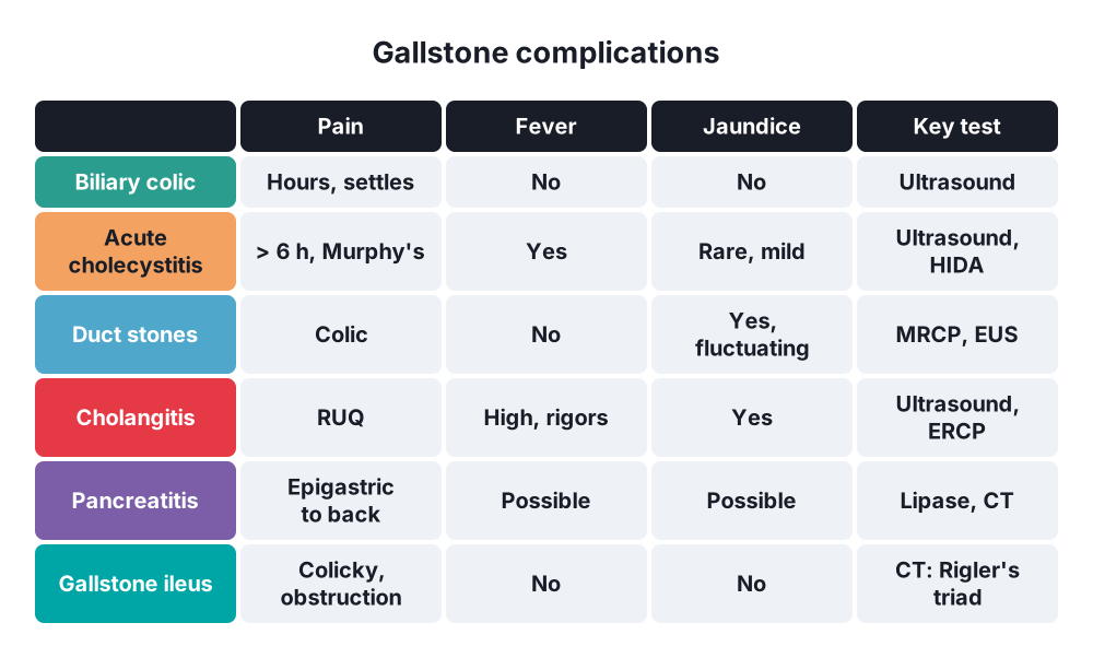
- Biliary colic:
- Constant (not truly colicky) pain in the right upper quadrant or epigastrium, lasting over 30 minutes and under 6 hours
- Often after fatty meals or at night
- Radiates to the right scapula or shoulder
- Nausea, vomiting
- No fever, settles completely
- Vague "flatulent dyspepsia" (bloating, belching) → not reliably due to stones
- CBD stones → colic, jaundice, dark urine, pale stools, itch; may come and go (ball valve)
- Gallstone ileus → elderly woman; intermittent small bowel obstruction ("tumbling" stone)
Signs
- Colic → mild RUQ tenderness during attacks; Murphy's sign negative (no inflammation)
- Jaundice → yellow sclera; gallbladder usually not palpable (Courvoisier's law: a palpable gallbladder with jaundice is unlikely to be from stones, because a stone-filled gallbladder is fibrotic)
- Mucocele → large, smooth, non-tender gallbladder
7. Diagnosis
- Ultrasound → first test (sensitivity over 95% for gallbladder stones):
- Echogenic foci with acoustic shadowing, move with position
- Gallbladder wall, CBD diameter (normal under 6–7 mm; larger after cholecystectomy and in the elderly)
- Less good for CBD stones (about 50%)
- Bloods → LFT (bilirubin, ALP, GGT, transaminases), FBC, CRP, amylase or lipase
- MRCP → CBD stones when LFT or ultrasound suggest them (sensitivity over 90%)
- Endoscopic ultrasound (EUS) → small CBD stones, microlithiasis; as accurate as MRCP
- ERCP → therapy, not diagnosis (pancreatitis risk)
- CT → complications (perforation, abscess, pancreatitis), gallstone ileus (Rigler's triad: small bowel obstruction, pneumobilia, ectopic stone)
- HIDA scan → acute cholecystitis (non-filling), biliary dyskinesia (ejection fraction under 35–40%)
- Intraoperative cholangiogram or laparoscopic ultrasound → CBD stones during surgery
- Risk of CBD stone (ASGE): high → stone seen in the CBD, cholangitis, bilirubin over 68 µmol/L with dilated CBD → straight to ERCP; intermediate → MRCP or EUS
8. Differential diagnosis
- Peptic ulcer, gastritis, functional dyspepsia
- Acute pancreatitis
- Myocardial infarction (inferior), pericarditis
- Right lower lobe pneumonia
- Hepatitis, liver abscess, Fitz-Hugh-Curtis syndrome
- Renal colic, pyelonephritis
- Irritable bowel syndrome, sphincter of Oddi dysfunction
9. Treatment
Conservative
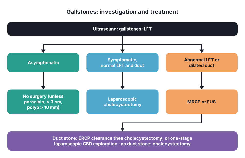
- Asymptomatic gallstones → no surgery (expectant management); exceptions:
- Porcelain gallbladder, stones over 3 cm, polyps over 10 mm (cancer risk)
- Sickle cell disease, during other operations in selected cases, before transplant
- Populations with high gallbladder cancer risk
- Biliary colic → analgesia (NSAIDs first, e.g. diclofenac; opioids), antiemetics; low-fat diet until surgery
- Ursodeoxycholic acid → dissolves small cholesterol stones in a functioning gallbladder; slow, recurrence 50% at 5 years; used after bariatric surgery to prevent stones
- ESWL (shock wave lithotripsy) → historical
- CBD stones:
- ERCP with sphincterotomy and stone extraction (balloon or basket) → over 90% clearance
- Large stones → mechanical lithotripsy, cholangioscopy with laser or electrohydraulic lithotripsy, large-balloon sphincteroplasty
- Failed clearance → plastic stent for drainage, then retry or surgery
- Unfit elderly → stent alone
Surgical
- Laparoscopic cholecystectomy → treatment of choice for symptomatic stones:
- Pneumoperitoneum; 4 ports (umbilical camera, epigastric, two right subcostal)
- Retract the fundus over the liver, Hartmann's pouch laterally
- Open the peritoneum over Calot's triangle
- Achieve the critical view of safety (Strasberg): Calot's triangle cleared of fat, lower third of the gallbladder off the liver bed, two and only two structures entering the gallbladder
- Clip and divide the cystic artery and cystic duct
- Dissect the gallbladder from the liver bed; remove in a bag
- Intraoperative cholangiogram → routine or selective (unclear anatomy, CBD stones suspected)
- Timing → elective; within 2 weeks for colic (NICE suggests soon after diagnosis to avoid readmission)
- Bail-out options when the critical view cannot be achieved → subtotal (fenestrating or reconstituting) cholecystectomy, fundus-first dissection, cholecystostomy, conversion to open
- Open cholecystectomy → Kocher subcostal incision; now for conversion, previous surgery, suspected cancer
- CBD stones found at surgery:
- Laparoscopic CBD exploration (transcystic or choledochotomy with T-tube or primary closure) → one-stage, shorter stay
- Or postoperative ERCP (two-stage); equal clearance
- Open CBD exploration → large stones, failed endoscopy; choledochoduodenostomy or hepaticojejunostomy for many primary duct stones
- Mirizzi → open or careful laparoscopic subtotal cholecystectomy; fistula → repair or hepaticojejunostomy
- Gallstone ileus → laparotomy, enterolithotomy (proximal to the stone), check for other stones; fistula repair later or never (elderly)
- After gallstone pancreatitis → cholecystectomy in the same admission (mild cases)
10. Complications
Of gallstones
- Acute cholecystitis, empyema, gangrene, perforation
- Obstructive jaundice, cholangitis, acute pancreatitis
- Mirizzi syndrome, cholecystoenteric fistula, gallstone ileus
- Gallbladder cancer
Of cholecystectomy
- Bile duct injury (0.3–0.5% laparoscopic; about twice the open rate) → most serious; Strasberg classification A–E
- Bile leak (cystic duct stump, ducts of Luschka) → ERCP and stent
- Bleeding (cystic or right hepatic artery, liver bed)
- Retained CBD stones, spilled stones (abscess)
- Bowel injury (port), port-site hernia
- Postcholecystectomy syndrome → persistent pain (retained stone, sphincter dysfunction, missed other cause)
- Diarrhoea (bile acids)
- ERCP → pancreatitis (3–5%), bleeding, perforation, cholangitis
Show as a list
Gallstones
- Types
- Cholesterol
- Mixed
- Black: haemolysis
- Brown: infection
- Risk
- Female, oestrogen
- Obesity
- Rapid weight loss
- Ileal disease
- Complications
- Colic
- Cholecystitis
- Duct stones, jaundice
- Cholangitis
- Pancreatitis
- Mirizzi
- Gallstone ileus
- Tests
- Ultrasound
- LFT
- MRCP, EUS
- ERCP: therapy
- Treatment
- Silent: leave
- Lap cholecystectomy
- Critical view of safety
- ERCP or CBD exploration
- Risks
- Bile duct injury
- Bile leak


