54. Femoral hernia
High yield
Hernia through the femoral canal below and lateral to the pubic tubercle; often strangulates, so all are repaired.
Exam Prep · State examSurgery · General surgerySurgery · Emergency and traumaSurgical Anatomy · Abdominal wall and hernia
1. Definition
- Femoral hernia → protrusion of abdominal contents through the femoral ring into the femoral canal, below the inguinal ligament
- Emerges at the saphenous opening and turns up over the inguinal ligament
2. Epidemiology
- About 5% of groin hernias; third commonest primary hernia
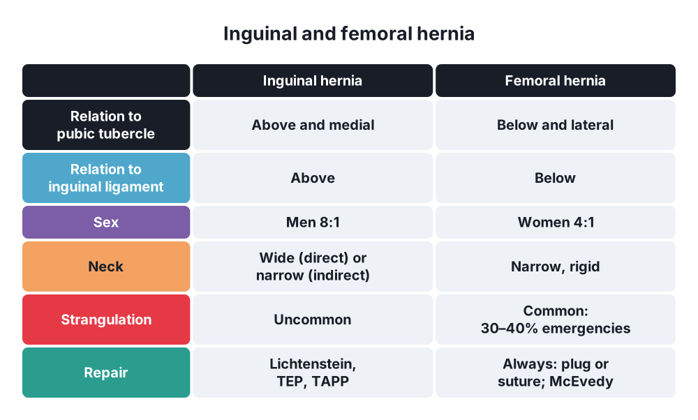
- Women : men about 4 : 1; still, inguinal hernia is more common than femoral hernia in women
- About 20% of hernias in women, 5% in men
- Older age, multiparous women
- Right side more common
- 30–40% present as an emergency (incarceration or strangulation); up to half of these need bowel resection
- Rare in children
3. Aetiology and risk factors
Anatomy (revision)
- Femoral sheath → three compartments: lateral (femoral artery), intermediate (femoral vein), medial (femoral canal)
- Femoral canal → about 1.25 cm long; contains fat, lymphatics and the lymph node of Cloquet
- Femoral ring (upper opening) → boundaries:
- Anterior → inguinal ligament
- Posterior → pectineal (Cooper's) ligament, pectineus fascia, pubic bone
- Medial → lacunar ligament
- Lateral → femoral vein
- Three sides are rigid → narrow, unyielding neck
- Abnormal (accessory) obturator artery → branch of the inferior epigastric crossing the lacunar ligament in about 25–30% → bleeding if the lacunar ligament is cut blindly ("corona mortis")
Causes
- Acquired → raised abdominal pressure on a weak area
- Wider female pelvis and femoral canal
- Pregnancy, obesity, constipation, chronic cough
- Age → loss of muscle bulk around the canal
- Previous inguinal hernia repair (missed or new femoral hernia)
4. Pathogenesis
- Extraperitoneal fat pushed into the femoral ring
- Fat drags peritoneum → sac
- Sac passes down the canal (medial to the femoral vein)
- Exits at the saphenous opening (cribriform fascia)
- Turns upwards and forwards over the inguinal ligament (looks like an inguinal hernia)
- Narrow rigid neck → contents trapped → strangulation
- Richter's hernia common → part of the bowel wall trapped; may strangulate without obstruction
5. Classification
- By course → typical; prevascular (in front of the vessels); retrovascular; Laugier's (through the lacunar ligament)
- EHS → F (femoral)
- By state → reducible, irreducible, obstructed, strangulated
- Special contents → appendix (De Garengeot's hernia), bladder, ovary
6. Clinical picture
Symptoms
- Small groin lump; often no symptoms until it complicates
- Many first present with bowel obstruction or a painful, tender lump
- Elderly woman with small bowel obstruction → always examine the groins
- Richter's → vague pain, vomiting, tender lump, but bowel may still work
Signs
- Lump below and lateral to the pubic tubercle, in the upper medial thigh
- Lies medial to the femoral pulse
- Small, firm, round; often irreducible (fat, narrow neck)
- Cough impulse often absent
- Strangulated → tender, tense, red, irreducible; signs of obstruction
7. Diagnosis
- Clinical
- Ultrasound → confirms and separates from nodes, varix, aneurysm
- CT → obstruction, obese patients, uncertain lump
- Emergency → FBC, U&E, lactate, group and save; abdominal X-ray (dilated small bowel)
- If in doubt → explore (risk of strangulation)
8. Differential diagnosis
- Inguinal hernia → above and medial to the pubic tubercle
- Inguinal (femoral) lymph node → Cloquet's node, lymphadenitis
- Saphena varix → soft, disappears on lying down, cough thrill, varicose veins
- Femoral artery aneurysm → pulsatile
- Psoas abscess → lateral to the femoral artery, fluctuant, back pain
- Lipoma
- Ectopic testis, hydrocele of a femoral sac (canal of Nuck in women)
9. Treatment
Conservative
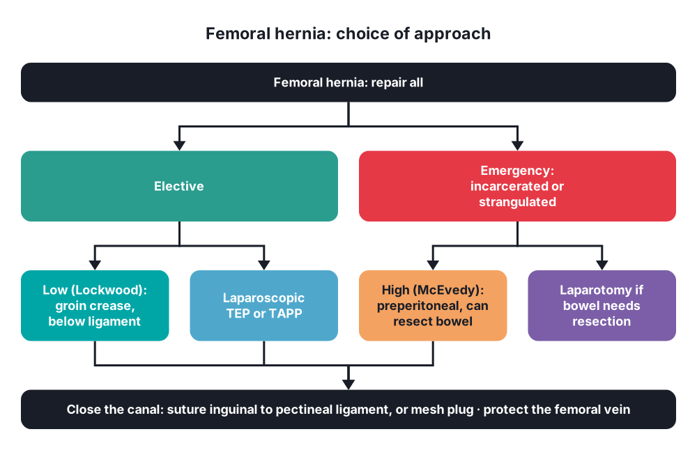
- No place → every femoral hernia is repaired (high strangulation risk); no truss
- Emergency → resuscitate first: IV fluids, NG tube, catheter, analgesia, antibiotics
Surgical
- Principles:
- Reduce or excise the sac, deal with the contents
- Close the femoral canal → suture the inguinal ligament to the pectineal ligament, or place a mesh plug
- Do not narrow the femoral vein (laterally)
- Low (infrainguinal, Lockwood) approach:
- Incision in the groin crease below the inguinal ligament
- Simple; good for elective repair
- Poor access for bowel resection
- High (inguinal, Lotheissen) approach:
- Through the inguinal canal, opening its posterior wall
- Access to the femoral ring from above; weakens the inguinal floor
- High (McEvedy) approach:
- Vertical or oblique incision over the femoral canal, extended above the inguinal ligament; preperitoneal approach
- Best for emergencies → can open the peritoneum and resect bowel
- Laparoscopic (TEP or TAPP) → elective repair; mesh covers the femoral ring with the inguinal orifices
- Strangulation:
- Do not let the contents slip back unseen → open the sac first
- Tight neck → divide the inguinal ligament or carefully the lacunar ligament (watch for an abnormal obturator artery)
- Non-viable bowel → resection via the preperitoneal space or laparotomy
- Contaminated field → sutured repair rather than mesh
10. Complications
- Strangulation, bowel gangrene, perforation, peritonitis; mortality higher than inguinal hernia
- Bleeding → abnormal obturator artery, femoral vein injury
- Femoral vein compression or thrombosis (repair too tight)
- Recurrence (about 2–10%)
- Wound infection, haematoma
- Bladder injury (medial)
Show as a list
Femoral hernia
- Anatomy
- Femoral canal
- Lacunar ligament medial
- Femoral vein lateral
- Cloquet's node
- Corona mortis
- Clinical
- Older women
- Below, lateral to tubercle
- Often irreducible
- Strangulation 30-40%
- Richter's
- Differential
- Inguinal hernia
- Lymph node
- Saphena varix
- Aneurysm
- Repair
- All repaired
- Low: Lockwood
- High: Lotheissen
- McEvedy: emergency
- Laparoscopic
- Plug or suture


