57. Diaphragmatic and internal hernias
High yield
Hiatus, congenital and traumatic diaphragmatic hernias, and hernias into peritoneal pockets that cause closed-loop obstruction.
Exam Prep · State examSurgery · General surgerySurgery · Upper GIAnatomy · Abdomen
1. Definition
- Diaphragmatic hernia → passage of abdominal organs into the chest through a defect in the diaphragm
- Hiatus hernia → through the oesophageal hiatus
- Congenital diaphragmatic hernia (CDH) → Bochdalek (posterolateral) or Morgagni (anterior, retrosternal)
- Traumatic → rupture after blunt or penetrating injury
- Internal hernia → bowel passes through a normal or abnormal opening inside the peritoneal cavity (no external lump)
2. Epidemiology
- Hiatus hernia → very common; increases with age (over 50% over 60 years on barium studies); obese, women
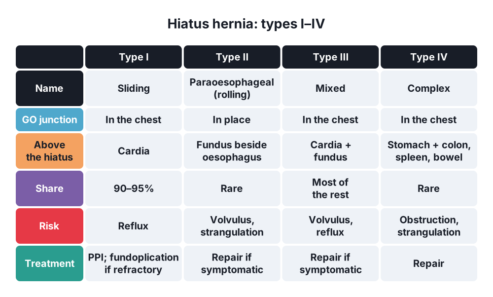
- Sliding (type I) → about 90–95%
- Paraoesophageal (types II–IV) → about 5–10%
- CDH → about 1 in 2,500–4,000 births; Bochdalek 85–90%, left side 80–85%; Morgagni 2–5%, usually right
- Traumatic rupture → 1–5% of major blunt trauma; left side in about 75% (liver protects the right)
- Internal hernia → under 1% of all hernias, but 0.5–5% of small bowel obstructions; common after Roux-en-Y gastric bypass (Petersen's)
3. Aetiology and risk factors
Anatomy (revision)
- Diaphragm → central tendon, muscle from sternum, ribs and lumbar spine (crura)
- Openings → T8 inferior vena cava, T10 oesophagus (and vagi), T12 aorta (and thoracic duct, azygos)
- Phreno-oesophageal ligament holds the gastro-oesophageal junction in the hiatus
- Foetal development → pleuroperitoneal membranes close at weeks 8–10; left side closes last → Bochdalek defects left
Causes
- Hiatus → age, obesity, pregnancy, raised abdominal pressure, weak phreno-oesophageal ligament, short oesophagus
- CDH → failure of the pleuroperitoneal canal to close; associated anomalies 30–40% (heart, chromosomes)
- Morgagni → defect at the sternocostal triangle (foramen of Morgagni, superior epigastric vessels)
- Traumatic → high-energy blunt trauma (car crash), stab wounds of the lower chest
- Internal hernia:
- Congenital pockets → paraduodenal (left and right), foramen of Winslow, pericaecal, intersigmoid, supravesical
- Acquired defects → mesenteric gaps after surgery: Petersen's space (behind the Roux limb), mesocolon, jejunojejunostomy, stomas, adhesive bands
4. Pathogenesis
- Sliding hernia → gastro-oesophageal junction moves up into the chest → loss of the lower oesophageal sphincter barrier → reflux
- Paraoesophageal (rolling) → junction stays in place; fundus rolls up beside the oesophagus → volvulus, obstruction, strangulation, bleeding (Cameron ulcers)
- CDH → bowel in the chest during foetal life → pulmonary hypoplasia (both lungs, worse on the side of the defect) and pulmonary hypertension → main cause of death
- Traumatic → sudden rise in abdominal pressure tears the diaphragm; it never heals (negative chest pressure pulls organs up); may present years later with strangulation
- Internal hernia → bowel enters a pocket → closed-loop obstruction → strangulation; symptoms come and go
5. Classification
Hiatus hernia
- Type I → sliding
- Type II → pure paraoesophageal (junction in place, fundus up)
- Type III → mixed (junction and fundus up)
- Type IV → other organs too (colon, spleen, small bowel)
Diaphragmatic hernia
- Congenital → Bochdalek, Morgagni, eventration (thin, high diaphragm with no defect), central tendon
- Acquired → hiatus, traumatic, iatrogenic
Internal hernia
- Paraduodenal → left (fossa of Landzert, behind the inferior mesenteric vein) more common than right (fossa of Waldeyer, behind the superior mesenteric artery)
- Foramen of Winslow (lesser sac)
- Transmesenteric, transmesocolic, Petersen's
- Pericaecal, intersigmoid, supravesical, broad ligament
6. Clinical picture
Symptoms
- Sliding → heartburn, regurgitation, worse lying down and bending
- Paraoesophageal → postprandial chest fullness, dysphagia, vomiting, breathlessness, anaemia; acute volvulus → severe pain
- Borchardt's triad (gastric volvulus) → epigastric pain, retching without vomiting, cannot pass an NG tube
- CDH (Bochdalek) → respiratory distress at birth, cyanosis
- Morgagni → often adults; vague chest or epigastric pain, found by chance
- Traumatic → breathlessness, chest pain; or late bowel obstruction
- Internal hernia → intermittent colicky pain, obstruction; after gastric bypass → pain out of proportion, often little vomiting
Signs
- CDH → scaphoid abdomen, barrel chest, bowel sounds in the chest, heart sounds shifted to the right
- Traumatic → reduced breath sounds, bowel sounds in the chest
- Internal hernia → few signs early; distension, tenderness later; peritonitis when strangulated
7. Diagnosis
- Hiatus hernia:
- Barium swallow → type and size
- Gastroscopy → oesophagitis, Barrett's, Cameron ulcers
- Manometry and 24-hour pH monitoring → before antireflux surgery
- CT → paraoesophageal and complicated hernias
- CDH:
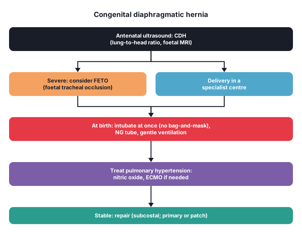
- Antenatal ultrasound (most diagnosed by 20–24 weeks); lung-to-head ratio and foetal MRI predict outcome
- Chest X-ray at birth → bowel loops in the chest, NG tube in the chest
- Echocardiography
- Traumatic → chest X-ray (raised hemidiaphragm, NG tube above it); CT; laparoscopy for left thoracoabdominal stab wounds (CT misses small tears)
- Internal hernia → CT: cluster of small bowel loops in an abnormal place, swirl sign of the mesentery, displaced vessels
8. Differential diagnosis
- Hiatus hernia → angina, peptic ulcer, achalasia, oesophageal cancer
- CDH → congenital lung cysts (CPAM), eventration, pneumothorax
- Traumatic → raised hemidiaphragm from phrenic nerve palsy, effusion, lung collapse
- Internal hernia → adhesive obstruction, volvulus, intussusception, mesenteric ischaemia
9. Treatment
Conservative
- Sliding hiatus hernia → treat the reflux: weight loss, raise the head of the bed, small meals, stop smoking and alcohol, proton pump inhibitors
- Small asymptomatic paraoesophageal hernia in an elderly patient → may be watched
- CDH → no emergency operation; stabilise first:
- Intubate at birth; no bag-and-mask ventilation (fills the bowel in the chest)
- NG tube to decompress
- Gentle ventilation, permissive hypercapnia
- Treat pulmonary hypertension (nitric oxide); ECMO if needed
- Foetal tracheal occlusion (FETO) in severe cases
Surgical: hiatus hernia
- Indications → reflux not controlled by drugs, complications, symptomatic paraoesophageal hernia
- Laparoscopic repair:
- Reduce the hernia and excise the sac
- Mobilise the oesophagus (at least 2–3 cm in the abdomen)
- Crural repair (posterior sutures, mesh in selected cases)
- Fundoplication → Nissen (360°) or partial (Toupet 270° posterior, Dor anterior)
- Short oesophagus → Collis gastroplasty
- Acute volvulus → decompress (NG or endoscope), emergency reduction; resect necrotic stomach; gastropexy in unfit patients
Surgical: congenital and traumatic
- CDH → repair when stable (days): abdominal (subcostal) approach; reduce, close primarily or with a patch; thoracoscopic repair in selected cases
- Morgagni → laparoscopic reduction and repair (sutures or mesh)
- Traumatic rupture → repair always (strangulation risk):
- Acute → laparotomy (other abdominal injuries common) or laparoscopy; non-absorbable sutures
- Late (chronic) → thoracotomy or thoracoscopy (adhesions in the chest)
Surgical: internal hernia
- Urgent operation (laparoscopy or laparotomy) → reduce the bowel, resect if dead, close the defect
- Paraduodenal → open the sac along the avascular side (avoid the inferior mesenteric vein or superior mesenteric artery in its edge)
- After gastric bypass → close Petersen's and mesenteric defects at the first operation (prevention)
10. Complications
- Hiatus hernia → oesophagitis, stricture, Barrett's oesophagus, bleeding, iron deficiency, gastric volvulus, strangulation
- Antireflux surgery → dysphagia, gas bloat, recurrence (up to 30% for large paraoesophageal hernias), slipped wrap
- CDH → survival about 70–90%; chronic lung disease, reflux, recurrence (patch), developmental delay
- Traumatic → strangulation of the stomach or colon, tension gastrothorax
- Internal hernia → strangulation, short bowel after resection
Show as a list
Diaphragmatic and internal hernias
- Hiatus
- Type I: sliding, reflux
- Type II-IV: rolling
- Volvulus
- Nissen, crural repair
- Congenital
- Bochdalek: back, left
- Morgagni: front, right
- Lung hypoplasia
- Stabilise, then repair
- Traumatic
- Left side
- Late strangulation
- Always repair
- Internal
- Paraduodenal
- Winslow
- Petersen's
- Closed loop
- CT swirl sign


