
85. Colon: congenital anomalies, diverticular disease, ulcerative colitis, tuberculosis and polyps
High yield
Benign colonic disease: malrotation and duplications, diverticulitis by Hinchey stage, ulcerative colitis, ileocaecal TB and polyps.
Exam Prep · State examSurgery · ColorectalSurgery · General surgerySurgery · Emergency and trauma
1. Definition
- Congenital anomalies of the colon → malrotation, duplication, Hirschsprung's disease (see question 106), anorectal malformations, colonic atresia, megacolon
- Diverticulosis → presence of diverticula (mucosal outpouchings through the muscle wall); diverticular disease → symptoms from them; diverticulitis → inflammation or infection of a diverticulum
- Ulcerative colitis (UC) → chronic relapsing mucosal inflammation that always starts in the rectum and spreads continuously up the colon
- Colonic tuberculosis → infection of the bowel by Mycobacterium tuberculosis, mainly the ileocaecal region
- Colonic polyp → any growth projecting from the mucosa into the lumen
2. Epidemiology
Congenital
- Malrotation → about 1 in 500 (symptomatic 1 in 6,000); most present in the first month
- Duplications → rare; ileum most common, colon about 15%
Diverticular disease
- Very common in the West → about 5% at 40, 30% at 60, over 60% at 80
- Sigmoid colon in 90–95% of Westerners; right-sided diverticula more common in Asia
- About 10–25% of people with diverticulosis develop diverticulitis (newer data: under 5%)
- About 15–20% of diverticulitis is complicated (abscess, perforation, fistula, stricture)
Ulcerative colitis
- Incidence 10–20 per 100,000 a year; peak 15–30 years and 50–70
- More in non-smokers and ex-smokers
- Extent at diagnosis → proctitis 30–50%, left-sided 30–40%, pancolitis 20%
- Colectomy in about 10–15% at 10 years
Colonic tuberculosis
- Abdominal TB → about 10% of extrapulmonary TB; ileocaecal region in 75–90% of GI TB
- Endemic regions (India, Africa, Asia), migrants, HIV
Polyps
- Adenomas in 25–40% of people over 50
- Hyperplastic polyps common in the rectosigmoid
- Most colorectal cancers arise from adenomas (adenoma-carcinoma sequence) or serrated lesions
3. Aetiology and risk factors
Congenital
- Malrotation → midgut fails to complete its 270° anticlockwise rotation around the superior mesenteric artery during weeks 5–12 → narrow mesenteric base + Ladd's bands across the duodenum
- Duplication → faulty recanalisation or diverticulum formation; on the mesenteric border, shares blood supply
Diverticular disease
- Low-fibre diet → small hard stools, high intracolonic pressure (segmentation)
- Age → weaker collagen and elastin in the wall
- Obesity, physical inactivity, smoking
- Drugs → NSAIDs, steroids, opioids (perforation)
- Connective tissue disease (Marfan's, Ehlers-Danlos), polycystic kidney disease
- Nuts, seeds and popcorn → no proven link (not banned)
Ulcerative colitis
- Genetic (family history 10–15%, HLA-B27 for spondylitis), dysregulated immune response (Th2)
- Non-smoking (smoking protective), appendicectomy before age 20 is protective
- Gut microbiota, Western diet
Colonic tuberculosis
- Swallowed infected sputum (active lung TB), unpasteurised milk (M. bovis), blood spread, nearby nodes
- Ileocaecal region → abundant lymphoid tissue (Peyer's patches), stasis, more absorption
- HIV, malnutrition, diabetes, immunosuppression (anti-TNF)
Polyps
- Age, family history, Western diet (red and processed meat), obesity, smoking, alcohol
- Polyposis syndromes → FAP (APC), MUTYH-associated polyposis, Lynch syndrome, Peutz-Jeghers (STK11), juvenile polyposis (SMAD4, BMPR1A), serrated polyposis
- Long-standing IBD (dysplasia)
4. Pathogenesis
Diverticula
- Pulsion "false" diverticula → mucosa and submucosa herniate through the muscle where the vasa recta penetrate (between the mesenteric and antimesenteric taeniae)
- Sigmoid → narrowest part, highest pressure (Laplace's law)
- Muscle thickening (myochosis) of the sigmoid
- Diverticulitis → micro-perforation of a diverticulum (no longer thought to be faecolith obstruction alone) → pericolic inflammation → phlegmon, abscess, free perforation
- Bleeding → vasa recta stretched over the dome of the diverticulum rupture → painless, large bleeding; often right-sided diverticula
- Fistula → abscess drains into a nearby organ: colovesical (most common), colovaginal (after hysterectomy), coloenteric, colocutaneous
- Stricture → repeated inflammation → fibrosis → obstruction
Ulcerative colitis
- Mucosal and submucosal inflammation only
- Crypt abscesses, goblet cell depletion, pseudopolyps (regenerating mucosa between ulcers)
- Continuous from the rectum (no skip lesions); backwash ileitis in pancolitis
- Chronic disease → shortened, featureless "lead pipe" colon (loss of haustra)
- Toxic megacolon → inflammation spreads to the muscle → paralysis → dilatation (transverse colon over 6 cm) → perforation
- Long-standing → dysplasia → colorectal cancer
Colonic tuberculosis
- Ulcerative → transverse (circumferential) ulcers along lymphatics; heal with strictures
- Hypertrophic → thick fibrotic mass in the caecum (mimics cancer)
- Ulcerohypertrophic → both
- Caseating granulomas, pulled-up caecum, incompetent ileocaecal valve
Polyps
- Adenoma-carcinoma sequence (Vogelstein) → APC loss → KRAS activation → 18q (SMAD4, DCC) loss → TP53 loss → carcinoma (about 10 years)
- Serrated pathway → BRAF mutation, CpG island methylation (CIMP), MLH1 silencing → right colon cancers
- Malignant risk rises with size (over 1 cm), villous histology, high-grade dysplasia, number (3 or more)
5. Classification
Congenital
- Malrotation (non-rotation, incomplete rotation, reversed rotation)
- Duplications (cystic, tubular)
- Atresia, Hirschsprung's disease, anorectal malformations, congenital megacolon
Diverticular disease
- Asymptomatic diverticulosis
- Symptomatic uncomplicated diverticular disease (SUDD)
- Acute diverticulitis → uncomplicated or complicated
- Chronic or recurrent diverticulitis
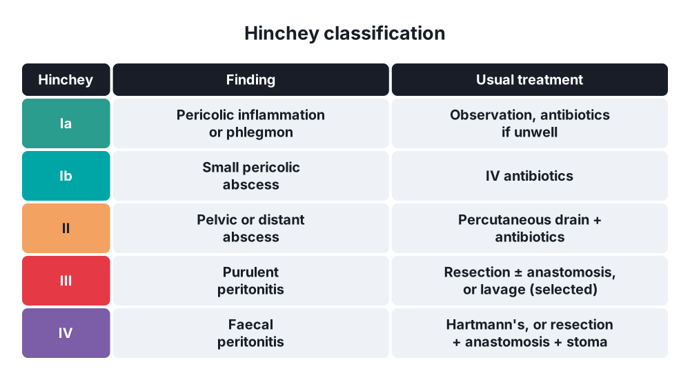
- Hinchey classification (complicated diverticulitis):
- Ia → pericolic inflammation or phlegmon
- Ib → pericolic abscess (small, confined)
- II → pelvic, distant intra-abdominal or retroperitoneal abscess
- III → generalised purulent peritonitis (abscess ruptured; no communication with the lumen)
- IV → generalised faecal peritonitis (open communication with the lumen)
- Modified Hinchey (CT-based) adds stage 0 (mild clinical diverticulitis)
Ulcerative colitis
- Montreal extent → E1 proctitis, E2 left-sided (to the splenic flexure), E3 extensive (pancolitis)
- Truelove and Witts severity → severe = 6 or more bloody stools a day plus one of: temperature over 37.8 °C, pulse over 90/min, haemoglobin under 105 g/L, ESR over 30 mm/h
- Mayo score (clinical and endoscopic)
Colonic polyps
- Neoplastic → adenomas: tubular (most common, lowest risk), tubulovillous, villous (highest risk); sessile serrated lesions, traditional serrated adenomas
- Non-neoplastic → hyperplastic, inflammatory (pseudopolyps), hamartomatous (juvenile, Peutz-Jeghers), lymphoid
- Shape (Paris) → pedunculated (Ip), sessile (Is), flat (IIa, IIb, IIc)
- Haggitt levels (invasion in a pedunculated malignant polyp) and Kikuchi levels (sessile: sm1–sm3)
6. Clinical picture
Malrotation
- Newborn → bilious vomiting (midgut volvulus until proven otherwise), distension, bloody stools, shock
- Older children and adults → intermittent pain, vomiting, malabsorption
Diverticulitis
- Left iliac fossa pain ("left-sided appendicitis"), constant
- Fever, anorexia, nausea
- Change in bowel habit (constipation or diarrhoea)
- Tender LIF, guarding, palpable mass (phlegmon or abscess)
- Peritonitis → generalised tenderness, rigidity, sepsis (Hinchey III–IV)
- Fistula → pneumaturia, faecaluria, recurrent urinary infections (colovesical); faeces through the vagina
- Stricture → large bowel obstruction
- Diverticular bleeding → painless, sudden, large fresh red or maroon rectal bleeding; stops on its own in about 75–80%
Ulcerative colitis
- Bloody diarrhoea with mucus, urgency, tenesmus
- Lower abdominal cramps before defecation
- Fever, weight loss, malaise in severe disease
- Proctitis → rectal bleeding, often with constipation
- Acute severe colitis → 6 or more bloody stools, tachycardia, fever, anaemia
- Toxic megacolon → distension, tenderness, sepsis, fewer stools
- Extraintestinal → primary sclerosing cholangitis (much more than in Crohn's), erythema nodosum, pyoderma gangrenosum, episcleritis, uveitis, arthritis, ankylosing spondylitis, VTE
Colonic tuberculosis
- Chronic RIF pain, weight loss, fever, night sweats, anorexia
- Altered bowel habit, subacute obstruction (strictures)
- RIF mass (doughy, matted)
- Ascites (peritoneal TB), lung TB signs
Polyps
- Usually no symptoms → found on screening
- Rectal bleeding, anaemia
- Villous adenoma (large, rectal) → mucus discharge, hypokalaemia (secretory diarrhoea)
- Intussusception (large polyp), prolapse (pedunculated rectal polyp)
7. Diagnosis
Malrotation
- Upper GI contrast study → duodenojejunal flexure to the right of the spine and below the pylorus; corkscrew duodenum in volvulus
- Ultrasound → whirlpool sign, reversed SMA and SMV position (SMV left of SMA)
Diverticulitis
- Bloods → WCC, CRP, U&E, lactate
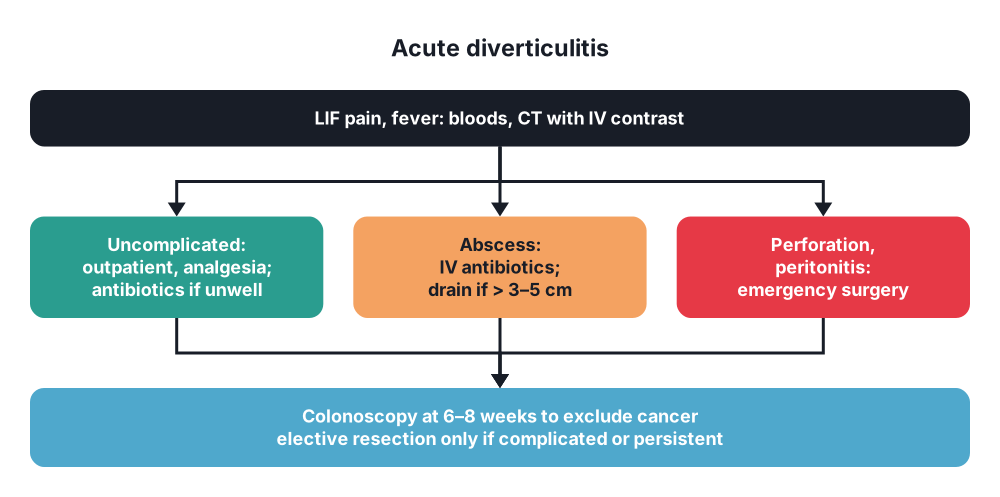
- CT with IV contrast → test of choice: diverticula, bowel wall thickening, fat stranding, abscess, free gas, fistula, Hinchey stage
- Ultrasound → alternative in experienced hands
- Colonoscopy 6–8 weeks after the attack → exclude cancer (especially after complicated disease, or if CT is unclear)
- Avoid colonoscopy during the acute attack (perforation risk)
- Fistula → CT (gas in the bladder), cystoscopy, poppy seed test
- Bleeding → CT angiography (active bleeding over 0.5 mL/min), colonoscopy after bowel prep, mesenteric angiography with embolisation, red cell scan
Ulcerative colitis
- Bloods → FBC, CRP, ESR, albumin, U&E, LFT (PSC)
- Stool cultures, C. difficile toxin, CMV (in steroid-resistant colitis)
- Faecal calprotectin
- Flexible sigmoidoscopy (acute severe colitis) or colonoscopy with biopsies → continuous inflammation from the rectum, loss of vascular pattern, friability, contact bleeding, ulcers, pseudopolyps
- Plain abdominal X-ray in acute severe colitis → colonic dilatation (over 5.5–6 cm), mucosal islands, thumbprinting, lead pipe colon
- Chest X-ray → perforation
- Surveillance colonoscopy from 8 years after onset (for extent beyond proctitis), then every 1–5 years by risk; yearly with PSC
Colonic tuberculosis
- Colonoscopy with biopsy → transverse ulcers, nodules, deformed ileocaecal valve; caseating granulomas, acid-fast bacilli, culture, PCR (GeneXpert)
- CT → ileocaecal thickening, necrotic nodes (low-density centres), ascites, omental thickening
- Barium (historical) → Stierlin sign (rapid emptying of an inflamed caecum), Fleischner sign (inverted umbrella: thick ileocaecal valve, narrow ileum), pulled-up conical caecum, goose-neck deformity
- Chest X-ray (active or old TB in about 25–50%), IGRA, ascitic fluid adenosine deaminase (ADA), HIV test
Polyps
- Colonoscopy → gold standard: find, remove and examine
- CT colonography → screening alternative, frail patients
- Faecal immunochemical test (FIT) → screening (positive → colonoscopy)
- Histology → type, dysplasia grade, margins, invasion depth (Haggitt, Kikuchi)
- Genetic testing → 10 or more adenomas (FAP, MAP), Lynch features
8. Differential diagnosis
- Diverticulitis → colorectal cancer (perforated or obstructing), ischaemic colitis, IBD, IBS, ovarian pathology, ureteric colic, appendicitis (redundant sigmoid on the right)
- Ulcerative colitis → Crohn's colitis, infective colitis (C. difficile, Campylobacter, Shigella, amoebiasis, CMV), ischaemic colitis, radiation proctitis, microscopic colitis
- Colonic TB → Crohn's disease (key), caecal carcinoma, lymphoma, amoeboma, actinomycosis, appendix mass
- Polyps → cancer, lipoma, submucosal tumours, inflammatory polyps
9. Treatment
Conservative
- Malrotation → none; it is a surgical condition
- Diverticulosis → high-fibre diet, fluids, exercise, weight loss, stop smoking; nuts and seeds are allowed
- Uncomplicated diverticulitis:
- Outpatient care in most → oral fluids, analgesia (paracetamol; avoid NSAIDs and opioids if possible)
- Antibiotics not routinely needed in immunocompetent patients (AVOD, DIABOLO trials)
- Antibiotics for: immunosuppression, sepsis, comorbidity, high CRP, pregnancy, failure to improve → co-amoxiclav, or cefuroxime with metronidazole
- Admit if → severe pain, cannot tolerate oral intake, sepsis, complicated disease, frailty
- Mesalazine and rifaximin → no proven benefit in prevention
- Diverticular abscess:
- Small (under 3–5 cm) → IV antibiotics alone
- Larger (over 3–5 cm) → percutaneous CT-guided drainage + antibiotics
- Diverticular bleeding → resuscitation; most stop; colonoscopic haemostasis (clips, adrenaline), angioembolisation
- Ulcerative colitis:
- Proctitis → topical mesalazine (suppositories); topical steroids
- Left-sided and extensive mild-moderate → oral + topical 5-ASA (mesalazine); add oral prednisolone if no response
- Maintenance → 5-ASA (also lowers cancer risk); thiopurines (steroid-dependent), biologics (infliximab, adalimumab, golimumab, vedolizumab, ustekinumab), JAK inhibitors (tofacitinib, upadacitinib), S1P modulators (ozanimod)
- Acute severe colitis (Truelove and Witts):
- Admit, joint medical-surgical care
- IV hydrocortisone 100 mg four times daily (or methylprednisolone)
- IV fluids, potassium, VTE prophylaxis (high risk), stool tests, avoid opioids, anticholinergics and NSAIDs
- Daily bloods, abdominal X-ray
- Day 3 assessment (Oxford/Travis index) → over 8 stools a day, or 3–8 stools with CRP over 45 mg/L → 85% will need colectomy
- Rescue therapy → infliximab or ciclosporin
- No response in 5–7 days → colectomy
- Colonic TB → antituberculous treatment for 6 months (2 months HRZE + 4 months HR); good response in most, even strictures may improve
- Polyps → endoscopic removal (polypectomy):
- Cold snare for small polyps
- Endoscopic mucosal resection (EMR) for large sessile polyps; endoscopic submucosal dissection (ESD) for selected lesions
- Surveillance colonoscopy by risk (BSG 2020): high-risk findings (2 or more premalignant polyps with 1 advanced, or 5 or more premalignant polyps) → colonoscopy at 3 years; low risk → back to screening
- Malignant polyp with favourable features (clear margin over 1 mm, well differentiated, no lymphovascular invasion, Haggitt 1–3, sm1) → endoscopic removal is enough
Surgical
- Malrotation → Ladd's procedure (urgent; emergency if volvulus):
- Untwist the volvulus counterclockwise
- Divide Ladd's bands over the duodenum
- Widen the mesenteric base
- Appendicectomy (caecum ends in the left upper quadrant)
- Place small bowel on the right, colon on the left (non-rotation position)
- Duplications → excision with the shared bowel segment
- Diverticulitis:
- Hinchey III (purulent peritonitis) → laparoscopic lavage (selected patients; higher reintervention) or sigmoid resection (primary anastomosis with or without a defunctioning ileostomy, preferred in stable patients) or Hartmann's procedure
- Hinchey IV (faecal peritonitis) → Hartmann's procedure (sigmoid colectomy, end colostomy, closed rectal stump) in unstable or high-risk patients; resection with primary anastomosis and ileostomy in stable patients (DIVERTI, LADIES trials)
- Damage control → resection with stapled ends, laparostomy, relook in 24–48 hours
- Elective sigmoid colectomy → individualised; not after every second attack; for complicated disease (fistula, stricture), persistent symptoms (smouldering disease), immunosuppressed patients after a severe attack, or when cancer cannot be excluded
- Resection margins → proximal on soft, non-thickened colon, distal on the upper rectum (below the taeniae; lowers recurrence)
- Colovesical fistula → sigmoid resection with primary anastomosis, repair of the bladder (simple closure), catheter for 7–10 days
- Massive bleeding not controlled → segmental colectomy if the site is known; subtotal colectomy if unknown
- Ulcerative colitis:
- Indications → acute severe colitis not responding to steroids and rescue therapy, toxic megacolon, perforation, massive bleeding, chronic disease not controlled by drugs or drug side effects, dysplasia or cancer, growth failure in children
- Emergency → subtotal colectomy with end ileostomy (rectal stump left, or brought out as a mucous fistula); no pouch in the emergency
- Elective (cure) → restorative proctocolectomy with ileal pouch-anal anastomosis (IPAA, J-pouch):
- Removes the whole colon and rectum; pouch made from the terminal ileum, joined to the anal canal
- Often staged (2 or 3 stages) with a loop ileostomy
- 4–7 bowel movements a day; continence good in most
- Panproctocolectomy with permanent end ileostomy → older patients, poor sphincters, patient choice
- Ileorectal anastomosis → selected patients with spared rectum (needs surveillance)
- Kock continent ileostomy → rare
- Colonic TB → surgery for complications:
- Obstruction → stricturoplasty (short strictures) or limited ileocaecal resection
- Perforation, abscess, fistula, massive bleeding
- Doubt with cancer → resection
- Continue antituberculous drugs after surgery
- Polyps:
- Surgical resection → polyps that cannot be removed endoscopically, malignant polyps with unfavourable features (positive margin, poor differentiation, lymphovascular invasion, Haggitt 4, deep submucosal sm3) → oncological colectomy with lymph nodes (see question 86)
- FAP → prophylactic restorative proctocolectomy with IPAA (most), or total colectomy with ileorectal anastomosis (few rectal polyps), usually in the late teens to early 20s; lifelong duodenal surveillance
- Lynch syndrome → colonoscopy every 1–2 years from age 25; colectomy for cancer (extended colectomy considered)
10. Complications
Malrotation
- Midgut volvulus → ischaemia of the whole midgut, short bowel syndrome, death
Diverticular disease
- Abscess, perforation (purulent or faecal peritonitis)
- Fistula (colovesical most common)
- Stricture → large bowel obstruction
- Bleeding (most common cause of major lower GI bleeding in adults)
- Recurrence after a first attack → about 15–30%
- Surgery → anastomotic leak, stoma complications, Hartmann's reversal never done in up to 40–50%
Ulcerative colitis
- Toxic megacolon, perforation, massive bleeding
- Colorectal cancer → risk rises with duration (over 8–10 years), extent (pancolitis), PSC, family history, ongoing inflammation; about 1–2% at 10 years, up to 8–18% at 30 years
- PSC and cholangiocarcinoma
- Pouchitis (most common pouch complication; 20–50%), pouch failure, cuffitis
- Anaemia, osteoporosis (steroids), VTE
Colonic TB
- Strictures, obstruction, perforation, fistula, bleeding, malabsorption
- Paradoxical reaction on treatment (strictures tighten)
Polyps
- Malignant change
- Bleeding, intussusception
- Polypectomy → post-polypectomy bleeding (immediate or up to 2 weeks), perforation, post-polypectomy syndrome (transmural burn: pain, fever, no free air)
Show as a list
Colon: benign disease
- Congenital
- Malrotation: Ladd's bands
- Bilious vomiting
- Ladd's procedure
- Duplications
- Diverticular disease
- Sigmoid, false diverticula
- Low fibre, age
- CT: Hinchey
- Ia-Ib: antibiotics
- II: drain
- III: lavage or resection
- IV: Hartmann's
- Colonoscopy at 6-8 weeks
- Ulcerative colitis
- Rectum upwards, continuous
- Mucosal
- Truelove and Witts
- 5-ASA, steroids
- Infliximab, ciclosporin
- Subtotal colectomy
- IPAA pouch
- Cancer, PSC
- TB
- Ileocaecal
- Caseating granulomas
- Transverse ulcers
- ATT 6 months
- Polyps
- Adenomas: tubular, villous
- Serrated
- Polypectomy
- FAP: proctocolectomy


