
86. Colon cancer
High yield
Adenocarcinoma from adenomas; right-sided tumours bleed, left-sided obstruct; CT staging, complete mesocolic excision and adjuvant chemotherapy.
Exam Prep · State examSurgery · ColorectalSurgery · Surgical oncology
1. Definition
- Colon cancer → malignant tumour of the colon, from the caecum to the rectosigmoid junction; over 90% adenocarcinoma
- Colorectal cancer (CRC) → colon and rectal cancer together; rectal cancer is covered in question 100
- Others → mucinous adenocarcinoma, signet ring carcinoma, neuroendocrine tumours, lymphoma, GIST, squamous (rare)
2. Epidemiology
- Third most common cancer worldwide and the second cause of cancer death
- About two-thirds colon, one-third rectum
- Age → 90% over 50; median about 70; rising in under-50s (early-onset CRC)
- Men slightly more than women
- Site → sigmoid and rectosigmoid most common in the colon; right-sided cancers increasing with age and in women
- About 20–25% have metastases at diagnosis (liver most often)
- 5-year survival → stage I about 90%, stage II 80%, stage III 60–70%, stage IV 10–15%
- Screening finds cancers earlier and removes polyps
3. Aetiology and risk factors
Lifestyle and environment
- Red and processed meat, low fibre, high fat
- Obesity, physical inactivity, diabetes
- Smoking, alcohol
- Protective → fibre, exercise, aspirin (long-term), calcium, vitamin D
Personal history
- Adenomatous polyps (size, villous, dysplasia, number) and sessile serrated lesions
- Previous colorectal cancer
- Inflammatory bowel disease (UC and Crohn's colitis; duration, extent, PSC)
- Ureterosigmoidostomy (cancer at the anastomosis)
- Pelvic radiotherapy, acromegaly, cholecystectomy (weak)
- Streptococcus bovis (gallolyticus) bacteraemia and endocarditis → look for CRC
Family history and hereditary syndromes (5–10%)
- First-degree relative → risk doubled (higher if diagnosed under 50)
- Lynch syndrome (HNPCC) → most common hereditary CRC (about 3%):
- Mismatch repair (MMR) genes → MLH1, MSH2, MSH6, PMS2, EPCAM
- Autosomal dominant; right-sided, younger (mean 45), mucinous, many lymphocytes, MSI-high
- Lifetime CRC risk up to 50–80%; endometrial, ovarian, gastric, small bowel, urinary tract, pancreatic cancers
- Amsterdam II criteria (3-2-1 rule): 3 relatives with Lynch-associated cancer (one a first-degree relative of the other two), 2 successive generations, 1 diagnosed under 50; FAP excluded
- Revised Bethesda criteria → which tumours to test for MSI
- Familial adenomatous polyposis (FAP) → APC gene (chromosome 5q); hundreds to thousands of adenomas by the teens; 100% cancer by 40 without surgery; duodenal adenomas, desmoids, osteomas (Gardner's), brain tumours (Turcot's), CHRPE (retinal pigment)
- Attenuated FAP (under 100 polyps), MUTYH-associated polyposis (autosomal recessive)
- Peutz-Jeghers, juvenile polyposis, serrated polyposis syndrome
4. Pathogenesis
Molecular pathways
- Chromosomal instability (CIN) → about 70–85%: APC → KRAS → SMAD4/DCC (18q) → TP53 (adenoma-carcinoma sequence, Vogelstein); mostly left-sided
- Microsatellite instability (MSI) → about 15%: MMR deficiency (Lynch: germline; sporadic: MLH1 promoter methylation); right-sided; better prognosis in early stage; responds to immunotherapy
- Serrated pathway / CIMP → BRAF V600E mutation; sessile serrated lesions; right-sided
Spread
- Direct → through the bowel wall into the pericolic fat, adjacent organs (bladder, uterus, small bowel, abdominal wall, duodenum, kidney)
- Lymphatic → epicolic → paracolic → intermediate → central (apical) nodes along the feeding arteries
- Haematogenous → portal vein → liver (most common; 50% develop liver metastases), then lung; rectal cancer also to the lung through systemic veins
- Transcoelomic → peritoneal metastases (10–15%), ovaries (Krukenberg), ascites
- Implantation → anastomosis, wounds, port sites
Right vs left colon cancer
- Right → exophytic, polypoid, large lumen with liquid stool → anaemia and bleeding, late diagnosis; more MSI, BRAF, older women; worse prognosis in advanced stage
- Left → annular "apple-core", narrow lumen with solid stool → obstruction, change in bowel habit; more CIN, KRAS
5. Classification
TNM 8th edition
- Tis → carcinoma in situ, intramucosal (lamina propria)
- T1 → submucosa
- T2 → muscularis propria
- T3 → through the muscularis propria into pericolic tissue
- T4a → visceral peritoneum; T4b → invades other organs
- N1 → 1–3 nodes (N1a 1, N1b 2–3, N1c tumour deposits without nodes); N2 → 4 or more (N2a 4–6, N2b 7+)
- M1a → one organ; M1b → more than one organ; M1c → peritoneum
- At least 12 lymph nodes must be examined for staging
Stage groups
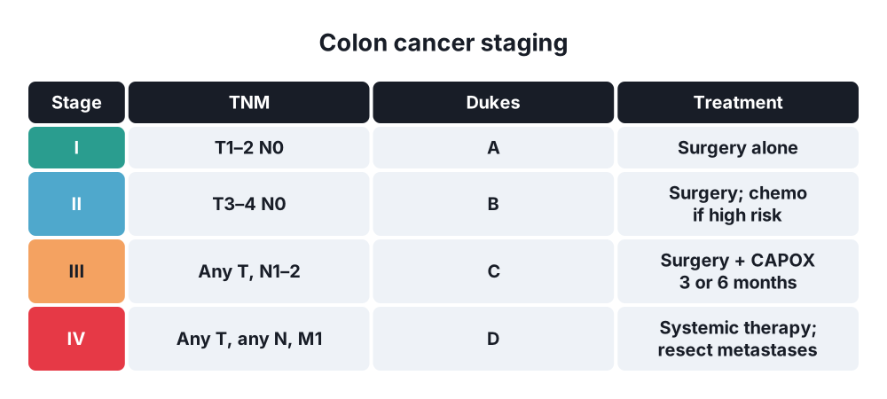
- Stage I → T1–T2 N0 M0
- Stage II → T3–T4 N0 M0
- Stage III → any T, N1–N2, M0
- Stage IV → any M1
Dukes' classification (historical, still asked)
- A → confined to the bowel wall (not beyond the muscularis propria) → 5-year survival about 90%
- B → through the bowel wall, nodes negative → about 70%
- C → lymph nodes positive → about 30–60% (C1 apical node free, C2 apical node involved)
- D → distant metastases (added by Turnbull) → under 10%
Histology
- Grade → well, moderately, poorly differentiated
- Mucinous (over 50% mucin), signet ring (poor)
- Lymphovascular invasion, perineural invasion, tumour budding, extramural venous invasion (EMVI) → adverse features
6. Clinical picture
Symptoms
- Change in bowel habit → looser or more frequent stools, or constipation, for weeks (left colon)
- Rectal bleeding → dark blood mixed with stool (left colon); occult in right colon
- Iron deficiency anaemia → tiredness, breathlessness (right colon)
- Abdominal pain, bloating, weight loss, anorexia
- Tenesmus, mucus (rectosigmoid)
- Presentation as an emergency in 20–30%:
- Large bowel obstruction (left-sided tumours; sigmoid most common)
- Perforation (at the tumour or proximal caecum from closed-loop obstruction)
- Bleeding (rarely massive)
- Fistula → pneumaturia (colovesical), faeculent vomiting (gastrocolic)
- Metastases → RUQ pain, jaundice, ascites
NICE urgent referral (2-week pathway) examples
- Age 40+ with unexplained weight loss and abdominal pain
- Age 50+ with unexplained rectal bleeding
- Age 60+ with iron deficiency anaemia or change in bowel habit
- Positive FIT (≥ 10 µg Hb/g faeces) with symptoms
- Abdominal or rectal mass
Signs
- Often none
- Pallor (anaemia)
- Abdominal mass (right iliac fossa in caecal cancer)
- Hepatomegaly (metastases), ascites, jaundice
- Rectal examination → low tumours, blood on the glove
- Distension, high-pitched bowel sounds (obstruction); peritonism (perforation)
- Virchow's node, Sister Mary Joseph nodule (advanced)
7. Diagnosis
Investigations to diagnose
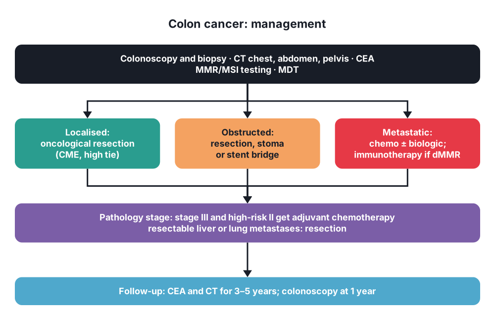
- Colonoscopy with biopsy → gold standard; checks the whole colon for synchronous tumours (3–5%) and polyps; tattoo the lesion (for laparoscopic surgery)
- CT colonography → frail patients, incomplete colonoscopy
- Flexible sigmoidoscopy → left-sided symptoms
- FIT → screening and triage of low-risk symptoms
Bloods
- FBC (microcytic anaemia), ferritin, U&E, LFT
- CEA → baseline before surgery (prognosis) and follow-up (recurrence); not for diagnosis or screening
Staging
- CT chest, abdomen and pelvis with contrast → T stage, nodes, liver and lung metastases, peritoneum
- MRI liver → characterise liver lesions, plan liver surgery
- PET-CT → selected (recurrence, before metastasectomy, rising CEA)
- Molecular testing of every tumour → MMR/MSI (Lynch screening, immunotherapy, prognosis), RAS (KRAS, NRAS) and BRAF (anti-EGFR treatment in metastatic disease), HER2
- Multidisciplinary team decision
Screening (examples)
- UK → FIT every 2 years from 50 to 74
- Europe and USA → FIT or colonoscopy every 10 years from 45–50
- Bulgaria → opportunistic screening; FIT programmes being introduced
- High risk → colonoscopy surveillance (Lynch every 2 years from 25; FAP yearly flexible sigmoidoscopy from 12–14)
- Faecal occult blood (guaiac) testing → older, less sensitive
8. Differential diagnosis
- Diverticular disease (stricture, mass), inflammatory bowel disease
- Ischaemic colitis, infectious colitis
- Haemorrhoids (do not blame bleeding on piles without examining the colon in older patients)
- Irritable bowel syndrome (younger, no alarm features)
- Appendix mass, ileocaecal TB, lymphoma
- Other causes of iron deficiency → gastric cancer, coeliac disease, menstrual loss
- Ovarian cancer (pelvic mass, ascites)
- Other causes of large bowel obstruction → volvulus, pseudo-obstruction (Ogilvie's), diverticular stricture
9. Treatment
Conservative and systemic
- Prehabilitation → exercise, nutrition, correct anaemia (IV iron), stop smoking
- ERAS (enhanced recovery) → no long fasting, carbohydrate drinks, minimal opioids, early feeding and mobilisation
- Bowel preparation → mechanical bowel preparation with oral antibiotics before elective colectomy lowers surgical site infection and leak (many guidelines)
- Adjuvant chemotherapy (after resection):
- Stage III → CAPOX (capecitabine + oxaliplatin) for 3 months (low-risk T1–3 N1) or 6 months (high risk: T4 or N2), or FOLFOX (IDEA collaboration)
- High-risk stage II (T4, perforation, obstruction, under 12 nodes, lymphovascular invasion, poor differentiation) → consider fluoropyrimidine ± oxaliplatin
- MSI-high stage II → no adjuvant 5-FU (no benefit; good prognosis)
- Older patients → capecitabine alone
- Neoadjuvant chemotherapy → locally advanced colon cancer (T4) on CT (FOxTROT trial: fewer incomplete resections)
- Neoadjuvant immunotherapy → dMMR / MSI-high tumours → very high response (NICHE-2) (emerging)
- Metastatic disease:
- Chemotherapy → FOLFOX, FOLFIRI, CAPOX, FOLFOXIRI
- Biologics → anti-EGFR (cetuximab, panitumumab) only in RAS wild-type and mainly left-sided tumours; anti-VEGF (bevacizumab)
- Immunotherapy (pembrolizumab) → first-line for MSI-high / dMMR
- BRAF V600E → encorafenib + cetuximab; HER2 → targeted therapy
- Conversion chemotherapy → may make liver metastases resectable
- Palliative care → symptom control, stents, best supportive care
Surgical: elective colectomy
- Principles of oncological resection:
- Resect the tumour with its lymphatic field → divide the feeding artery at its origin (high tie)
- Complete mesocolic excision (CME) with central vascular ligation (CVL) → dissect in the embryological plane and remove the mesocolon intact (Hohenberger); more nodes, possibly better survival
- Clear margins → at least 5 cm of bowel on each side; en bloc resection of any adherent organ (T4b; adhesions may be tumour)
- At least 12 nodes
- No-touch technique (debated), protect the wound
- Laparoscopic surgery → same cancer results as open (CLASICC and COST trials), faster recovery; robotic in some centres
- Operations by tumour site:
- Caecum, ascending colon, hepatic flexure → right hemicolectomy (ileocolic, right colic and right branch of the middle colic vessels); ileocolic anastomosis
- Transverse colon → extended right hemicolectomy (middle colic at origin) or transverse colectomy
- Splenic flexure → extended right hemicolectomy or left hemicolectomy (segmental splenic flexure resection in selected cases)
- Descending colon → left hemicolectomy (inferior mesenteric artery or left colic artery)
- Sigmoid colon → sigmoid colectomy / high anterior resection (IMA ligated at origin, often with splenic flexure mobilisation)
- Multiple tumours, Lynch syndrome, FAP → subtotal or total colectomy (± proctectomy)
- Anastomosis → stapled or hand-sewn; tension-free, good blood supply (ICG fluorescence angiography in some centres), no distal obstruction
- Defunctioning stoma → rarely needed in colon surgery (more in low rectal anastomoses)
Surgical: emergency presentations
- Obstructing right-sided cancer → right or extended right hemicolectomy with primary anastomosis (or end ileostomy if unstable)
- Obstructing left-sided cancer:
- Resection with primary anastomosis ± defunctioning ileostomy in fit, stable patients (with or without on-table lavage)
- Hartmann's procedure → unstable, frail, faecal peritonitis
- Subtotal colectomy with ileorectal anastomosis → caecal ischaemia or perforation, or synchronous tumours
- Self-expanding metal stent → bridge to surgery (convert emergency to elective; risk of perforation and tumour seeding) or palliation in incurable disease
- Loop colostomy or ileostomy → palliation or to allow neoadjuvant treatment
- Perforation → resection of the tumour (Hartmann's commonly), washout; worse prognosis (seeding)
- Bleeding → resection once stable; embolisation rarely
Surgical: metastatic and recurrent disease
- Liver metastases → resection if all disease can be removed with clear margins and enough future liver remnant (5-year survival 40–50%); synchronous or staged (liver-first) approaches; ablation for small lesions; portal vein embolisation
- Lung metastases → resection (metastasectomy) in selected patients
- Peritoneal metastases → cytoreductive surgery (± HIPEC) in selected patients with low peritoneal cancer index
- Primary tumour in incurable disease → resect only if symptomatic (obstruction, bleeding, perforation)
Follow-up
- CEA every 3–6 months and CT chest, abdomen and pelvis at intervals for 3–5 years
- Colonoscopy at 1 year after surgery (or within 6 months if the colon was not fully seen before), then at 3 and 5 years by findings
10. Complications
Of the cancer
- Large bowel obstruction → caecal perforation if the ileocaecal valve is competent (closed loop)
- Perforation, peritonitis, abscess
- Bleeding, anaemia
- Fistula (colovesical, colovaginal, gastrocolic, enteric)
- Invasion of nearby organs; ureteric obstruction
- Metastases → liver failure, jaundice, ascites, lung
- Cachexia
Of surgery
- Anastomotic leak → about 2–4% (right side), 5–10% (left side and low anastomoses); days 3–7; sepsis, peritonitis, ileus
- Surgical site infection, intra-abdominal abscess
- Bleeding, ileus (most common), small bowel obstruction
- Ureteric injury (left colectomy, sigmoid), splenic injury (splenic flexure), duodenal injury (right hemicolectomy)
- Incisional hernia, parastomal hernia
- VTE, pneumonia, cardiac events
- Bowel dysfunction (diarrhoea after right hemicolectomy, bile acid malabsorption)
- Low anterior resection syndrome (after sigmoid and rectal resections)
Of chemotherapy
- Oxaliplatin → peripheral neuropathy (cold-induced), may be permanent
- Capecitabine and 5-FU → hand-foot syndrome, diarrhoea, mucositis, DPD deficiency (test before starting: severe toxicity)
- Irinotecan → diarrhoea, neutropenia
- Cetuximab → acneiform rash; bevacizumab → bleeding, perforation, poor wound healing
Show as a list
Colon cancer
- Risk
- Age, diet, obesity
- Adenomas
- IBD
- Lynch: MMR, 3-2-1
- FAP: APC
- Pathways
- CIN: APC, KRAS, p53
- MSI: MMR
- Serrated: BRAF
- Clinical
- Right: anaemia, mass
- Left: obstruction, bleeding
- Emergency 20-30%
- Tests
- Colonoscopy + biopsy
- CT chest, abdomen, pelvis
- CEA baseline
- MMR, RAS, BRAF
- Staging
- TNM: I-IV
- Dukes A-D
- 12 nodes
- Surgery
- Right hemicolectomy
- Extended right
- Left hemicolectomy
- Sigmoid colectomy
- CME, high tie
- Laparoscopic
- Emergency
- Hartmann's
- Stent as bridge
- Chemotherapy
- Stage III: CAPOX, FOLFOX
- High-risk stage II
- Metastatic: anti-EGFR if RAS wild
- MSI-high: pembrolizumab
- Metastases
- Liver resection
- Lung resection
- CRS +/- HIPEC


